Kaempferol inhibited VEGF and PGF expression
and
in vitro
angiogenesis of HRECs under
diabetic-like environment
X.H. Xu
1,2*, C. Zhao
1*, Q. Peng
2, P. Xie
1and Q.H. Liu
11
Department of Ophthalmology, The First Affiliated Hospital of Nanjing Medical University, Nanjing City, Jiangsu Province, China 2
People’s Liberation Army 454 Hospital, Nanjing City, Jiangsu Province, China
Abstract
Diabetic retinopathy (DR) is one of the common and specific microvascular complications of diabetes. This study aimed to investigate the anti-angiogenic effect of kaempferol and explore its underlying molecular mechanisms. The mRNA expression level of vascular endothelial growth factor (VEGF) and placenta growth factor (PGF) and the concentrations of secreted VEGF and PGF were measured by qTR-PCR and ELISA assay, respectively. Human retinal endothelial cells (HRECs) proliferation, migration, and sprouting were measured by CCK-8 and transwell, scratching wound, and tube formation assays, respectively. Protein levels were determined by western blot. High glucose (25 mM) increased the mRNA expression levels of VEGF and PGF as well as the concentrations of secreted VEGF and PGF in HRECs, which can be antagonized by kaempferol (25mM). Kaempferol (5–25mM) significantly suppressed cell proliferation, migration, migration distance and sprouting of HRECs under high glucose condition. The anti-angiogenic effect of kaempferol was mediated via downregulating the expression of PI3K and inhibiting the activation of Erk1/2, Src, and Akt1. This study indicates that kaempferol suppressed angiogenesis of HRECs via targeting VEGF and PGF to inhibit the activation of Src-Akt1-Erk1/2 signaling pathway. The results suggest that kaempferol may be a potential drug for better management of DR.
Key words: Diabetic retinopathy; Kaempferol; VEGF; PGF; Anti-angiogenic
Introduction
Diabetic retinopathy (DR) is one of the common and specific microvascular complications of diabetes and is also the leading cause of new-onset blindness in the developed world (1,2). Retinal neovascularization is considered an important factor in the pathogenesis of DR (3). Vascular endothelial growth factor (VEGF) is a signal protein pro-duced by cells that stimulate vasculogenesis and angiogen-esis. The levels of VEGF were found to be increased in the aqueous humour of DR patients and in endothelial cells exposed to high glucose (4–6). In the rat diabetic animal model, increased levels of VEGF and upregulation of VEGF receptor-1 (VEGFR-1) and VEGF receptor-2 (VEGFR-2) were also detected in the retina (7). Activation of these receptors by VEGF promotes angiogenesis by inducing mig-ration and sprouting of endothelial cells. Therefore, VEGF has been suggested to be an important mediator in the progression of DR, and anti-VEGF agents have been used as a strategy for the management of DR.
Placental growth factor (PGF) is a protein that is en-coded by thePGFgene, and it is a member of the VEGF superfamily (8). Like VEGF, PGF has also been found to play an important role in angiogenesis, and especially in embryogenesis (9). PGF not only activates its own sig-naling via VEGFR-1 that is independent of VEGFA, but also enhances VEGF signaling by displacing VEGFA from VEGFR-1 to VEGFR-2, which subsequently amplifies VEGFR-2 signaling (10). Increased levels of PGF have also been found in the vitreous of DR patients (11), which may indicate that PGF is involved in the progression of DR. Therefore, targeting multiple VEGF family members may provide therapeutic strategies for the treatment of DR.
Kaempferol (3,40,5,7-tetrahydoxyflavone), one the most
commonly found dietaryflavonoids, has been isolated from grapefruit, tea, broccoli, and other plant sources (12). The anti-cancer effects of kaempferol have been demonstrat-ed in various studies. Its anti-cancer functions have been
Correspondence: Q.H. Liu:<liuqh@njmu.edu.cn>
*These authors contributed equally to this study.
proposed to be via inhibiting DNA synthesis including nuclear DNA degradation, and inhibiting kinase activities (13,14). A recent study also suggested an anti-angiogenic role of kaempferol by inhibiting VEGF in ovarian cancer via the ERK-NFkB-cMyc-p21 pathway (15). In a trans-genic-zebrafish model, kaempferol suppressed angiogen-esis through inhibiting VEGFR2 expression, which can be enhanced by FGF inhibition (16). In addition, studies also indicate that oxidative stress may play a central role in the pathogenesis of diabetic retinopathy (17), and several types of natural products with anti-oxidative property have been shown to be beneficial to diabetic retinopathy. For example, anthocyanins with potential properties in protecting against oxidative stress in some degenerative diseases also showed potential beneficial effects on dia-betic retinopathy (18). Another study also demonstrated that adults with diabetes consuming more flavonoid-rich fruits and vegetables have reduced odds of diabetic retinopathy. A further study suggests that polyphenols with anti-oxidative properties may target Nrf2 as a ther-apeutic strategy for diabetic retinopathy (19). Kaempferol, as a natural flavonol, may be also beneficial to diabetic retinopathy due to its anti-oxidative property (20). Also, kaempferol exerts an anti-inflammatory function by mod-ulation of gene expression as well as pro-inflammatory enzyme activities (12,21). In addition, studies have dem-onstrated the cardio-protective, anti-bacterial and anti-viral effects of kaempferol (12). However, whether kaempferol has an anti-angiogenic effect on endothelial cells is unknown.
In the present study, we investigated the anti-angio-genic effect of kaempferol on HRECs under high glucose condition.
Material and Methods
Cell culture
HRECs were purchased from ATCC, cultured in a human microvascular endothelial medium (Cell Applica-tions, Inc., USA) and maintained at 37°C in a humidified 5% CO2 incubator. To maintain uniform conditions, all experiments were carried out using passage 3–6 HRECs.
Treatment of HRECs
For the time-dependent study, HRECs were treated with 25 mM glucose for 12, 24, or 48 h; HRECs that received 5 mM normal glucose were used as negative control. For the kaempferol treatment study, HRECs were divided into 5 mM normal glucose, 25 mM glucose and 30 mM glucose plus different concentrations of kaemp-ferol (5–25mM; Sigma, USA). The HRECs were split at 90% confluence and sub-cultured in 9well plates or 6-well plates according to the appropriate assay conditions. After cultured in the above media for 12, 24, or 48 h, HRECs were used for the subsequent experiments.
RNA preparation and qRT-PCR analysis
Total RNAs were isolated from cells using Trizol re-agent (Invitrogen, USA). The TaqMan Reverse Transcrip-tion Kit (Takara, China) was used to obtain cDNA for mRNA detection. For VEGF and PGF mRNA, qRT-PCR was performed using SYBR Green PCR Kit (Takara) according to manufacturer instructions. GAPDH was used as internal control. The primers for VEGF mRNA were: for-ward, 50-TGCCATCCAATCGAGACCCTG-30 and reverse,
50-GGTGATGTTGGACTCCTCAGTG-30; the primers for
PGF mRNA were: forward, 50-AAGATGCCGGTCATGAG
GC-30and reverse, 50-CTGCATGGTGACATTGGC-30. Data
are reported as fold changes relative to GAPDH calculated based on the following formula: RQ=2-DDCt.
ELISA for VEGF and PGF
The concentrations of secreted VEGF and PGF in the media were measured by the human VEGF165 and PGF enzyme-linked immunosorbent assay (ELISA) kits (R&D Systems, USA) according to manufacturer instructions (22).
Cell proliferation assay
HRECs were seeded onto each well of a 96-well plate, allowed to adhere for 24 h, and were cultured in serum-free media for starvation for 24 h. The cells were then treated with different concentrations of glucose (5 and 30 mM) with or without kaempferol (5–25 mM) for 24 h, and the pro-liferative activity was determined by CCK-8 assay according to manufacturer instructions. In brief, 10 mL CCK-8 was added to each well followed by incubation for an additional 2–4 h, and the absorbance at a 450 nm wavelength was detected.
Transwell assay
The bottom of 24-well transwell inserts (8 mm pore size, Costar, USA) was coated with 2mg/mL offibronectin at 4°C overnight. The following day, 105HRECs/mL were seeded on the upper chamber of inserts with serum-free medium. After being cultured with different media for 24 h, HRECs on the upper side of the insert were scraped off gently with a cotton swab, and HRECs on the bottom side of the insert were washed with PBS, fixed with 4% paraformaldehyde and then stained with hematoxylin and eosin. The number of cells that migrated to the bottom side of the insert was counted under a microscope.
Scratching wound assay
migration monolayer was photographed at 0 and 24 h, and the migration distance was measured.
Tube formation assay
For the tube formation assay, the 2:1 mixture of chilled Matrigel (Beckton Dickinson Biosciences, UK) and endothelial growth medium were dispensed into pre-chilled wells of a 12-well plate, and then incubated at 37°C in a 5% CO2 incubator for 30 min to solidify the Matrigel. HRECs that had been cultured under appropriate media for 48 h were seeded on the solidified Matrigel at a density of 106cells/well. The plates were then placed in a humidified atmosphere of 5% CO2and 95% air at 37°C for 8 h to allow the formation of a capillary-like structure. The plates were photographed, and the number of capillaries formed was qualitatively assessed using Image-Pro Plus 6.0 software (Media Cybernetics, USA).
Western blot
HRECs were lysed in lysis buffer containing protease inhibitor, and the protein concentration was measured using a Bio-Rad Protein Assay Kit (Bio-Rad Laboratories, USA) according to manufacturer instructions. Proteins were then separated by sodium dodecyl sulfate-poly-acrylamide gel electrophoresis, then transferred to a polyvinylidene fluoride membrane. The following primary antibodies were used: rabbit anti-PI3K (1:1000; Abcam, USA), rabbit Erk1/2 (1:1000; Abcam), rabbit anti-phosphorylated Erk1/2 (p-Erk1/2, 1:1500; Santa Cruz Biotechnology, USA), rabbit anti-Src (1:500; Abcam), bit anti-phosphorylated Src (p-Scr, 1:1000; Abcam), rab-bit anti-Akt1 (1:2000; Abcam), rabrab-bit anti-phosphorylated Akt1 (p-Akt1, 1:1500; Abcam), and mouse anti-b-actin
(1:6000; Santa Cruz Biotechnology). Membranes were then incubated with the horseradish peroxidase-conju-gated secondary antibodies (1:4000; Abcam). The mem-branes were exposed using a ChemoDoc XRS detection system (Bio-Rad, Italy).
Statistical analysis
Statistical analyses were performed using SPSS 15.0 software (SPSS Inc., USA). Data are reported as means± SD, and the differences among treatment groups were compared by one-way ANOVA, followed by Dunnett’s multiple comparison test, as appropriate. Differences were considered to be significant when Po0.05.
Results
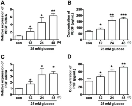
Effect of high glucose on VEGF and PGF expression The results of qRT-PCR showed that high glucose treatment significantly increased the mRNA expression levels of VEGF and PGF in a time-dependent manner when compared to control group (Figure 1A and B). The concentrations of secreted VEGF and PGF were meas-ured by ELISA kit, and high glucose treatment also increased protein concentrations of VEGF and PGF in the media in a time-dependent manner when compared to control group (Figure 1C and D).
Effect of kaempferol on cell proliferation, migration and tube formation of HRECs under high glucose condition The in vitro CCK-8 assay showed that high glucose treatment had no significant effect on the proliferation ability of HRECs compared to 5 mM glucose treatment group; the proliferative ability of HRECs was inhibited by
treatment with high glucose 25 mM plus kaempferol (Figure 2A).
To further examine if kaempferol affected the angio-genesis process of HRECs under high glucose condition, we performed transwell migration and wound scratching assays to assess the effect of keampferol on HREC migration. For the transwell migration assay, the number of cells that migrated through the transwell under 25 mM high glucose condition was more than those under 5 mM normal glucose condition, and this migration was signi-ficantly suppressed by the treatment with 25mM kaemp-ferol for 24 h (Figure 2B). Similar results were observed in the wound scratching assay: HRECs under 25 mM high glucose condition showed an enhanced ability to migrate with accelerated wound closure, while the wound area remained wide in HRECs under 5 mM normal glucose, and high glucose plus 25 mM kaempferol conditions (Figure 2C). Our results indicated that kaempferol inhib-ited glucose-induced migration of HRECs.
To examine the effect of high glucose and kaempferol on angiogenesis, we also performed Matrigel assay to measure the tube formation of HRECs. Compared to 5 mM normal glucose, high glucose significantly increased the number of capillary-like structures, while co-treatment with 25 mM high glucose and 25mM kaempferol produced a lower number of capillary-like structures compared to 25 mM high glucose treatment group (Figure 2D). The
results suggested that kaempferol inhibited glucose-induced tube formation.
Effect of kaempferol on mRNA expression level of VEGF and PGF and on secreted VEGF and PGF induced by high glucose
qRT-PCR results showed that co-treatment with 25 mM high glucose and 25mM kaempferol suppressed the mRNA expression levels of VEGF and PGF compared to treatment with 25 mM high glucose alone (Figure 3A and B). ELISA assay results showed that the protein concentrations of secreted VEGF and PGF in the media were significantly suppressed by co-treatment with 25 mM high glucose and 25mM kaempferol compared to treatment with 25 mM high glucose alone (Figure 3C and D).
Effect of kaempferol on high glucose-induced PI3K expression and Erk1/2, Src, and Akt1 activation
of PI3K and the activation of Erk1/2, Src, and Akt1 induced by high glucose (Figure 4).
Discussion
In the present study, we demonstrated for the first time that kaempferol suppressed cell proliferation, migra-tion and tube formamigra-tion of HRECs under high glucose condition, and the anti-angiogenic effect of kaemp-ferol may be mediated via suppressing mRNA and protein expression levels of VEGF and PGF. Further studies demonstrated that kaempferol suppressed high glucose-induced PI3K expression and Erk1/2, Src, and Akt1 activation.
VEGF has been considered to be an important factor in the pathogenesis of DR in response to high glucose or other stimulus, and it can activate VEGFR-1 and -2, which triggers a signaling cascade promoting endothelial cells migration and sprouting (1,7). PGF that upregulates the expression of VEGFA and other angiogenic factors has been shown to be associated with ischemia or wound stimulated angiogenesis and tumor angiogenesis (23). Therefore, VEGF and PGF may be useful targets for the treatment of DR. In the present study, we observed that high glucose stimulation of HRECs increased the mRNA expression of VEGF and PGF as well as the concentra-tions of secreted VEGF and PGF protein, which is con-sistent with previousfindings (10,11). Similar to previous reports (6), the in vitrofunctional studies further showed that high glucose induced migration and tube formation of
HRECs. After establishment of thein vitrocell model, we further investigated the anti-angiogenic effects of kaemp-ferol and its underlying molecular mechanisms.
A recent study suggested an anti-angiogenic role of kaempferol in cancer progression. For instance, kaempferol inhibited angiogenesis and VEGF expression through both hypoxia-inducible factor dependent and independent path-ways in human ovarian cancer cells (24). In the present study, kaempferol had an anti-angiogenic effect on HRECs, which suppressed migration and tube formation induced by high glucose. The anti-angiogenic effect of kaempferol in HRECs may be mediated via down-regulation of VEGF and PGF, as kaempferol treatment suppressed the expres-sion levels of both VEGF and PGF under high glucose condition.
Pharmacologically, kaempferol has been shown to be an estrogen-related receptor a (ERRa) inverse agonist, which inhibits the activity of ERRa(25), which could cause a decrease in VEGF expression (26). Collectively, it is likely that kaempferol suppressed VEGF expression via targeting ERRa signaling in HRECs, and further studies should be performed to confirm this hypothesis. In future studies, more doses of kaempferol can be tested to further confirm its beneficial effects on DR. Also of scientific interest is the comparison between the beneficial effects of kaempferol and other natural products on DR.
Angiogenesis is a complex and multistep process. Endothelial cell migration is the initial step in angiogen-esis, followed by the endothelial cells differentiation into a capillary-like network (27). In the angiogenesis process, Figure 3. Effect of kaempferol on the mRNA expression level of (A) vascular endothelial growth factor (VEGF) and (C) placenta growth factor (PGF) in human retinal endothelial cells (HRECs) treated with high glucose (25 mM) alone or high glucose plus kaempferol (25mM) for 24 h by qRT-PCR. Protein concentrations of VEGF (B) and PGF (D) in the media were measured by ELISA. Data are reported as means±SD (n=3). *Po0.05 compared to 5 mM;#P
interactions of VEGF and PGF with its receptors trigger intracellular signal pathways via activating Src, PI3K/ Akt1and Erks (28,29), which in turn affects the angiogen-esis of endothelial cells. For the first time, our results demonstrated that kaempferol treatment inhibited high glucose-induced expression of PI3K and the phosphor-ylation of Src, Akt1 and Erk1/2, which suggested that the anti-angiogenic effect of kaempferol on HRECs under high glucose condition is mediated by downregulation of PI3K and inactivation of Src, Akt1 and Erk1/2 signaling.
In conclusion, our study indicates that kaempferol suppressed angiogenesis of HRECs by targeting VEGF
and PGF to inhibit the activation of Src-Akt1-Erk1/2. These results suggest that kaempferol may be a potential drug for better management of DR. Furtherin vivo studies are necessary to confirm the anti-angio-genic effect of kaempferol and to explore its underlying mechanisms.
Acknowledgments
This study was supported by Science and Tech-nology Research Program of Nanjing City (#NJ2014Z 09347923).
References
1. Hampton BM, Schwartz SG, Brantley MA Jr, Flynn HW Jr. Update on genetics and diabetic retinopathy.Clin Ophthal-mol2015; 9: 2175–2193.
2. Lee R, Wong TY, Sabanayagam C. Epidemiology of diabetic retinopathy, diabetic macular edema and related vision loss.
Eye Vis2015; 2: 17, doi: 10.1186/s40662-015-0026-2.
3. Behl T, Kotwani A. Exploring the various aspects of the pathological role of vascular endothelial growth factor (VEGF) in diabetic retinopathy. Pharmacol Res 2015; 99: 137–148, doi: 10.1016/j.phrs.2015.05.013.
Rev Diabet Stud 2015; 12: 159–195, doi: 10.1900/RDS. 2015.12.159.
5. Li JK, Wei F, Jin XH, Dai YM, Cui HS, Li YM. Changes in vitreous VEGF, bFGF and fibrosis in proliferative diabetic retinopathy after intravitreal bevacizumab.Int J Ophthalmol
2015; 8: 1202–1206.
6. Jiang Q, Zhao F, Liu X, Li R, Liu J. Effect of miR-200b on retinal endothelial cell function under high glucose environ-ment.Int J Clin Exp Pathol2015; 8: 10482–10487. 7. Gong CY, Lu B, Hu QW, Ji LL. Streptozotocin induced
diabetic retinopathy in rat and the expression of vascular endothelial growth factor and its receptor.Int J Ophthalmol
2013; 6: 573–577.
8. Maglione D, Guerriero V, Viglietto G, Delli-Bovi P, Persico MG. Isolation of a human placenta cDNA coding for a protein related to the vascular permeability factor.Proc Natl Acad Sci U S A1991; 88: 9267–9271, doi: 10.1073/pnas. 88.20.9267.
9. De Falco S. The discovery of placenta growth factor and its biological activity.Exp Mol Med2012; 44: 1–9, doi: 10.3858/ emm.2012.44.1.025.
10. Tjwa M, Luttun A, Autiero M, Carmeliet P. VEGF and PlGF: two pleiotropic growth factors with distinct roles in develop-ment and homeostasis.Cell Tissue Res2003; 314: 5–14, doi: 10.1007/s00441-003-0776-3.
11. Khaliq A, Foreman D, Ahmed A, Weich H, Gregor Z, McLeod D, et al. Increased expression of placenta growth factor in proliferative diabetic retinopathy.Lab Invest1998; 78: 109–116.
12. Calderon-Montano JM, Burgos-Moron E, Perez-Guerrero C, Lopez-Lazaro M. A review on the dietaryflavonoid kaemp-ferol.Mini Rev Med Chem2011; 11: 298–344, doi: 10.2174/ 138955711795305335.
13. Kim SH, Choi KC. Anti-cancer effect and underlying mecha-nism(s) of kaempferol, a phytoestrogen, on the regulation of apoptosis in diverse cancer cell models.Toxicol Res2013; 29: 229–234, doi: 10.5487/TR.2013.29.4.229.
14. Sak K. Cytotoxicity of dietaryflavonoids on different human cancer types. Pharmacogn Rev 2014; 8: 122–146, doi: 10.4103/0973-7847.134247.
15. Luo H, Rankin GO, Juliano N, Jiang BH, Chen YC. Kaempferol inhibits VEGF expression andin vitro angiogen-esis through a novel ERK-NFkappaB-cMyc-p21 pathway.
Food Chem2012; 130: 321–328, doi: 10.1016/j.foodchem. 2011.07.045.
16. Liang F, Han Y, Gao H, Xin S, Chen S, Wang N, et al. Kaempferol identified by zebrafish assay andfine fractiona-tions strategy fromDysosma versipellisInhibits angiogen-esis through VEGF and FGF pathways.Sci Rep 2015; 5: 14468, doi: 10.1038/srep14468.
17. Kowluru RA, Kowluru A, Mishra M, Kumar B. Oxidative stress and epigenetic modifications in the pathogenesis of
diabetic retinopathy.Prog Retin Eye Res2015; 48: 40–61, doi: 10.1016/j.preteyeres.2015.05.001.
18. Nabavi SF, Habtemariam S, Daglia M, Shafighi N, Barber AJ, Nabavi SM. Anthocyanins as a potential therapy for diabetic retinopathy. Curr Med Chem 2015; 22: 51–58, doi: 10.2174/0929867321666140815123852.
19. Mahoney SE, Loprinzi PD. Influence offlavonoid-rich fruit and vegetable intake on diabetic retinopathy and diabetes-related biomarkers. J Diabetes Complications 2014; 28: 767–771, doi: 10.1016/j.jdiacomp.2014.06.011.
20. Saric A, Balog T, Sobocanec S, Kusic B, Sverko V, Rusak G, et al. Antioxidant effects offlavonoid from Croatian Cystus incanus L. rich bee pollen.Food Chem Toxicol2009; 47: 547–554, doi: 10.1016/j.fct.2008.12.007.
21. Devi KP, Malar DS, Nabavi SF, Sureda A, Xiao J, Nabavi SM, et al. Kaempferol and inflammation: From chemistry to medicine. Pharmacol Res 2015; 99: 1–10, doi: 10.1016/ j.phrs.2015.05.002.
22. Medinger M, Halter J, Heim D, Buser A, Gerull S, Stern M, et al. Angiogenic markers in plasma cell myeloma patients treated with novel agents. Anticancer Res 2015; 35: 1085–1090.
23. Marcellini M, De Luca N, Riccioni T, Ciucci A, Orecchia A, Lacal PM, et al. Increased melanoma growth and metastasis spreading in mice overexpressing placenta growth factor.
Am J Pathol2006; 169: 643–654, doi: 10.2353/ajpath.2006. 051041.
24. Luo H, Rankin GO, Liu L, Daddysman MK, Jiang BH, Chen YC. Kaempferol inhibits angiogenesis and VEGF expression through both HIF dependent and independent pathways in human ovarian cancer cells. Nutr Cancer 2009; 61: 554–563, doi: 10.1080/01635580802666281.
25. Wang J, Fang F, Huang Z, Wang Y, Wong C. Kaempferol is an estrogen-related receptor alpha and gamma inverse agonist. FEBS Lett 2009; 583: 643–647, doi: 10.1016/ j.febslet.2009.01.030.
26. Zhang K, Lu J, Mori T, Smith-Powell L, Synold TW, Chen S, et al. Baicalin increases VEGF expression and angiogenesis by activating the ERR(alpha)/PGC-1(alpha) pathway. Car-diovasc Res2011; 89: 426–435, doi: 10.1093/cvr/cvq296. 27. Griffioen AW, Molema G. Angiogenesis: potentials for
phar-macologic intervention in the treatment of cancer, cardio-vascular diseases, and chronic inflammation. Pharmacol Rev2000; 52: 237–268.
28. Huang Q, Sheibani N. High glucose promotes retinal endothelial cell migration through activation of Src, PI3K/ Akt1/eNOS, and ERKs. Am J Physiol Cell Physiol 2008; 295: C1647–C1657, doi: 10.1152/ajpcell.00322.2008. 29. Xin X, Khan ZA, Chen S, Chakrabarti S. Extracellular