w w w . e l s e v i e r . c o m / l o c a t e / b j i d
The Brazilian Journal of
INFECTIOUS DISEASES
Original article
Predominance of
Leishmania major
and rare
occurrence of
Leishmania tropica
with haplotype
variability at the center of Iran
Amir Hossein Zahirnia
a,b, Ali Bordbar
a, Sahar Ebrahimi
a, Adel Spotin
c,
Somayeh Mohammadi
a, Seyedeh Maryam Ghafari
a, Setareh Ahmadvand
a,
Negar Jabbari
a, Ahmad Reza Esmaeili Rastaghi
a, Parviz Parvizi
a,∗aPasteur Institute of Iran, Parasitology Department, Molecular Systematics Laboratory, Tehran, Iran
bHamadan University of Medical Sciences, School of Medicine, Department of Medical Entomology, Hamadan, Iran cTabriz University of Medical Sciences, Immunology Research Center, Tabriz, Iran
a r t i c l e
i n f o
Article history:
Received 11 March 2018 Accepted 11 July 2018
Available online 13 August 2018
Keywords:
Leishmaniasis
Leishmania major Leishmania tropica
Nuclear gene Iran
a b s t r a c t
Background: Leishmania majoris a causative agent of zoonotic cutaneous leishmaniasis in the center of Iran, Abarkouh district. Molecular characterization and precise incrimination ofLeishmaniaspecies was carried out to perform controlling measurements and to design treatment programs for zoonotic cutaneous leishmaniasis.
Methods:All smears isolated from ulcers of suspected patients were examined under a light microscope and graded for amastigotes frequency. Extraction of DNA, PCR, RFLP and sequencing of ITS-rDNA genotype were done to increase the efficacy ofLeishmaniaparasites identification at their species-specific level and to detect anyLeishmaniainfections within.
Results:Humans were found to be infected withL. majorwith high infection frequency and alsoLeishmania tropicawas identified with low occurrence for the first time as non-native species using molecular analyses. The rates of infections was considerable with microscopic observation (n= 65, 73%) out of 89 smears prepared from suspected patients. Molecular anal-yses showed that the density ofL. majorwas significantly higher (n= 48, 53.93%) thanL. tropica
(n= 4, 4.49%) (Mann–WhitneyUtest:p< 0.05) and two samples (2.25%) remained ambiguous after several sequencing.L. majordid not have diversity with two common haplotypes but
L. tropicawere found to exhibit high diversity with three novel haplotypes.
Conclusion: L. majorwas considered the causative agent of leishmaniasis in the region, but the identification of a non-nativeL. tropica revealed the importance of further isolation of
Leishmaniaparasites following molecular analyses and confirmation, and also revealed the importance of further isolation ofLeishmaniaparasites from patients of the field areas who do not have easily access to health care centers for specialized treatment strategies.
© 2018 Sociedade Brasileira de Infectologia. Published by Elsevier Editora Ltda. This is an open access article under the CC BY-NC-ND license (http://creativecommons.org/licenses/ by-nc-nd/4.0/).
∗ Corresponding author.
E-mail address:parp@pasteur.ac.ir(P. Parvizi).
https://doi.org/10.1016/j.bjid.2018.07.005
b r a z j i n f e c t d i s .2 0 1 8;2 2(4):278–287
279
Introduction
Cutaneous leishmaniasis (CL) is a noticeable tropical disease which has lots of adverse effects on human health in 98 countries on 5 continents in the world as well as Iran.1,2The
mammalian host(s) of CL in the Old World including Rodentia, Carnivora and human has been infected by different species of single-celled Leishmania parasites. Humans are naturally infected by the bite of female sand flies from vertebrate ani-mals in the form of zoonotic cutaneous leishmaniasis (ZCL) with the exception ofLeishmania tropicawhich is often known as an anthroponotic transmissible disease from humans to vertebrate animals.3,4 An epidemiological study of ZCL in 11
provinces of Iran showed thatL. majorwas the causative agent of CL in rural areas of Iran with 95% prevalence rate whereas
L. tropicahad a distribution rate of 65% in urban districts.4
Although, papers have been published on the transmission and epidemiological circulation of Leishmania parasites in vectors,5 reservoir hosts6,7 and humans from different ZCL
foci of Iran,2,8there is no considerable and sufficiently
investi-gation on leishmaniasis in humans at the center of Iran (Yazd province). Nevertheless ZCL is considered as an endemic dis-ease in Isfahan province adjacent to Yazd, it has also become endemic in several areas in Yazd province at the center of Iran during the last 10 years.9,10Therefore, the constant
appear-ance of infectious agents of Leishmaniasis within a human population of this focus highlights the direct significant atten-tion to identifying transmission cycles ofLeishmaniaparasites and their clinical manifestations in humans of Yazd district.
Leishmania majoris one of the leading-off agents causing rural, zoonotic and vector-borne disease and the digenetic form ofLeishmanialife cycle is completed in different species of wild rodents and phlebotomine sandflies as reservoir hosts and vectors respectively in many geographical locations where it occurs.10In factL. majoris known as the causative
agent of ZCL and more frequent than the otherLeishmania
parasites in Iran which induces Th2 immune response in case of exacerbation with disfigured cutaneous patterns.11
In addition, another principal agent of cutaneous leishman-iasis,L. tropica, has been implicated in occasional cases of recidivans or viscerotropic cases.12 Recently, L. tropica has
stimulated many interests because of its potential traits to visceralize and/or classical visceralize in humans,13
dis-seminating of cutaneous leishmaniasis along with visceral leishmaniasis14and developing of mucosal leishmaniasis in
Iran.15 An epidemiological study of ZCL in 11 provinces of
Iran.15Accordingly, finding non-native species ofL. tropicais
of great importance in studied region.
However, three species ofLeishmaniaparasites have been incriminated as the causative agents of human leishmaniasis in Iran. They areL. major,L. tropicaandL. infantum.16Also, some
other mammals’Leishmaniasuch asL. turanica,L. gerbilliand
L.close togerbillihave been reported from Iran.2and isolated
from reservoir hosts and sand flies vectors in different CL foci of Iran.6,16
The popular and practical method for identification of Leishmania species is Giemsa stained smears prepared from patients’ ulcer with the examination of Leishmania
amastigotes under microscopic observation.17 In addition,
incrimination of Leishmania parasites was previously based on clinical symptoms, vector assessments, and pathogeni-city in laboratory animals and growth in media.9,18In recent
years,Leishmaniaspecies have firmly been identified because of high sensitivity, more rapid determination and character-ization after amplifying targeted genes using PCR, RFLP and sequencing.15,19
In our investigations, PCR amplification, RFLP digestion and sequencing of ITS-rDNA genotype were employed to identify all Leishmaniaspecies isolated from the lesion of suspected patients having serous exudate.
The motivation of this work arises from increasing prob-lems of leishmaniasis in the center district of Iran. In fact, this investigation was conducted to monitor the effectiveness of control measures, to accurate incrimination ofLeishmania
parasites at their species level with molecular analyses, to characterize the species-specific parasites isolated from humans residing in the natural field working regions and also to improve our knowledge about allLeishmaniaspecies those readily maintain their ecological circulation in the endemic focus of Yazd province.
Methods
Origin and sampling of Leishmania parasites from suspected patients
Within the ZCL focus, prepared samples of suspected patients were collected from 16 surveyed villages of Abarkouh city in Yazd province (Fig. 1). Yazd district is geographically located between 31◦ 07′ 44′′ N and 53◦ 16′ 57′′E of central Iran. The
city proper is situated at an altitude of 1510 m (4,954 ft) above sea level (a.s.l.) and all villages of this region were screened and sampled from suspected patients from 2014 to 2016. The smears firstly isolated from patients’ lesion, prepared on the slide (slide fixation) and froze within the days of collection. Samples were then transferred on ice to the Pasteur Institute of Iran, Tehran, for microscopic identification and molecular experiments.
The occurrence of leishmaniasis was very low in four con-secutive months (May–August). Whereas we found a sharp increasing rate of CL lesions during the 3rd Quarter 2014 (October–December), sampling was carried out at the time of lesions’ appearance in Abarkouh district (Fig. 1). The smears were prepared from the active lesions of patients residing in different rural regions of leishmaniasis throughout Abarkouh district. The personal information, lesion duration, type and the number of lesion, ulcer(s)’ location, patients’ travelling to endemic areas and also drug consumption were recorded for each patient separately. Expanded set of tools (conven-tional and then molecular methods) were exerted to recognize samples including presumptive CL parasites based on Evans protocol17and were then smeared on two microscopic slides,
Fig. 1 – Lesions of patients with Old-World zoonotic cutaneous leishmaniasis caused byLeishmania majorin Abarkouh District, Central Iran. A, primary nodule formation; B, classic wet-lesion; C, classic wet-lesion (typical); D, non-classic wet-lesion.
(30 min. per slide) by ocular micrometer (×100 objective = 1m per unit space) and Dino Capture 2.0. The positive smears of each patient were scored forLeishmaniaamastigotes density and frequency from 1+ to 6+.2The distribution of our
statisti-cal population was asymmetric and not normal; therefore we required using a non-parametric statistical Mann–WhitneyU
test in SPSS 16:p< 0.05.
DNA extraction of genomic DNA
All Giemsa-stained slide washed with absolute ethanol for eliminating immersion oil on the smears and covered with 300l lysis buffer (50 mM NaCl, 50 mM Tris, 10 mM EDTA, pH 7.4, 1% (v/v) Triton x-100) for the extraction of total DNA from graded slides. After a short time the smears completely removed and transferred to a 1.5 ml reaction tube. Then, 15l proteinease K (20 mg/ml, fermentase) was added to each tube. Cell lyses was accomplished after incubation for 4–6 h at 56◦C.
The lysate was extracted by phenol–chloroform followed by ethanol precipitation.18
The DNA was re-suspended in 30l TE1X and stored at −20◦C. The genomic DNA of each smear from suspected
patient and any parasite within was extracted in the Molec-ular Systematics Laboratory suite in a room where amplified and cloned DNAs were never processed.
PCR amplification and molecular identification of Leishmania parasites
Each PCR assay was executed to screenLeishmaniainfection by the nested PCR of a fragment of ITS (nuclear ribosomal) gene. For the first stage of nested PCR, forward primer IR1 with the reverse primer IR2 and for the second-stage forward primer ITS1F with the reverse primer ITS2R4 were used.16
Dou-ble distilled water and DNA fromL. majorandL. tropicawere used as negative and positive controls for each batch of PCR (primers were produced at CinnaGen Co.). The reactions were performed in an Eppendorf Mastercycler Gradiant device. PCR products were subjected to electrophoresis in 1.5% agarose gel at 150 V in 0.5×TBE (0.045 M Tris-borate,1 mM EDTA) (Merck
b r a z j i n f e c t d i s .2 0 1 8;2 2(4):278–287
281
Co.) buffer and monitored under ultraviolet light after stain-ing for 15 min with (0.5 g/ml) safe stain; in order to see an approximately 480 bp (including primers) showing ITS-rDNA gene fragment forLeishmaniainfections. The reactions were performed in an Eppendorf Mastercycler Gradiant device.
Identification of the species described by the molecular analyses (RFLP and sequencing)
To select the suitable enzyme for digesting the PCR product in RFLP method, the sequences of standard strains (L. major,L. tropica,L. infantum,L. turanicaandL. gerbilli) registered in Gen-Bank were employed for sequence analysis. Standard strains sequencing ofLeishmaniawere examined byin silicoanalysis, CLC DNA Workbench 5.2 software (CLC bio A/S, Aarhus, Den-mark). A suitable restriction enzymeBsuRI (HaeIII) (ParsGene Co.) was selected with different cut sites GG↓CC as an appro-priate restriction enzyme digestion to incriminate different CL species.
Endonuclease digestion was done in a volume of 30l consisting of 10l of PCR product, 2l of BsuRI (HaeIII) enzyme (Fermentas), 2l of 10× buffer and 16l of dis-tilled water for 4 h (rapid digestion) at 37◦C for ITS-rDNA
gene fragment (Fig. 3). Also, standard strains of WHO were employed as positive controls as fallow: L. major
(MHOM/SU/73 /5ASKH), L. tropica (MHOM/PS/2008/344Jn SF53) and L. donovani (MHOM /TN/80 /IPT1). Two negative controls were utilized, one of which did not have restriction enzyme and the other one did not have PCR product. After endonuclease digestion of PCR product with BsuRI (HaeIII) enzyme, the fragments were analysed using electrophoresis on agarose gel 3% containing safe stain and ladder 50 bp (Fermentas) (Fig. 3).
Sequencing and phylogenetic analysis
To reconfirm the results of PCR-RFLP, the numbers of PCR products were sequenced by targeting ITS-rDNA in both direc-tions using the ABIPRISMTM 3130 Genetic Analyzer automated sequencer (Applied Biosystem, USA). Individual sequences were justified, edited and aligned in consensus positions with GenBank sequences of all regional species in case of homology and similarity. The SequencherTMv. 4.1.4 Software for PC was used to incriminate unique haplotypes (PC, Gene Codes Corpo-ration). The MEGA v5.05 software was applied for phylogenetic analysis.20To permit grouping of the new species, the
phyloge-netic maximum likelihood (PhyML)21program was run to get
maximum likelihood tree based on the Kimura 2-parameter model of nucleotide substitution search by stepwise addition of 100 random replicates, and nonparametric ML bootstrap was calculated with 1000 replicates.
Results
Microscopic observation and molecular experiments
The presence ofL. tropicawas proved as a non-native species where there was no report of this species before. To acquire an appropriate statistical results, all patients (n= 89) having
dis-tinctive, fresh, acute and/or chronic lesions (wet, dry and/or mixed) examined in all 16 important villages from different sites of Abarkouh (12500 populations) (Fig. 1).L. major and
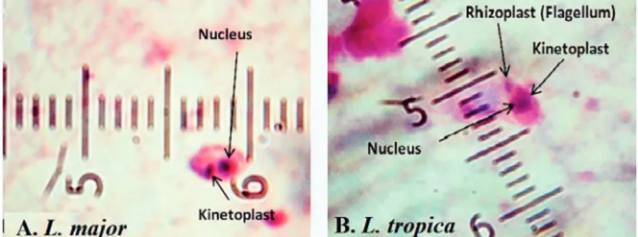
L. tropicawere firmly found and molecularly typed from the active lesion of patients. L. majorwas identified with regu-lar amastigotes’ shapes (round or oval with a size of 2–4m) whereas appearances ofL. tropicaamastigotes were irregular (pear, spindle or cigarette with more than 4m) (Fig. 2). Infec-tions were found from the lesion of individuals (ranging from <1 to >25), residents of villages (Abarkouh region) who hadL. major more in face, hand and foot isolated from wet lesion respectively and,L. tropicain face and hand isolated from dry lesion respectively (Table 1). Of 89 samples, 65 (73%) Giemsa stained slides were observed positive microscopically for Leish-mania amastigotes and the specified score was designated to each prepared slide smear (Table 1). Results showed that the median prevalence of ZCL was considerable in Abark-ouh district (approximately 71.2 cases per 10,000 inhabitants). All 65Leishmaniapositive samples were applied to screen by PCR assay that only 54 samples were successfully identified
Leishmaniapositive using ITS-rDNA gene fragment and then subjected to RFLP following sequencing. To identify species-specific Leishmania parasites, molecular experiments were carried out on all microscopically observed grading numbers (n= 65) using PCR. The positive PCR samples are as follows: 1+ (n= 6 out of 15), 2+ (n= 5 out of 7), 3+ (n= 14), 4+ (n= 14), 5+ (n= 11) and 6+ (n= 4). This inclusive inquiry revealed that the number of patients those who had Leishmania lesions were more in Chahgir (n= 17) and Abarkouh (n= 16) than the other villages. The samples of +1 grading number exhibited (n= 15, 16.8%) at the highest rate and the +6 grading num-ber remained at the lowest rate (n= 4.5%) isolated from the suspected patients of collected sites (Table 1).
Our investigation showed the readily maneuver ofL. major
parasites through expanding distribution in the broad ranges of geographical confines at the center of Iran (Yazd province). All examined patients had typical lesions in Abarkouh dis-trict and the serous was taken from fresh lesions (Fig.1). The grade numbering of amastigotes is illustrated according to the WHO instruction inTable 1. The overall ratio ofL. major
infections were mainly higher thanL. tropicawith low den-sity (Mann–WhitneyUtest:p< 0.05) based on the asymmetric distribution and non parametric analysis of the samples.
Leishmania parasites identification with RFLP and sequencing of ITS-rDNA fragment
braz
j
infect
dis.
2018
;
2
2(4)
:278–287
Table 1 – Microscopic observations; MO, on Giemsa-stained slides prepared from human lesion and molecular analyses onLeishmaniapositive of MO using ITS-rDNA
genotype, Abarkouh district, Yazd province.
Collection site Total samples
Microscopic observation Personal information based on age (years) and lesion type
Molecular analysis
Province District Villages of Abarkouh
Positive grading numbers ofLeishmania amastigotes based on Giemsa-stained smears
Sex <1 1-5 5-10 10-1515-25 >25 PCR using ITS-rDNA geno-type + ve
RFLP withBsuRI & Sequencing
+ve (%) −ve (%) Total +ve F M WDWD W DW D W D W D L.
major +ve
L. trop-ica +ve
NI +ve
1 2 3 4 5 6
Yazd Abarkouh Chahgir 17 2 0 1 4 2 0 8 9 9 8 1 1 2 3 5 1 4 8 6 2 0
Nosrat Abad 6 2 1 1 1 1 0 0 6 1 5 1 1 3 1 4 3 0 1
Taghi Abad 2 0 0 0 0 0 1 1 1 0 2 2 1 1 0 0
Esfand Abad 14 4 0 2 3 2 0 3 11 6 8 1 1 2 4 4 2 10 9 1 0
Ardi 3 1 0 0 2 0 0 0 3 0 3 1 2 2 1 0 1
Asad Abad Sofla 4 1 0 0 1 1 0 1 3 2 2 1 2 1 2 1 1 0
Mehr Abad 8 0 3 1 1 0 1 2 6 2 6 1 1 1 3 2 6 6 0 0
Harooni 7 0 0 1 0 2 2 2 5 3 4 1 1 1 1 1 2 5 5 0 0
Asad Abad-Esfand Abad
2 1 0 0 0 1 0 0 2 0 2 2 1 1 0 0
Hossein Abad 1 0 0 1 0 0 0 0 1 0 1 1 1 1 0 0
Raeis Abad 2 1 0 0 0 0 0 1 1 0 2 1 1 0 0 0 0
Kolpeh 1 0 0 0 0 1 0 0 1 0 1 1 0 0 0 0
ShahreAsb 4 1 0 2 1 0 0 0 4 1 3 2 2 3 3 0 0
Harook 1 0 0 1 0 0 0 0 1 1 0 1 1 1 0 0
KhoramAbad 1 0 1 0 0 0 0 0 1 0 1 1 1 1 0 0
Abarkouh 16 2 2 4 1 1 0 6 10 4 12 1 2 3 3 1 3 1 2 9 9 0 0
Total (%) 89 15
(16.8) 7 (7.9)
14 (15.7)
14 (15.7)
11 (12.4)
4 (4.5)
24 (27)
65 (73)
29604 8 11 19 31 16 54 48 (53.93)
4 (4.49)
2 (2.25) Total Ve+ 65(73) 89 89 Total Ve+ 54(60.67)
b r a z j i n f e c t d i s .2 0 1 8;2 2(4):278–287
283
RFLP analysis was done in CLC DNA Workbench 5.2 (CLC bio A/S, Aarhus, Denmark), two fragments of 120 and 310 bp belong toL. major, four (30, 40, 50 and 310 bp) for L. tropica
andL. turanicaand three (50, 70 and 310 bp) forL. Infantum. The pattern ofBsuRI (HaeIII) enzyme confirmed as DNA frag-ments’ digestion in agarose gel after electrophoresis (Fig. 3). In addition, the two sequence of ITS-rDNA PCR products were compared to the GenBank sequences in case of similarity and homology. All sequences ofLeishmaniaspecies (approx-imately 480 bp, including primers) were positioned in two different groups at which 52Leishmaniasequences had sig-nificant homology to each other and found asL. major(n= 48, 53.93%),L. tropica(n= 4, 4.49%) and two were unidentified (n= 2, 2.25) (Table 1).
Molecular analyses exhibited 48 positive L. major in a total of 65 microscopically positive lesions. 48 samples of
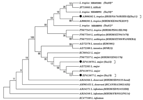
L. major had two common haplotypes for L. major using the ITS-rDNA gene fragment GenBank access nos. EF413078 (Yaz32) and EF413077 (Yaz19) along with a common haplo-type of standard strain (IROS/NA/76/ROSSI-II) form GenBank forL. tropica, accession no. AJ000302 (Yaz11). Also, three novel haplotypes were identified forL. tropicawhich had high vari-ation in their nucleotide sequences GenBank accession nos.
MH488992 (Yaz58); MH488993 (Yaz61) and MH492002 (Yaz60) (Fig. 4). Besides, the transition event of nucleotide substitution occurred in several positions of open reading frame (ORFs).
Phylogenetic analyses
Phylogenic tree showed that allLeishmaniaspecies were placed in their own exacting clade (Fig. 4). The topology of tree showed that gene duplication was occurred subsequent to speciation resulting from in-paralog event in a way thatL. tropicaandL. killickiwere positioned in a Taxon of monophyletic clade.
48 L. major had common haplotypes incriminated pre-viously in sandflies, rodents and humans in central and other places of Iran GenBank accession nos. EF413078 (Yaz32); EF413077 (Yaz19). But we found these only among the popula-tion of sandflies in Abarkouh district.2,5,6
Discussion
This investigation has been kept evidential proof aboutL. major
distribution with notable emerging ofL. tropicain patients of Yazd foci, Iran. Current workup turned out that the speciesL.
Fig. 3 – (a)Leishmaniapositive results isolated from freshly ulcers of suspected patients by PCR targeting ITS-rDNA genotype without RFLP. (b) PCR-RFLP observation in Yazd isolates based on ITS-rDNA genotype. Lane 1 (−ve, negative control
containingBsuR1 without PCR product), lane 2 (−ve: negative control containing PCR product withoutBsuR1), lane 3 (+ve,
positive control forL. major), lanes 4, 5; Yaz32 and Yaz19 (L. major, common haplotypes): isolated from wet lesions of suspected patients in Esfand-Abad and Chahgir, respectively. Lanes 6–8; MH488992 (Yaz58), MH492002 (Yaz60), and
Fig. 4 – Maximum parsimony tree of the haplotypes of the ITS1-5.8SrRNA gene fragment for the isolates ofL. majorandL.
tropicain humans of Yazd province with those submitted in GenBank using MEGA 5.05 software (details were shown in
Table 1). *, Unique haplotypes; , Common haplotypes ofLeishmaniaspecies identified in Abarkouh district, Central Iran; the standard reference strains of the World Health Organization are shown in the parenthesis.
majorhad low genetic variation with predominant and high circulation rate whilst the speciesL. tropica exhibited high genetic diversity with different novel haplotypes and constant phenotypic features among the patients of Abarkouh district. Previously, we found more diversity inL. majorusing the ITS-rDNA genotype isolated from parasites in Iran.2,6Although,L.
majoris well-known as a causative agent of ZCL in Iran,2,5,8,22
L. tropica(n= 4; 4.49%) was isolated from the patients as a non-native parasite in the region. In addition,L. major was also found with high density (n= 48; 53.93%) in Abarkouh district.
The smears isolated from the chronic lesions of suspected patients had low amastigotes located more on hands, arms and foot respectively and they scored as +1 to +2 under micro-scopic observation. Although prepared smears from chronic old lesions had low amastigotes number microscopically, they had noLeishmaniapositive results after PCR and molecular analyses. In fact after doing molecular experiments on 22 microscopically positive samples from low grading numbers of amastigotes (n= 15, +1 andn= 7, +2), only 11 (n= 11/22) sam-ples were confirmed positive and the rest (n= 11) were negative that this may be resulted from chronic lesion or DNA degrada-tion in extracdegrada-tion procedures (Table 1). Also, smears isolated from freshly acute lesions were scored from +1 to +6 which they all were positive after molecular experiments. Freshly or acute lesions were sampled more from faces and hands respectively.17
Isoenzyme analyses were previously used as a gold standard-method for characterizingLeishmaniaspecies partic-ularly in the old world23but requiring the great numbers of
live parasites in the culture and consequently the risk of being
infected with other agents and/or fastest growth of any con-queror microbe was probable. Today, there is no single widely accepted method to specifyLeishmaniaspecies stoutly. Thus, a wealth of molecular techniques is conducted to incriminate
Leishmaniaparasites in primary infections.8,24 recent reports
on vector and reservoir hosts of ZCL showed that onlyL. major
was detected in femaleP. papatasi(15.8%) and within the differ-ent species of roddiffer-ents in the villages of Abarkouh district.5,25
Likewise, Spotin and colleagues have recently reported the dif-ferent morphologies ofL. majoramastigotes’ shapes but no molecular diversity observed in ZCL area of Iran.26P. papatasi
is a proven vector of ZCL even in Isfahan (central) and Golestan (North east) provinces of Iran.16,27Although, noL. tropicawas
previously detected in vectors of Abarkouh district,5there was
a striking difference in the detection of Leishmania species from principal vectors ofL. tropica(Phlebotomus sergenti) and from human’s lesion as mammalian host in the same region. Molecular identification of L. tropica from skin lesions has shown thatL. tropica is present in a considerable region at the center of Iran, Abarkouh district, and for the first time in the areas formerly known only asL. majorfoci.19,28Hence,
it is likely that Anthroponotic CL (ACL) has been considerably underreported in this district. However, because of existence of canines, stray dogs, sheep dogs and particularly domestic dogs as main reservoir hosts ofL. tropica, more humans and dogs need to be monitored before concluding whether they are principal reservoir hosts ofL. tropicain central Iran (Yazd province).29
south-b r a z j i n f e c t d i s .2 0 1 8;2 2(4):278–287
285
ern Iran.13,14 In addition, two provinces of Isfahan and Fars
are adjacent to Yazd province (sympatric areas) and must be considered in case of patients travelling, epidemiological dis-tribution of reservoirs and vectors, biodiversity ofLeishmania
parasites and their potential as neglected emergence disease. Nonetheless, two PCR amplicons ofLeishmania parasites had a noticeable incongruity between their sequences and were not readable even after several resequencing. These two unreadable sequences were isolated from lesions of infected patients in two villages of Abarkouh (Nosrat Abad and Ardi) (Table 1). Two unreadable sequences could be arise from inadequacy of DNA, mixed infections with other ZCL and/or non-pathogenic parasites, or containing two loci that PCR primers have greater tendency or homology to one of the loci in this case.31Anyhow, Oryan and colleagues (2013) have
observed the high genetic variation ofL. majorrelating to their different clinical manifestations of cutaneous leishmaniasis based on minicircle kDNA in Fars province, southern Iran.32
But some issues including the probable existence of other
Leishmaniaspecies might have been failed to notice if the PCR primers demonstrated haplotype specificity.
To determine intra and inter-species relationships and even to identify separation between strains, applying mis-cellaneous (nucleus, e.g. ITS-rDNA and/or mitochondrial, e.g. Cytb) genes with different degrees of diversity needs to be done on the basis of different metabolic enzyme through sys-tematic multilocus sequence analysis (MLSA) through.33MLST
approaches need to be applied as a precious tool to determine similarities betweenLeishmaniaspecies34and to screen more
patients in different ZCL foci, of Iran.
Also we have already assessed the sensitivity of two meth-ods of semi-nested for minicircle kinetoplast (kDNA) and nested PCR of ITS-rDNA to detectLeishmaniaparasites in Phle-botomus papatasiwhich revealed the high infection rates of
L. major in Isfahan villages close to Yazd province.9 Apart
from geographical habitats, the isolated L. tropicaparasites in this study found to be diverged from their ancestors with three novel haplotypes. All haplotypes ofL. tropicaparasites were placed in a monophyletic cluster of a unique group and this can be resulted from more nucleotide discrepancies in a few copy numbers of ITS-rDNA (nuclear) genotype of ortholo-gous loci for this species-specific phylogenetics. Also, Paralog mechanism (tandem duplication) of homologous recombina-tion (HR) in extra chromosomal circles may have occurred between direct repeated sequences35,36to expand their gene
families. Accordingly,Leishmania parasites would be able to increase their gene copy number37and therefore survive and
adapt in a different conditions.
Recent studies have highlighted that aneuploidy appears as a pervasive belief inLeishmania37,38 and some eukaryotic
pathogens likeLeishmania develop their rapid adaptation to environment changes associated with aneuploidy as a new model of adaptation in parasites with asexual or unicellu-lar reproduction.39 Changes in chromosome number may
not necessarily be present in all individual cells.Leishmania
parasites are generally considered as a diploid organism with their own manifestation of genome plasticity.40,41
Whereas, increasing in copy number of the Leishmania
genes can be expected in response to the conditions of
environmental changes,42 genetic variation of L. tropica is
perceived as a constitutive feature and/or effective strategy in parasite–vector–host interplay to maintain parasites’ life cycles in their human hosts in the region of our study.
In addition, mixed infections of the same macrophage with different species of Leishmania parasites were shown to be experimentally possible.42Concomitant infection ofL. tropica
andL. majorhas already isolated from an Iranian patient with mucosal leishmaniasis (ML) in Fars province (Southern Iran) next to Yazd province.15The recent investigation showed that
inP. sergenti(the vector ofL. tropica),L. turanicapromastigotes were present until the defection of blood meal remnants and also proved thatL. turanicadevelops late-stage infections inP. papatasi43the proven vector ofL. majorin Iran.
Maybe it is possible that had we investigated hard on sand-flies as vectors or rodents as reservoir hosts, more elucidation ofLeishmaniaspecies might have found asL. tropicaorL. turan-icabut these, are outside the scope of this paper.
To confirm the precise identification ofLeishmaniaspecies in unknown, concomitant or mixed infection in vectors or reservoir hosts, the DNA samples are needed to be cloned.44
To determine the distribution of all species ofLeishmania
parasites that infect humans particularly in areas where foci of two species overlap and also to clarify the role of reservoir hosts (rodents or dogs) in maintaining ofLeishmaniaparasites as co-infectors, molecular characterization and phylogenetic analyzes may have a pivotal approach to identify species-specific parasites in their potential sandflies as vectors.24
Therefore, simultaneous identification ofLeishmaniaparasites in their vectors and reservoirs provides a route for more differ-ent comparative molecular studies prior to ancidiffer-ent methods.
Conflicts of interest
The authors declare no conflicts of interest.
Acknowledgments
The fund of this work was supported by Pasteur Institute of Iran and the grant was awarded to Prof. Parviz Parvizi by the Pasteur Institute of Iran. The authors would like to thank Abarkouh Health Care authorities and the personnel of National Institute of Health Research (NIHR) in Yazd for their assistance and support to facilitate easier communica-tion with indigenous people from rural areas of Abarkouh district during this investigation. We would like to express our deep sorrow over the passing away of Mehdi Baghban and thank him for his kind assistance with the field work and laboratory experiments.
r e f e r e n c e s
1. Alvar J, Vélez ID, Bern C, et al. Leishmaniasis worldwide and global estimates of its incidence. PLoS ONE. 2012;7:e35671. 2. Bordbar A, Parvizi P. High density ofLeishmania majorand
3. WHO. In: Crompton DWT, editor. First WHO report on neglected tropical diseases: working to overcome the global impact of neglected tropical diseases. Geneva, Switzerland: World Health Organization; 2010. p. 91–6. Available from
http://www.who.int/neglecteddiseases/2010report/en
4. Mahmoudzadeh-Niknam H, Ajdary S, Riazi-Rad F, et al. Molecular epidemiology of cutaneous leishmaniasis and heterogeneity ofLeishmania majorstrains in Iran. Trop Med Int Health. 2010;11:1335–44.
5. Jafari R, Najafzadeh N, Sedaghat MM, Parvizi P. Molecular characterization of sandflies andLeishmaniadetection in main vector of zoonotic cutaneous leishmaniasis in Abarkouh district of Yazd province, Iran. Asian Pac J Trop Med. 2013;6:792–7.
6. Mirzaei A, Rouhani S, Kazerooni P, Farahmand M, Parvizi P. Molecular detection and conventional identification of
Leishmaniaspecies in reservoir hosts of zoonotic cutaneous leishmaniasis in Fars province, south of Iran. Iran J Parasitol. 2013;8:280–8.
7. Rouhani S, Mirzaei A, Spotin A, Parvizi P. Novel finding of
Leishmania majorinHemiechinus auritusand molecular detection of this parasite inMeriones libycusfrom important foci of zoonotic cutaneousleishmaniasisin Iran. J Infect Public Health. 2014;7:210–7.
8. Bordbar A, Parvizi P. High infection frequency, low diversity of
Leishmania majorand first detection ofLeishmania turanicain human in northern Iran. Acta Trop. 2014;133:69–72. 9. Parvizi P, Mauricio I, Aransay AM, Miles MA, Ready PD. First
detection ofLeishmania majorin peridomesticPhlebotomus papatasifrom Isfahan province, Iran: comparison of nested PCR of nuclear ITS ribosomal DNA and semi-nested PCR of minicircle kinetoplast DNA. Acta Trop. 2005;93:75–83. 10. Ready PD. Biology of phlebotomine sandflies as vectors of
disease agents. Annu Rev Entomol. 2013;58:227–50.
11. Alimohammadian MH, Jones SL, Darabi H, et al. Assessment of interferon-␥levels and leishmanin skin test results in persons recovered for leishmaniasis. Am J Trop Med Hyg. 2012;87:70–5.
12. Jacobson RL.Leishmania tropica(Kinetoplastida:
Trypanosomatidae) – a perplexing parasite. Folia Parasitol. 2012;50:241–50.
13. Alborzi A, Rasouli M, Shamsizadeh A.Leishmania tropica-isolated patient with visceral leishmaniasis in southern Iran. Am J Trop Med Hyg. 2006;74:306–7. 14. Alborzi A, Pouladfar GR, Fakhar M, et al. Isolation of
Leishmania tropicafrom a patient with visceral leishmaniasis and disseminated cutaneous leishmaniasis, southern Iran. Am J Trop Med Hyg. 2008;79:435–7.
15. Shirian S, Oryan A, Hatam G-R, Daneshbod K, Daneshbod Y. Molecular diagnosis and species identification of mucosal leishmaniasis in Iran and correlation with cytological findings. Acta Cytol. 2012;56:304–9.
16. Parvizi P, Ready PD. Nested PCRs and sequencing of nuclear ITS-rDNA fragments detect threeLeishmaniaspecies of gerbils in sandflies from Iranian foci of zoonotic cutaneous
leishmaniasis. Trop Med Int Health. 2008;13:1159–71. 17. Evans D. Hand book on isolation, characterization and
cryopreservation of leishmania. Diag Microbial Infect Dis. 1989;47:349–58.
18. Schönian G, Nasereddin A, Dinse N, et al. PCR diagnosis and characterization ofLeishmaniain local and imported clinical samples. Diagn Microbiol Infect Dis. 2003;47:349–58. 19. Spotin A, Rouhani S, Parvizi P. The associations ofLeishmania
majorandLeishmania tropicaaspects by focusing their morphological and molecular features on clinical appearances in Khuzestan province, Iran. BioMed Res Int. 2014.
20. Tamura K, Peterson D, Peterson N, Stecher G, Nei M. MEGA5: molecular evolutionary genetics analysis using maximum likelihood, evolutionary distance, and maximum parsimony methods. Mol Biol Evol. 2011;10:2731–9.
21. Guindon S, Gascuel O. A simple, fast and accurate algorithm to estimate large phylogenies by maximum likelihood. Syst Biol. 2003;52:696–704.
22. Parvizi P, Alaeenovin E, Kazerooni PA, Ready PD. Low diversity ofLeishmaniaparasites in sandflies and the absence of the great gerbil in foci of zoonotic cutaneousleishmaniasisin Fars province, southern Iran. Trans R Soc Trop Med Health. 2013;107:356–62.
23. Pratlong F, Dereure J, Ravel C, et al. Geographical distribution and epidemiological features of old world cutaneous leishmaniasis foci, based on the isoenzyme analysis of 1048 strains. Trop Med Int Health. 2009;14:1071–85.
24. Schönian G, Kuhls K, Mauricio I. Molecular approaches for a better understanding of the epidemiology and population genetics of Leishmania. Parasitology. 2011;138:405–25. 25. Najafzadeh N, Sedaghat MM, Sultan SS, et al. The existence of
only one haplotype ofLeishmania majorin the main and potential reservoir hosts of zoonotic cutaneous leishmaniasis using different molecular markers in a focal area in Iran. Rev Soc Bras Med Trop. 2014;47:599–606.
26. Spotin A, Rouhani S, Ghaemmaghami P, et al. Different morphologies ofLeishmania majoramastigotes with no molecular diversity in a neglected endemic area of zoonotic cutaneous leishmaniasis in Iran. Iran Biomed J.
2015;19:149–59.
27. Killick-Kendrick R. Phlebotomine vectors of the leishmaniases. A review. Med Vet Entomol. 1990;4:1–24. 28. Parvizi P, Baghban N, AlaeeNovin E, Absavaran A. Detection,
identification and molecular typing ofLeishmania majorin
Phlebotomus papatasifrom a focus of zoonotic cutaneous leishmaniasis in central of Iran. Exp Parasitol. 2010;124: 232–7.
29. Mohebali M, Hajjaran H, Hamzavi Y, et al. Epidemiological aspects of canine visceral leishmaniasis in the Islamic Republic of Iran. Vet Parasitol. 2005;15:243–51.
30. Azizi HR, Hejazi SH, Borjian Boroujeni A, Jafari M, Taghizadeh N. Detection and identification ofLeishmaniaisolates from patients with cutaneous leishmaniasis (CL) in Isfahan (central region of Iran) by PCR method. Arch Razi Inst. 2013;68:153–8. 31. Parvizi P, Assmar M. Nuclear elongation factor-1␣gene a
molecular marker for Iranian sandfly identification. Iran J Publ Health. 2007;36:25–37.
32. Oryan A, Shirian S, Tabandeh MR, Hatam GR, Randau G, Daneshbod Y. Genetic diversity ofLeishmania majorstrains isolated from different clinical forms of cutaneous leishmaniasis in southern Iran based on minicircle kDNA. Infect Genet Evol. 2013;19:226–31.
33. Boite MC, Mauricio IL, Miles MA, Cupolillo E. New insights on taxonomy, phylogeny and population genetics ofLeishmania vianniaparasites based on multilocus sequence analysis. PLoS Negl Trop Dis. 2012;6:e1888.
34. Schönian G, Mauricio I, Cupolillo E. Is it time to revise the nomenclature of Leishmania. Trends Parasitol. 2010;26:466–9. 35. Grondin K, Papadopoulou B, Ouellette M. Homologous
recombination between direct repeat sequences yields P-glycoprotein containing amplicons in arsenite resistant Leishmania. Nucleic Acids Res. 1993;21:1895–901. 36. Grondin K, Roy G, Ouellette M. Formation of extra
chromosomal circular amplicons with direct or inverted duplications in drug-resistantLeishmania tarentolae. Mol Cell Biol. 1996;16:3587–95.
b r a z j i n f e c t d i s .2 0 1 8;2 2(4):278–287
287
between species and strains of Leishmania. Genome Res. 2011;21:2129–42.
38. Sterkers Y, Lachaud L, Crobu L, Bastien P, Pages M. FISH analysis reveals aneuploidy and continual generation of chromosomal mosaicism inLeishmania major. Cell Microbiol. 2011;13:274–83.
39. Mannaert A, Dowing T, Imamura H, Dujardin JC. Adaptive mechanisms in pathogens: universal aneuploidy in Leishmania. Trends Parasitol. 2012;28:370–6.
40. Ubeda JM, Legare D, Raymond F, et al. Modulation of gene expression in drug resistantLeishmaniais associated with gene amplification, gene deletion and chromosome aneuploidy. Genome Biol. 2008;9:R115.
41. Mukhrejee A, Langston LD, Ouellette M. Intrachromosomal tandem duplication and repeat expansion during attempts to inactivate the subtelomeric essential gene GSH1 in
Leishmania. Nucleic Acids Res. 2011:1–13.
42. Abdullah SM, Flath B, Presber W. Mixed infection of human U-937 cells by two different species of Leishmania. Am J Trop Med Hyg. 1998;59:182–8.
43. Chajbullinova A, Votypka J, Sadlova J, et al. The development ofLeishmania turanicain sand flies and competition withL. major. Parasites Vectors. 2012;5:219.