ORIGIN
AL RESEAR
CH
196
Corresponding address: Mariana Callil Voos – Universidade Ibirapuera – Interlagos Avenue, 1329 – São Paulo (SP), Brazil – Zip Code: 04661100 – E-mail: [email protected] – Financing source: Nothing to declare – Conflict of interests: Nothing to declare – Presentation: Sept. 16th, 2017 – Accepted for publication: Mar. 4th, 2018– Approved by the Ethics
Committee of Universidade Santa Cecília under protocol no 13555913.7.0000.5513.
Functional independence of one- to four-year-old
children with myelomeningocele
Independência funcional de crianças de um a quatro anos com mielomeningocele
Independencia funcional de niños de un a cuatro años con mielomeningocele
Fabiane Ramos Ferreira1, Fernanda Pinheiro Bexiga2, Vivian Vargas de Moraes Martins3,
Francis Meire Favero4, Cristina Dallemole Sartor4, Mariana Cunha Artilheiro4, Mariana Callil Voos4
Study presented at the 1st São Paulo Congress of Functional Performance and Rehabilitation of the Universidade Ibirapuera, in March 2017. 1Academic of the Physical Therapy Course of the Universidade Ibirapuera (Unib) – São Paulo (SP), Brazil.
2Academic of the Physical Therapy Course of the Universidade Santa Cecília – São Paulo (SP), Brazil. 3Professor of the Physical Therapy Course of the Universidade Santa Cecília – São Paulo (SP), Brazil. 4Professors of the Physical Therapy Course of the Universidade Ibirapuera – São Paulo (SP), Brazil. ABSTRACT | Myelomeningocele is caused by neural
tube closure defects and represents the second cause of locomotion disability in children. Functional independence depends on level of spine injury and assessment is important to determine proper therapeutic approaches. We aimed to describe functional Independence and level of injury in 15 children, aged one to four years and with complete spinal cord injury caused by myelomeningocele. This is an observational transversal study developed in the Ibirapuera University and Santa Cecília University. The Pediatric Evaluation of Disability Inventory (PEDI) was used to ask parents about the functional independence of children in daily life activities. The International Standards for Neurological Classification of Spinal Cord Injury of the American Spinal Injury Association was used to determine the motor and sensory levels. Nine girls and six boys were assessed (27.0 ±11.8 months of age). Three children showed thoracic level, nine showed high lumbar level, two were classified as low lumbar, and one as sacral level. PEDI scores varied from 15 to 60% on the self-care area, from 10 to 15% on mobility, and from 19 to 58% on social function. High variability was observed on the functional independence of children with myelomeningocele, detected by self-care and social function areas of PEDI. Mobility was the most affected domain.
Keywords | Myelomeningocele; Neural Tube Defects;
Spinal Dysraphism; Disability Evaluation; Physical Therapy Modalities; Rehabilitation.
pelos domínios autocuidado e função social da PEDI. As crianças apresentaram grande prejuízo no domínio mobilidade.
Descritores | Mielomeningocele; Defeitos do Tubo Neural;
Disrafismo Espinhal; Avaliação da Deficiência; Fisioterapia; Reabilitação.
RESUMEN | El mielomeningocele es causado por defecto en el cierre del tubo neural. La enfermedad representa la segunda causa de deficiencia crónica en el aparato locomotor en niños. La independencia funcional depende del nivel de la lesión medular y su evaluación es importante para determinar enfoques terapéuticos adecuados. El objetivo fue describir la independencia funcional y el nivel de lesión de 15 niños de seis meses a cuatro años con lesión medular completa causada por mielomeningocele. Se realizó un estudio observacional del tipo transversal en las Universidades Ibirapuera y Santa Cecilia. Se aplicó el Inventario de Evaluación Pediátrica de Discapacidad (Pediatric Evaluation of Disability Inventory, PEDI) con los padres para evaluar la independencia funcional en las actividades de
vida diaria de los niños. La escala de Estándares Internacionales para la Clasificación Neurológica de la Lesión Medular de la Asociación Americana de la Lesión Medular (International Standards for Neurological Classification of Spinal Cord Injury of the American Spinal Injury Association) fue utilizada para determinar el nivel motor y sensitivo de la lesión. Se evaluaron seis niños y nueve niñas (27,0 ± 11,8 meses de edad). Tres niños presentaron lesión torácica, nueve presentaron lesión lumbar alta, dos presentaron lesión lumbar baja y una presentó lesión en el sacro. Las puntuaciones en la PEDI variaron del 15 al 60% en el dominio autocuidado, del 10 al 15% en el ámbito de la movilidad y del 19 al 58% en el ámbito de la función social. Hubo gran variabilidad en el desempeño funcional de los niños con mielomeningocele, detectada por los campos autocuidado y función social de la PEDI. Los niños presentaron gran daño en el dominio de la movilidad.
Palabras clave | Meningomielocele; Defectos del Tubo Neural;
Disrafia Espinal; Evaluación de la Discapacidad; Fisioterapia; Rehabilitación.
INTRODUCTION
Myelomeningocele is a form of spinal dysraphism, caused by a defect in the neural tube closure during the fourth week of gestation. In Brazil, the incidence is 2.28 for each 1000 childbirths1,2. It results in tetraparesis or
paraparesis, neurogenic bladder, and cognitive changes. Many children have difficulty in performing daily life activities. The decreased mobility may affect the cardiovascular system and generate difficulties in social relationships3-6. The injury occurs during the embryonic
development of the nervous system. The neural tube, which later becomes the central nervous system, remains open in the cranial and caudal ends around the 24th day of gestation1.
Myelomeningocele is the second most common cause of chronic disability in the locomotor system in children. Recent studies indicate the influence of genetic, environmental, and nutritional factors, such as folic acid deficiency at the beginning of gestation1,5.
Patients can also present other complications, like Arnold-Chiari II malformation and hydrocephalus. The Arnold-Chiari II malformation involves cerebellum, medulla oblongata, and the cervical part of the spinal cord. Herniation of the posterior lobe of cerebellum by the foramen magnum results in caudal displacements
of brainstem structures1. The fourth ventricle is
obstructed by these abnormally located structures of the foramen magnum, and the flow of cerebrospinal fluid is stopped. Hydrocephalus is characterized by the abnormal accumulation of cerebrospinal fluid due to excessive production, circulatory obstruction, or failed absorption1,6.
Incapacity severity and degree depends in the location where the spinal cord injury occurred, as well as on other neurological factors, mainly the hydrocephalus. It may occur in any region of the spinal cord, but most injuries (about 75%) are located in the lumbar area, which results in difficulty in standing up, prowling, and bladder and bowel controlling5,7,8. The
diagnosis can be done still in intrauterine stage, which increase the chances of treatment. In such cases, the option is made for a caesarean section, to reduce the risk of myelomeningocele rupture and infection. After birth, a simple radiograph, computerized tomography, and magnetic resonance imaging are complementary examinations that identify anatomic malformations1,6.
Sirzai et al. (2014) investigated the functional performance in children with different levels of injury, using the Pediatric Evaluation Disability Inventory (PEDI)12. They considered the functional
performance low in most children and concluded that lower-level injuries partially preserved muscle strength and mobility12.
The aim of this study was to describe the functional independence in children with myelomeningocele from one to four years of age. Our hypothesis is that those with higher-level lesions would present less functional independence.
METHODOLOGY
This study was conducted in accordance with the Resolution 196/96 of the National Health Council and approved by the Ethics Committee in Research with Human Beings of the Santa Cecília University – Santos/ SP, under the protocol No. 13555913.7.0000.5513.
Criteria for inclusion were: children between one and four years of age, with complete spinal cord injury according to the International Standards for Neurological Classification of Spinal Cord Injury of the American Spinal Injury Association, and an informed consent form signed by one of the responsible. The exclusion criterion was having presented clinical complications in the last six months (bladder infection, changes in the functioning of the ventriculoperitoneal shunt, etc.).
The evaluation was performed through PEDI, with questions directed to the patients’ parents, to assess functional independence. Parents were informed about the research procedures and signed the informed consent form prior to participation. The PEDI measures the functional skills and the need for assistance during daily living activities. It has three areas: self-care, mobility, and social function3,12,13, totaling 197 items with scores from
0 to 1 (0: Incapable of limited in performing the item in most situations; 1: Able to perform the item in most situations, or the item has been already mastered and the functional skills have progressed beyond this level).
The International Standards for Neurological Classification of Spinal Cord Injury scale of the American Spinal Injury Association was used to evaluate the motor and sensory level of injury and to classify it (complete or incomplete). The sensory examination consisted of testing 28 dermatomes in the right and left
sides of the body, for soreness (tested with a disposable needle) and light touch (tested with a brush). Motor examination tested ten muscles, bilaterally. The strength of each muscle was scored in an ordinal scale from zero (total paralysis) to five (normal active movement, full range of motion against total resistance). Scores of myotomes on both sides of the body are combined to generates a single total motor score (TM). The rectal examination involved sensation evaluation and contraction of the anal external sphincter A professional trained in this evaluation and classification method performed these examinations14.
Injury classification is based on the motor and sensory scores. It determines whether the injury is complete or incomplete. Classification A means a complete loss of sensory and motor function below the level of injury. B means that the sensitivity is preserved below the level of the injury but the motor function is lost. C denotes that the motor function is preserved below the level of injury, with more than half of the main muscles reaching less than 3 in the motor score. D means that the motor function below the level of injury is preserved, with more than half of the main muscles scoring at least 3 or more in the motor score. E is attributed when both motor and sensory functions are preserved. The complete spinal injury occurs when there is complete loss of motor and sensory function below the level of injury. A complete injury affects both sides of the body equally. In incomplete injuries, some functions and sensations remain below the level of injury.14
RESULTS
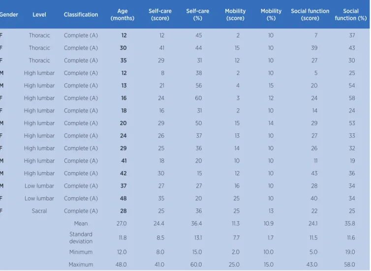
15 children were evaluated, six boys and nine girls, aged from 1 to 4 years and diagnosed with myelomeningocele. All of them were subjected to surgery to fix the injury within 48 hours after birth. According the evaluation by the International Standards for Neurological Classification of Spinal Cord Injury scale, of the American Spinal Injury Association, three children presented a thoracic level, nine showed high lumber injury, two had low lumber injury, and one featured a sacral level. They all featured complete spinal injury.
and the higher the injury, the greater the functional dependence detected. However, there was great variability and some exceptions to this general rule
(Table 1). PEDI scores ranged from 15 to 60% in the area self-care, from 10 to 15% in mobility, and from 19% to 58% in the social function.
Table 1. Level of injury, classification, age, and score in the Pediatric Evaluation of Disability Inventory
Gender Level Classification Age
(months)
Self-care (score)
Self-care (%)
Mobility (score)
Mobility (%)
Social function (score)
Social function (%)
F Thoracic Complete (A) 12 12 45 2 10 7 37
F Thoracic Complete (A) 30 41 44 15 10 39 43
F Thoracic Complete (A) 35 29 31 12 10 27 30
M High lumbar Complete (A) 12 8 38 2 10 5 25
M High lumbar Complete (A) 13 21 56 4 15 20 54
F High lumbar Complete (A) 16 24 60 3 12 24 58
F High lumbar Complete (A) 18 16 31 2 10 14 24
M High lumbar Complete (A) 20 29 50 15 14 29 53
F High lumbar Complete (A) 24 26 37 13 10 27 33
F High lumbar Complete (A) 29 25 36 14 10 26 32
M High lumbar Complete (A) 41 18 20 10 10 11 19
M High lumbar Complete (A) 42 30 15 12 10 43 36
M Low lumbar Complete (A) 37 27 27 16 10 28 34
F Low lumbar Complete (A) 48 35 20 25 10 40 34
F Sacral Complete (A) 28 25 36 25 13 22 25
Mean 27.0 24.4 36.4 11.3 10.9 24.1 35.8
Standard
deviation 11.8 8.5 13.1 7.7 1.7 11.5 11.6
Minimum 12.0 8.0 15.0 2.0 10.0 5.0 19.0
Maximum 48.0 41.0 60.0 25.0 15.0 43.0 58.0
DISCUSSION
This study described the functional independence and the level of injury of children with myelomeningocele from one to four years of age, evaluated by the Pediatric Evaluation of Disability Inventory and by the International Standards for Neurological Classification of Spinal Cord Injury scale of the American Spinal Injury Association.
PEDI scores ranged from 15 to 60% in the area self-care, from 10 to 15% in mobility, and from 19% to 58% in the social function. Therefore, mobility was the most affected aspect. There are few studies on the functional independence of children with myelomeningocele3,12,15,16. Overall, the higher the level
of the lesion, the greater the functional impairment – which was the hypothesis of this study. These findings are not in accordance with this study since some children with thoracic injury showed higher scores than those with lumbar injury in the areas self-care and social function.
Physical therapists have a fundamental role in stimulating the functional independence of children, through the continuous monitoring with the multidisciplinary team15-18. It is essential to guide
Physical therapy should promote the learning of motor skills, postural adjustments, independent locomotion (if necessary, with resources such as crutches or a wheelchair), to ensure the maximum functional independence possible3,5,9,12. Muscle stretching and
strengthening should be continuously carried out to assure the development and maintenance of strength and mobility. Consequently, is part of the physical therapist’s job to promote quality of life during all phases of development5,19.
According to Lundberg (2011), the higher the neurological level of injury, the worse will the quality of life of these patients be14. This can be explained by
the fact that higher levels tend to cause greater motor sensory, and cognitive damage; hence, the impact of the disease is greater, especially on mobility and
self-care4,5,11,20. In this study, though patients with thoracic
injury have had, indeed, low mobility scores, they varied greatly in self-care and social function. Given these findings, we emphasize the importance of encouraging early the used of wheelchairs in children with higher lesions, as well as the independence in self-care and social function11,19.
The study of Seitzber et al. (2008) indicates that a significant factor for the ambulance prognosis in children with myelomeningocele is age21. In this study,
the level of injury was more influential than age on the PEDI area of mobility. It is important to note that, overall, the children had low scores on mobility. That is, they did not show great mobility acquisition over the years. This did not occur for self-care and social function, in which there was greater independence since the first year of life.
In addition, the study shows that, even among complete injuries, the outcomes may vary, especially regarding self-care and social function. It is essential to use not only compensatory strategies but also stimulate motor gains, including the control below the level of injury, even in children with complete injuries.22
Functional independence should be always optimized so there is better quality of life.23
CONCLUSION
The level of injury in patients with myelomeningocele interferes with their mobility but has a smaller impact on self-care and social function in the age group from one to four years. It is essential to always used scores corrected by
age when evaluating this group because it has a significant interference on functional acquisitions. The monitoring with the multidisciplinary team is of paramount importance, especially including physical therapists to promote the neuropsychomotor performance.
REFERENCES
1. Zambelli H, Carelli E, Honorato D, et al. Assessment of neurosurgical outcome in children prenatally diagnosed with myelomeningocele and development of a protocol for fetal surgery to prevent hydrocephalus. Childs Nerv Syst. 2007;23:421-5. doi: 10.1007/s00381-006-0261-x
2. Rocco FM, Saito ET, Fernandes AC. Acompanhamento da locomoção de pacientes com mielomeningocele da Associação de Assistência à Criança Deficiente (AACD) em São Paulo, Brasil. Acta Fisiatr. 2007;14(3):126-9.
3. Tsai PY, Yang TF, Chan RC, Huang PH, Wong TT. Functional investigation in children with spina bifida, measured by the Pediatric Evaluation of Disability Inventory (PEDI). Childs Nerv Syst. 2002;18:48-53. doi: 10.1007/s00381-001-0531-6 4. Norrlin S, Strinnholm M, Carlsson M, Dahl M. Factors of
significance for mobility in children with myelomeningocele. Acta Paediatr. 2003;92:204-10. doi: 10.1111/j.1651-2227.2003. tb00527.x
5. Danielsson AJ, Bartonek A, Levey E, McHale K, Sponseller P, Saraste H. Associations between orthopaedic findings, ambulation and health-related quality of life in children with myelomeningocele. J Child Orthop. 2008;2:45-54. doi: 10.1007/s11832-007-0069-6
6. Warf BC. Hydrocephalus associated with neural tube defects: characteristics, management, and outcome in sub-Saharan Africa. Childs Nerv Syst. 2011;27:1589-94. doi: 10.1007/ s00381-011-1484-z
7. Schoenmakers MA, Uiterwaal CS, Gulmans VA, Gooskens RH, Helders PJ. Determinants of functional independence and quality of life in children with spina bifida. Clin Rehabil. 2005;19:677-85. doi: 10.1191/0269215505cr865oa
8. Roebroeck ME, Hempenius L, van Baalen B, Hendriksen JG, van den Berg-Emons HJ, Stam HJ. Cognitive functioning of adolescents and young adults with meningomyelocele and level of everyday physical activity. Disabil Rehabil. 2006;28:1237-42. doi: abs/10.1080/09638280600551716 9. Hetherington R, Dennis M, Barnes M, Drake J, Gentili F.
Functional outcome in young adults with spina bifida and hydrocephalus. Childs Nerv Syst. 2006;22:117-24. doi: 10.1007/ s00381-005-1231-4
10. Verhoef M, Barf HA, Post MW, van Asbeck FW, Gooskens RH, Prevo AJ. Functional independence among young adults with spina bifida, in relation to hydrocephalus and level of lesion. Dev Med Child Neurol. 2006;48:114-9. doi: 10.1017/ S0012162206000259
12. Sirzai H, Doqu B, Demir S, Yilmaz F, Kuran B. Assessment on self-care, mobility and social function in children with spina bifida in Turkey. Neural Regen Res 2014;15:1234-40. doi: 10.4103/1673-5374.135332
13. Mancini CM, Silva PC, Gonçalves SC, Martins MS. Comparação do desempenho funcional de crianças portadoras de síndrome de Down e crianças com desenvolvimento normal aos 2 e 5 anos de idade. Arq Neuropsiquiatr. 2003; 61(2-B):409-15. doi: 10.1590/S0004-282X2003000300016 14. Lundberg C. Validade e confiabilidade do questionário
de qualidade de vida de pessoas com espinha bífida [dissertação]. São Paulo: Faculdade de Medicina da Universidade de São Paulo; 2011.
15. Betz RR, Chafetz RS, Vogel LC, Samdani AF, Mulcahey MJ. Description of sensory preservation in children and adolescents with incomplete spinal cord injury. J Spinal Cord Med. 2011;34(3):297-300. doi: 10.1179/2045772311Y.0000000009 16. Brandão DA, Fujiwasa SD, Cardoso RJ. Características
de crianças com mielomeningocele: implicações para a fisioterapia. Fisioter Mov. 2009;22:69-75.
17. Collange AL, Franco CR, Esteves NR, Collange ZN. Desempenho funcional de crianças com mielomeningocele. Fisioter Pesqui. 2008;15:58-63. doi: 10.1590/S1809-29502008000100010 18. Medeiros MRD, Teixeira LL, Saraiva OL, Costa CSD, Nascimento
CGL. Plano terapêutico multidisciplinar para crianças com
mielomeningocele em um Hospital Universitário no Interior do Rio Grande do Norte. Rev Bras de Ciênc Saúde. 2011;15-2:219-22. doi: 10.4034/RBCS.2011.15.02.13
19. Ramos FS, Macedo LK, Scarlato A, Herrera G. Fatores que influenciam o prognóstico deambulatório nos diferentes níveis de lesão da mielomeningocele. Rev Neurociênc. 2005(13):80-6.
20. Bartonek A. Motor development toward ambulation in preschool children with myelomeningocele--a prospective study. Ped Phys Ther. 2010;22:52-60. doi: 10.1097/ PEP.0b013e3181cc132b
21. Seitzber A, Lind M, Biering-Sorensen F. Ambulation in adults with myelomeningocele. Is it possible to predict the level of ambulation in early life? Child Nerv Syst. 2008;(24):231-7. doi: 10.1007/s00381-007-0450-2
22. Aizawa CY, Morales MP, Lundberg C, Soares de Moura MCD, Pinto FCG, Voos MC, et al. Conventional physical therapy and physical therapy based on reflex stimulation showed similar results in children with myelomeningocele. Arq Neuro-Psiquiatr. 2017;5(3):160-6. doi: 10.1590/0004-282x20170009 23. Luz CL, Soares de Moura MCD, Becker KK, Teixeira RAA,