Schwann cells for spinal cord repair
1The Miami Project to Cure Paralysis, University of Miami, School of Medicine,
Miami, FL, USA
2Laboratório de Neuroregeneração, Departamento de Anatomia,
Instituto de Ciências Biomédicas, Universidade de São Paulo, São Paulo, SP, Brasil M. Oudega1,
L.D.F. Moon1 and
R.J. de Almeida Leme2
Abstract
The complex nature of spinal cord injury appears to demand a multi-factorial repair strategy. One of the components that will likely be included is an implant that will fill the area of lost nervous tissue and provide a growth substrate for injured axons. Here we will discuss the role of Schwann cells (SCs) in cell-based, surgical repair strategies of the injured adult spinal cord. We will review key studies that showed that intraspinal SC grafts limit injury-induced tissue loss and promote axonal regeneration and myelination, and that this response can be improved by adding neurotrophic factors or anti-inflammatory agents. These results will be compared with several other approaches to the repair of the spinal cord. A general concern with repair strategies is the limited functional recovery, which is in large part due to the failure of axons to grow across the scar tissue at the distal graft-spinal cord interface. Consequently, new synaptic connections with spinal neu-rons involved in motor function are not formed. We will highlight repair approaches that did result in growth across the scar and discuss the necessity for more studies involving larger, clinically relevant types of injuries, addressing this specific issue. Finally, this review will reflect on the prospect of SCs for repair strategies in the clinic.
Correspondence
M. Oudega The Miami Project to Cure Paralysis University of Miami School of Medicine 1095 NW 14th Terrace, R-48 Miami, FL 33136
USA
Fax: +1-305-243 3921 E-mail: [email protected]
Presented at the XIX Annual Meeting of the Federação de Sociedades de Biologia Experimental, Águas de Lindóia, SP, Brazil, August 25-29, 2004.
Publication supported by FAPESP.
Received May 27, 2005 Accepted February 24, 2005
Key words
•Spinal cord injury •Implantation •Regeneration •Neurotrophic factors •Clinical trial •Rehabilitation •Gene profiling
Introduction
Shortly after injury to the adult mamma-lian spinal cord, polymorphonuclear granu-locytes including neutrophils and, later on, lymphocytes, macrophages, and Schwann cells (SCs) invade the damaged area and local resident microglial cells become acti-vated (1). These inflammatory events in con-cert with other cytotoxic events result in progressive loss of spinal tissue, i.e., second-ary injury (2). In the case of a contusion injury, the resulting cavity often extends across the diameter of the cord leaving only a rim of spinal white matter (3-5). Damaged axons die back from the injury site (6,7) and
those that are spared may become demyeli-nated due to death of oligodendrocytes (8). Cellular and molecular changes at the injury site result in the formation of a growth-in-hibitory scar (9), which frustrates the axonal regeneration response often seen immedi-ately after injury (10). Consequently, axons do not grow beyond the lesion to form new synaptic connections with target neurons. Injury-induced partial or complete paralysis is permanent.
chroni-cally damaged cord will require implants to fill (bridge) the injury gap. Such implants will likely contain cells that need to be se-lected for any of the following criteria: com-patibility with spinal tissue, replacement of lost neurons or glial cells, promotion of axon regeneration, provision of a substrate/guid-ance for these axons, myelination of new sprouts and demyelinated axons, and ability to migrate into spinal tissue. The choice of implant will likely be determined by the nature and extent of the injury at the time of grafting. Cellular implants that have been explored include peripheral nerve, olfactory ensheathing glia (OEG), genetically engi-neered fibroblasts, fetal spinal tissue, stimu-lated macrophages, stem cells, and SCs. For
clarity and focus we will review here mainly the repair potential of SCs and compare their effects with some key approaches that in-volved other types of cells or tissue.
Spinal cord injury models
Human spinal cord injuries are very het-erogeneous (3-5). About 27% of human spi-nal cord injuries are lacerations caused by penetrating objects that tear the dura (‘open’ injuries) and spinal tissue, resulting in a discontinuity of the cord. This usually causes massive tissue loss, cyst formation, and a significant invasion of meningeal cells. The majority of the clinical cases are the result of a temporary compression of the cord that leaves the cord surface intact (‘closed’ ries; 73%). Three types of compression inju-ries are described: massive compression, contusion, and solid cord injury. A massive compression (44% of all compression inju-ries) causes substantial destruction/loss of spinal tissue. A contusion injury (31%) re-sults in the gradual formation of a central fluid-filled cyst and a minimal invasion of connective tissue. In case the contusion cyst progressively enlarges, it is referred to as syringomyelia, which is present in a limited number of cases. With a solid cord injury (25%) the shape of the cord is largely re-tained and there is no central hematomyelia and cyst formation, and mostly white matter tracts are damaged. Anatomically, compres-sion injuries are typically incomplete and present clinically as “central cord syndrome” with variable sensory and partial motor loss. Often anatomically incomplete compression injuries do result in complete paralysis.
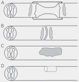
In the laboratory, a complete/partial dis-continuity of the spinal cord can be modeled using a surgical microknife or microscissors (Figure 1A,B,D). A complete transection causes major damage to spinal tissue and its blood supply, as well as the formation of a relatively large growth-inhibitory scar, which includes meningeal fibroblasts (11).
Consid-Figure 1. Schematic representation of various spinal cord repair models. In all cords rostral is on the left and dorsal is up. A, Completely transected spinal cord with tissue removed and a Schwann cell cable encased by a polymer tube implanted between the cord stumps. Note that only the back half of the tube is visible and that the cord ends are within the polymer tube (model used in Ref. 16). The dark gray areas close to the rostral and caudal graft-host spinal cord border repre-sent injected olfactory ensheathing glia (model used in Ref. 28). B, Completely transected spinal cord without removal of tissue and with olfactory ensheathing glia (dark gray) implanted in the rostral and caudal cord ends (model used in Ref. 29). C, Contused spinal cord with implantation of cells (darker gray; model used in Refs. 15,19,35,36,40,42). D, Dorsal column transec-tion and implantatransec-tion of a peripheral nerve bridge as used in Ref. 57.
B
C
erable practice of the surgery techniques and a well-organized and skilled animal mainte-nance team are essential before reliable sci-entific outcome and a tolerable survival rate can be achieved. A complete transection model allows for an unambiguous reading of the axonal regeneration response.
A contusion injury (Figure 1C) can be modeled by temporarily compressing the spinal cord (12,13), for which the New York University device is most frequently used. With this device a blunt 10 g weight is dropped onto the exposed cord from differ-ent heights resulting in injuries of graded severity. The electromagnetic spinal cord injury device from the Ohio State University (13) is also available; an impounder placed onto the cord compresses the cord over a precise distance in a short time period. If used appropriately both devices result in reproducible contusion injuries. With a con-tusion injury, the analysis of the axonal re-generation response is complex due to spared axons and their collateral sprouts.
Schwann cells in transection injuries
Early in the last century, Santiago Ramon y Cajal (14) documented the axonal growth-promoting abilities of peripheral nerve grafts in the injured central nervous system. Over the last decades many studies have explored the use of SCs, the major cellular component of peripheral nerve, for spinal cord repair either focusing on axonal regeneration (15-19) or remyelination (20-22).
Acute implantation of an SC/Matrigel cable contained within a polymer tube in the completely transected adult rat spinal cord (Figure 1A) promoted regeneration of prop-riospinal and sensory axons of which typi-cally about 25% were surrounded by SC myelin (16). The contribution of endoge-nous SCs that invade the implant from nearby roots (16) on the axonal growth/myelination response has not yet been properly deter-mined.
In this particular model, without addi-tional interventions, supraspinal axons did not regenerate into the SC bridge. Also, axons that grew into the implant failed to exit and grow into the spinal tissue beyond. Both these responses are essential for restoration of motor recovery controlled by the brain (23). It was clear that SC implantation needs to be combined with interventions to modify the permissiveness of the graft and/or graft-spinal cord interfaces that would then elicit supraspinal growth into and beyond the SC bridge. This was confirmed, at least in part, by increasing the levels of the neurotrophic factors, brain-derived neurotrophic factor (BDNF) and neurotrophin-3 (NT-3) within the SC graft environment (24) and by sys-temic administration of a high dose of the corticosteroid and anti-inflammatory agent, methylprednisolone shortly after SC graft implantation (25), which both resulted in supraspinal growth into the SC bridge. The elevation of neurotrophic levels caused a specific supraspinal response, i.e., the re-sponding axons derived from neurons that express tyrosine kinase receptor B and/or tyrosine kinase receptor C, the high affinity receptors for BDNF and NT-3, respectively (24).
The use of high doses of methylpredniso-lone following spinal cord injury has shown promise in the laboratory, but clinically its benefits have been a subject of disagreement (26). One of the concerns is that the reported functional improvements do not outweigh the possible secondary clinical complica-tions. The search for effective and clinically relevant neuroprotective agents is still going on.
further into the spinal cord. When an SC bridge in the completely transected cord was combined with adeno-associated viral vec-tor-mediated elevation of the levels of BDNF and NT-3 just caudal to the implant, axons still failed to grow into the caudal spinal tissue (27). This failure may reflect the fact that the levels of neurotrophic factors were not high enough to lure the axons through the graft-cord interface.
A different type of manipulation of the interfaces proved to be effective in promot-ing axon growth into the caudal cord. Graft-ing of OEG into the graft-cord interfaces combined with implantation of an SC bridge (Figure 1A) (28), and also in a transection only model (Figure 1B) (29), caused axons to exit the caudal bridge-cord interface and extend caudally. Why does OEG grafting into the cord near an injury result in such a response? At present, the underlying mechan-isms are not fully known, but it has been proposed that the OEG prevent (‘mask’) growing axons from recognizing inhibitory molecules in the scar tissue. Also, OEG are able to migrate within spinal nervous tissue (28,29), i.e., mingle with astrocytes (30), thereby accompanying growing axons. The migratory ability of OEG has been disputed (31) but it may set them apart from SCs, which fail to mingle with astrocytes.
There are several other strategies, fol-lowing a complete transection of the spinal cord, that result in growth of supraspinal axons into and/or axons out of the graft. And, similar to the experiments described above that involved SC grafting, all of these approaches have in common the fact that this response could only be achieved when graft-ing of cells/tissue into the injury site was accompanied by interventions that were de-signed to increase the levels of neurotrophins in the graft and/or to modify the graft-cord interfaces. Delayed (2-4 weeks) grafting of fetal spinal tissue promoted supraspinal growth, and, with the addition of BDNF and NT-3 at the implant site, axons grew beyond
the implant (32). Another study showed that bridging white to gray matter in a transection gap with peripheral nerves, which were sta-bilized using fibrin glue with acidic fibro-blast growth factor and compressive wiring of the nearby vertebrae, caused growth of corticospinal axons across the grafts and into the caudal cord (33).
In general, implantation of cells/tissue alone into an injury site is not sufficient to promote an axonal response that would lead to biologically significant functional recov-ery. Additional treatments are needed. It is difficult if not impossible to compare differ-ent existing combinatorial strategies for their axonal growth promoting abilities. Different groups perform the assessment of these re-sponses and of the functional improvements differently. Only a direct comparison be-tween strategies by one group would be sat-isfactory but this has not been done. Argu-ably, grafting of OEG has resulted in an impressive growth response of supraspinal axons beyond the injury site, which at 7 months after implantation had resulted in improved functional outcome (29). With the combination of an SC bridge and OEG im-plantation (28) hind limb motor improve-ments were not evaluated at earlier times. It is important to consider that for clinical use the harvest of OEG for grafting in the injured spinal cord does involve complicated and delicate surgical techniques. The results ob-tained by Cheng et al. (33) have been diffi-cult to reproduce by other groups, perhaps for technical reasons. Ethical issues compli-cate the use of fetal spinal tissue (32) in the clinic.
(34). As injury sites in the spinal cord can be extensive it will be an advantage if the grafted tissue can not only promote regeneration but also myelination of axons. This would im-prove conduction, i.e., functioning, of the regenerated axons. OEG naturally are en-sheathing cells and do not form myelin in situ, although following grafting of purified cultures of OEG a low degree of myelination is observed in the implantation site (35). For these myelination-related reasons, and other reasons mentioned above, SCs are a strong candidate for future surgical cell-based strat-egies to repair the spinal cord.
Schwann cells in contusion injuries
Purified adult rat SCs injected into the contused adultrat spinal cord (Figure 1C) limited injury-induced tissue loss (15,19,34). In addition, SC grafting into a contusion lesion promoted myelination and supraspi-nal and spisupraspi-nal axon sparing/regeneration, and improved hind limb motor function (35). Many different cell types have been grafted into the contused spinal cord, among them OEG (35,36), genetically modified fibro-blasts (37), stem cells (38), and macrophages (39), but only seldom have cell types been properly compared for their regenerative ef-fects. In one such comparative study, im-plantation of OEG was found to result in similar but slightly less strong improvements compared to those observed (and mentioned above) after SC grafting in the contused rat thoracic spinal cord (35). The main differ-ence between the groups was the superior ability of SCs to myelinate the responding axons.
In general, cell implantation into the con-tusion lesion results in a neuroprotective effect, which could indirectly be responsible for the observed improvements in the axonal and behavioral response. Recently, in an effort to profit more from neuroprotective effects, in the moderately contused spinal cord acute administration of two well-known
neuroprotective agents, methylprednisolone and interleukin-10, was combined with a 7-day delayed implantation of SC and/or OEG (40). This study demonstrated that the com-bination improved tissue sparing over the individual treatments but the overall func-tional improvements were largely similar between treatments or in some cases worse. In this study, it was not determined whether the approaches individually or in combina-tion caused either axonal sparing and/or ax-onal regeneration. The incompleteness of contusive injuries makes it difficult to distin-guish between spared and regenerated axons. The only reliable approaches ought to in-volve double-neuronal tracing techniques or time course studies, which both are techni-cally demanding and labor intensive.
The survival of cells implanted within a contusion environment may be compromised because of the ongoing immune, inflamma-tory, excitotoxic, proteolytic, and anoxic events (1,2). The optimal time for implanta-tion is not known and may be different for different types of cells. Grafting of SCs has been delayed up to 7-10 days, mainly to avoid the first wave of the inflammatory response (15,19,35). It is imperative to in-vestigate the fate of each type of cell after grafting into a contused spinal cord, and to determine the best time of grafting, to opti-mally benefit from the regeneration promot-ing effects of the implanted cell, especially when these cells have been genetically al-tered to secrete regeneration-supporting molecules.
interventions need to accompany cell graft-ing to increase, through either spargraft-ing or true regeneration, the number of supraspinal axons involved in motor control present in the caudal cord. A promising avenue to in-crease the overall regeneration response is the addition of cyclic adenosine monophos-phate (cAMP) analogs and/or preventing cAMP breakdown with phosphodiesterase inhibitors, such as Rolipram, which has been explored alone (41) or combined with cell transplantation (42-44) in various spinal cord repair models. It has been known for some time that increasing cAMP levels promote axonal extension (45), but the mechanisms are not fully known. Increased cAMP levels block axonal growth inhibition exerted by myelin-associated inhibitors, possibly through the protein kinase A/Rho pathway (46). It was demonstrated in vitro that the cAMP-mediated axonal growth response can be caused by a direct effect on axons rather than by the environment (47). With an SC graft into a moderately contused spinal cord, administration of cAMP and Rolipram en-hanced tissue sparing, axonal regeneration and functional outcome. Overall, the regen-erative response was larger than in any other combination approach that involved SCs. Further studies are necessary to elucidate the mechanisms behind these improvements.
Schwann cell - spinal cord interfaces
Regardless of the type of injury, the graft-host spinal cord interfaces are obstructive to axonal regeneration. Following an injury, reactive glial fibrillary acidic protein-posi-tive astrocytes, meningeal cells, and micro-glial cells form the micro-glial scar, a structural and chemical barrier for axon growth (8). The scar contains axonal growth inhibitory mol-ecules such as chondroitin sulfate proteogly-cans (CSPGs) (48) and semaphorins (11,49) and other myelin-associated proteins (50). So far, it is largely unknown how much different cell types contribute to the
forma-tion of the scar following injury/implanta-tion. Several observations suggest that ac-tual implantation of cells may increase the non-permissiveness of the interface. With an SC/Matrigel graft within a PAN/PVC tube implanted in the transected spinal cord an increased CSPG staining was found in both interfaces, but more so in the caudal one, at 3 weeks post-implantation (51). After a mod-erate contusion injury, a rim of CSPGs can be observed around the contused area, which persists for many months post-injury (35). Implantation of SCs one week after contu-sion injury increased the CSPG immuno-staining intensity at 8 weeks post-injury com-pared to control culture medium injections (35), but this has not been properly quanti-fied yet.
Overcoming the inhibitory nature of the graft-spinal cord interface
It is imperative to develop strategies to obtain axonal growth from grafts into the adjacent spinal nervous tissue. Approaches to obtain such a response in the injured adult spinal cord include: 1) decreasing the inhib-itory nature of the scar, 2) preventing axons from recognizing inhibitory molecules, and 3) enhancing the intrinsic growth ability of axons. The permissiveness of the scar for axons can be increased by preventing recep-tor-ligand (inhibitor) binding (52), by ob-structing the synthesis of inhibitors (53), or by degrading biologically active components of the inhibitors (54). The intrinsic growth ability of axons can be increased by targeting molecules downstream in the intracellular pathways that promote neurite extension or prevent growth cone collapse (55).
transec-tion/SC bridge model, a ‘larger size’ injury, with more severe scar formation, such ap-proaches have had no success in promoting axonal growth beyond the implant. As men-tioned above, grafting of OEG close to a large injury with (28) or without (29) an SC graft promoted axonal growth into the distal cord.
In several ‘smaller size’ injury/repair models, continuous infusion of neurotrophic factors a short distance away from a graft-spinal cord interface using an osmotic minipump resulted in axonal growth through scar tissue (Figure 1D) (57). However, com-bining an SC/fibrin bridge in the completely transected cord with adeno-associated viral vector-mediated elevation of the levels of BDNF and NT-3 just caudal to the bridge did not result in axonal growth beyond the graft, despite the observation that local spinal cells were infected with virus up to 16 weeks post-injury/treatment (27). With an SC/ Matrigel implant placed in the laterally hemisected spinal cord, continuous infusion with a minipump of BDNF and NT-3 cau-dally to the graft resulted in axonal growth into the caudal cord segments (58). In this model, some of the responding axons dis-played bouton-like structures (58), but it remains to be investigated whether this im-plied actual synaptic contacts that exhibit normal electrophysiological properties and/ or whether these axons were responsible for the behavioral improvements.
Delivering cAMP to dorsal root ganglia promotes growth of sensory axons beyond a dorsal column lesion (41). Also, with a dor-sal column injury, sensory axons were shown to exit a stromal cell implant, but only when combined with elevation of the levels of cAMP within dorsal root ganglia and admin-istration of NT-3 just rostral to the implanted area (43). The axonal regeneration responses seen after administration of cAMP, or eleva-tion of cAMP levels through inhibieleva-tion of their breakdown, have generated enthusiasm about its application in repair strategies for
the spinal cord (42-44). Although in some models the responses were interesting and promising, it is clear that more studies are necessary to fully understand and then profit from the actions of this drug. One aspect that needs attention is whether systemic delivery would result in unwanted side effects. Also, will this approach be successful in more severe injuries or, importantly, in chronic injuries?
The ability of grafted cells to promote central nervous system repair ultimately de-pends on the molecules they express after grafting. Similarly, the ability of a neuron to regenerate an axon into cellular environment depends on the molecules expressed. One may therefore envisage the possibility of engineering transplants and/or neurons to induce specific interactions, for example, to promote regeneration of specific axons into the transplants. Laser capture microdissec-tion and microarray profiling (59) now allow profiling of transcripts and proteins expressed by the grafted cells and neurons after spinal injury and transplantation. Specific neuron-transplant interactions can be engineered to remedy deficiencies using appropriate knock-up or knock-down genetic or pharmacologi-cal therapies. The concept of “tract-targeted repair” is attractive because “magic bullet” therapies could induce undesired side ef-fects, and therapies could be tailored to spe-cific injuries.
re-generation across scar tissue, we have not yet fully confronted the next crucial element of a successful repair strategy, i.e., the for-mation of synaptic connections by the regen-erating axons with spinal cord neurons.
Feasibility of clinical application of Schwann cells
In the clinic, treatment of spinal cord injury relies on preventing further damage using neuroprotective approaches and surgi-cal interventions such as decompression, sta-bilization, and detethering. Even under the best circumstances, the time between injury and these treatments is not short enough to prevent the onset of many acute events that result in (progressive) loss of tissue and neu-rological deficits. Thus, interventions aim-ing at replacaim-ing lost tissue and restoraim-ing lost axonal connections and motor and sensory function need to be developed. Could SCs be a component of such repair strategies?
From many experimental studies it has become clear that in case of a compression injury a combination strategy will be needed that includes the implantation of a cellular substrate to fill the cavity in order to promote axonal growth across and beyond the injury towards the lower areas of the cord that contain the neurons involved in motor func-tion. For optimal functioning of these cir-cuits, some regenerated axons may need to be myelinated. It is very likely that these newly formed axonal circuits are not identi-cal to the original ones (60), and additional rehabilitative therapies will be required to obtain biologically significant motor recov-ery that involves these new connections.
As discussed above, in the experimen-tally contused spinal cord, implantation of SCs fills the cavity, limits further tissue loss, and promotes regeneration of severed axons (15,19,35). Currently, additional interven-tions are being explored for their added ef-fects to the SC-mediated regeneration re-sponse. SCs are capable of myelinating
cen-tral axons that have grown into the graft. However, it has to be kept in mind that implanted SCs do not migrate into the sur-rounding adult spinal nervous tissue and can therefore not be expected to myelinate axons that have regenerated through and beyond the graft. It is imperative to also develop strategies that will rescue oligodendrocytes from dying following spinal cord injury. An important advantage of SCs over other cell types for implantation approaches is their ability to myelinate central axons. That plus the neuroprotective and regenerative abili-ties of SCs have established them as strong candidates for clinical cell-based repair strat-egies. However, as mentioned above, other cell types have also shown great promise for repair of the spinal cord.
Several issues need careful consideration before SC implantation strategies will be a legitimate option for repair strategies of the injured human spinal cord. The benefits of the procedure should outweigh the risks and the grafting technique should be safe and not exacerbate the neurological deficit. For this, visualization of the shape and dimensions of the lesion using magnetic resonance imaging before implantation could be advantageous. In general, any implantation strategy should not evoke immunological and/or inflamma-tory reactions. With SCs this can be accom-plished since they would permit autologous implantation thereby avoiding such reactions as well as the use of immunosuppressant drugs, such as cyclosporine-A, to prevent graft rejection. Autologous implantation can be performed by harvesting the SCs from a piece of peripheral nerve from the patient. The removal of a piece of a sensory nerve, such as the sural or saphenous nerve, may be preferred over removal of a piece of a motor nerve to avoid additional loss of motor func-tion. Also, the option for autologous implan-tation avoids ethical issues raised by the use of fetal tissue or embryonic stem cells.
necessary. Because SCs can be cultured in vitro with the help of mitogens large enough numbers can be obtained for implantation. One should be aware, though, that many divisions can induce the formation of malig-nant cells. This has not been observed fol-lowing multiple divisions of SCs in vitro. Another concern that has not yet been fully addressed in experimental studies is that mitogen-induced division may change the neuroprotective and regenerative abilities of SCs.
Taking all this in consideration,
autolog-ous implantation of SCs into the injured spinal cord has a future in the clinic. From experimental studies, many of which pre-sented in this review, it has become clear that grafting SCs alone will not result in substan-tial functional recovery. Additional inter-ventions and/or rehabilitative treatments need to be part of an SC-based repair strategy. These additional treatments need to be iden-tified before autologous SC implantation aimed at repairing the injured spinal cord will become a reality.
References
1. Schnell L, Fearn S, Klassen H, Schwab ME & Perry VH (1999). Acute inflammatory responses to mechanical lesions in the CNS: differ-ences between brain and spinal cord. European Journal of Neuro-science, 11: 3648-3658.
2. Anderson DK & Hall ED (1993). Pathophysiology of spinal cord trauma. Annals of Emergency Medicine, 22: 987-992.
3. Kakulas BA (1999). A review of the neuropathology of human spinal cord injury with emphasis on special features. Journal of Spinal Cord Medicine, 22: 119-124.
4. Bunge RP, Puckett WR, Becerra JL, Marcillo A & Quencer RM (1993). Observations on the pathology of human spinal cord injury. A review and classification of 22 new cases with details from a case of chronic cord compression with extensive focal demyelination.
Advances in Neurology, 59: 75-89.
5. Tuszynski MH, Gabriel K, Gerhardt K & Szollar S (1999). Human spinal cord retains substantial structural mass in chronic stages after injury. Journal of Neurotrauma, 16: 523-531.
6. McPhail LT, Stirling DP, Tetzlaff W, Kwiecien JM & Ramer MS (2004). The contribution of activated phagocytes and myelin degen-eration to axonal retraction/dieback following spinal cord injury.
European Journal of Neuroscience, 20: 1984-1994.
7. Oudega M, Vargas CG, Weber AB, Kleitman N & Bunge MB (1999). Long-term effects of methylprednisolone following transection of adult rat spinal cord. European Journal of Neuroscience, 11: 2453-2464.
8. Beattie MS, Hermann GE, Rogers RC & Bresnahan JC (1999). Cell death in models of spinal cord injury. Progress in Brain Research, 137: 37-47.
9. Fawcett JW & Asher RA (1999). The glial scar and central nervous system repair. Brain Research Bulletin, 49: 377-391.
10. Sharp FR & Sagar SM (1994) Alterations in gene expression as an index of neuronal injury: heat shock and immediate early gene response. Neurotoxicology, 15: 51-59.
11. De Winter F, Oudega M, Lankhorst AJ, Hamers FP, Blits B, Ruitenberg MJ, Pasterkamp RJ, Gispen WH & Verhaagen J (2002). Injury-induced class 3 semaphorin expression in the rat spinal cord.
Experimental Neurology, 175: 61-75.
12. Young W (2002). Spinal cord contusion models. Progress in Brain
Research, 137: 231-255.
13. Behrmann DL, Bresnahan JC, Beattie MS & Shah BR (1992). Spinal cord injury produced by consistent mechanical displacement of the cord in rats: behavioral and histologic analysis. Journal of Neurotrauma, 9: 197-217.
14. Ramon Y Cajal S (1928). Degeneration and Regeneration of the Nervous System (May RM, translator). Oxford University Press, London, UK.
15. Martin D, Robe P, Franzen R, Delree P, Schoenen J, Stevenaert A & Moonen G (1996). Effects of Schwann cell transplantation in a contusion model of rat spinal cord injury. Journal of Neuroscience Research, 45: 588-597.
16. Xu XM, Chen A, Guénard V, Kleitman N & Bunge MB (1997). Bridging Schwann cell transplants promote axonal regeneration from both the rostral and caudal stumps of transected adult rat spinal cord. Journal of Neurocytology, 26: 1-16.
17. Tuszynski MH, Weidner N, McCormack M, Miller I, Powell H & Conner J (1998). Grafts of genetically modified Schwann cells to the spinal cord: survival, axon growth, and myelination. Cell Transplan-tation, 7: 187-196.
18. Weidner N, Blesch A, Grill RJ & Tuszynski MH (1999). Nerve growth factor-hypersecreting Schwann cell grafts augment and guide spi-nal cord axospi-nal growth and remyelinate central nervous system axons in a phenotypically appropriate manner that correlates with expression of L1. Journal of Comparative Neurology, 413: 495-506. 19. Azanchi R, Bernal G, Gupta R & Keirstead HS (2004). Combined demyelination plus Schwann cell transplantation therapy increases spread of cells and axonal regeneration following contusion injury.
Journal of Neurotrauma, 21: 775-788.
20. Franklin RJ, Gilson JM & Blakemore WF (1997). Local recruitment of remyelinating cells in the repair of demyelination in the central nervous system. Journal of Neuroscience Research, 50: 337-344. 21. Blakemore WF & Crang AJ (1985). The use of cultured autologous
Schwann cells to remyelinate areas of persistent demyelination in the central nervous system. Journal of Neurological Sciences, 70: 207-223.
transplanted in the adult mouse spinal cord. Journal of Neuroimmu-nology, 40: 235-242.
23. Basso DM, Beattie MS & Bresnahan JC (2002). Descending sys-tems contributing to locomotor recovery after mild or moderate spinal cord injury in rats: experimental evidence and a review of literature. Restorative Neurology and Neuroscience, 20: 189-218. 24. Xu XM, Guénard V, Kleitman N, Aebischer P & Bunge MB (1995). A
combination of BDNF and NT-3 promotes supraspinal axonal regen-eration into Schwann cell grafts in adult rat thoracic spinal cord.
Experimental Neurology, 134: 261-272.
25. Chen A, Xu XM, Kleitman N & Bunge MB (1996). Methylpredniso-lone administration improves axonal regeneration into Schwann cell grafts in transected adult rat thoracic spinal cord. Experimental Neurology, 138: 261-276.
26. Gunnarsson T & Fehlings MG (2003). Acute neurosurgical manage-ment of traumatic brain injury and spinal cord injury. Current Opin-ion in Neurology, 16: 717-723.
27. Blits B, Oudega M, Boer GJ, Bartlett Bunge M & Verhaagen J (2003). Adeno-associated viral vector-mediated neurotrophin gene transfer in the injured adult rat spinal cord improves hind-limb function. Neuroscience, 118: 271-281.
28. Ramon-Cueto A, Plant GW, Avila J & Bunge MB (1998). Long-distance axonal regeneration in the transected adult rat spinal cord is promoted by olfactory ensheathing glia transplants. Journal of Neuroscience, 18: 3803-3815.
29. Ramon-Cueto A, Cordero MI, Santos-Benito FF & Avila J (2000). Functional recovery of paraplegic rats and motor axon regeneration in their spinal cords by olfactory ensheathing glia. Neuron, 25: 425-435.
30. Lakatos A, Franklin RJ & Barnett SC (2000). Olfactory ensheathing cells and Schwann cells differ in their in vitro interactions with astrocytes. Glia, 32: 214-225.
31. Ruitenberg MJ, Plant GW, Christensen CL, Blits B, Niclou SP, Harvey AR, Boer GJ & Verhaagen J (2002). Viral vector-mediated gene expression in olfactory ensheathing glia implants in the le-sioned rat spinal cord. Gene Therapy, 9: 135-146.
32. Coumans JV, Lin TT, Dai HN, MacArthur L, McAtee M, Nash C & Bregman BS (2001). Axonal regeneration and functional recovery after complete spinal cord transection in rats by delayed treatment with transplants and neurotrophins. Journal of Neuroscience, 21: 9334-9344.
33. Cheng H, Cao Y & Olson L (1996). Spinal cord repair in adult paraplegic rats: partial restoration of hind limb function. Science, 273: 510-513.
34. Imaizumi T, Lankford KL & Kocsis JD (2000). Transplantation of olfactory ensheathing cells or Schwann cells restores rapid and secure conduction across the transected spinal cord. Brain Re-search, 854: 70-78.
35. Takami T, Oudega M, Bates ML, Wood PM, Kleitman N & Bunge MB (2002). Schwann cell but not olfactory ensheathing glia trans-plants improve hindlimb locomotor performance in the moderately contused adult rat spinal cord. Journal of Neuroscience, 22: 6670-6681.
36. Plant GW, Christensen CL, Oudega M & Bunge MB (2003). Delayed transplantation of olfactory ensheathing glia promotes sparing/re-generation of supraspinal axons in the contused adult rat spinal cord. Journal of Neurotrauma, 20: 1-16.
37. Liu Y, Murray M, Tessler A & Fischer I (2000). Grafting of genetically modified fibroblasts into the injured spinal cord. Progress in Brain Research, 128: 309-319.
38. McDonald JW, Liu XZ, Qu Y, Liu S, Mickey SK, Turetsky D, Gottlieb
DI & Choi DW (1999). Transplanted embryonic stem cells survive, differentiate and promote recovery in injured rat spinal cord. Nature in Medicine, 5: 1410-1412.
39. Franzen R, Schoenen J, Leprince P, Joosten E, Moonen G & Martin D (1998). Effects of macrophage transplantation in the injured adult rat spinal cord: a combined immunocytochemical and biochemical study. Journal of Neuroscience Research, 51: 316-327.
40. Pearse DD, Marcillo AE, Oudega M, Lynch MP, Wood PM & Bunge MB (2004). Transplantation of Schwann cells and olfactory en-sheathing glia after spinal cord injury: does pretreatment with meth-ylprednisolone and interleukin-10 enhance recovery? Journal of Neurotrauma, 21: 1223-1239.
41. Qiu J, Cai D, Dai H, McAtee M, Hoffman PN, Bregman BS & Filbin MT (2002). Spinal axon regeneration induced by elevation of cyclic AMP. Neuron, 34: 895-903.
42. Pearse DD, Pereira FC, Marcillo AE, Bates ML, Berrocal YA, Filbin MT & Bunge MB (2004) cAMP and Schwann cells promote axonal growth and functional recovery after spinal cord injury. Nature Medi-cine,10: 610-616.
43. Lu P, Yang H, Jones LL, Filbin MT & Tuszynski MH (2004). Combina-torial therapy with neurotrophins and cAMP promotes axonal regen-eration beyond sites of spinal cord injury. Journal of Neuroscience, 24: 6402-6409.
44. Nikulina E, Tidwell JL, Dai HN, Bregman BS & Filbin MT (2004). The phosphodiesterase inhibitor rolipram delivered after a spinal cord lesion promotes axonal regeneration and functional recovery. Pro-ceedings of the National Academy of Sciences, USA, 101: 8786-8790.
45. Kilmer SL & Carlsen RC (1987). Chronic infusion of agents that increase cyclic AMP concentration enhances the regeneration of mammalian peripheral nerves in vivo. Experimental Neurology, 95: 357-367.
46. Cai D, Shen Y, De Bellard M, Tang S & Filbin M (1999). Prior exposure to neurotrophins blocks inhibition of axonal regeneration by MAG and myelin via a cAMP-dependent mechanism. Neuron, 22: 89-101.
47. Shearer MC, Niclou SP, Brown D, Asher RA, Holtmaat AJ, Levine JM, Verhaagen J & Fawcett JW (2003). The astrocyte/meningeal cell interface is a barrier to neurite outgrowth which can be over-come by manipulation of inhibitory molecules or axonal signaling pathways. Molecular and Cellular Neurosciences, 24: 913-925. 48. Properzi F & Fawcett JW (2004). Proteoglycans and brain repair.
News in Physiological Sciences,19: 33-38.
49. Lindholm T, Skold MK, Suneson A, Carlstedt T, Cullheim S & Risling M (2004). Semaphorin and neuropilin expression in motoneurons after intraspinal motoneuron axotomy. NeuroReport, 15: 649-654. 50. Filbin MT (2003). Myelin-associated inhibitors of axonal
regenera-tion in the adult mammalian CNS. Nature Reviews. Neuroscience, 4: 703-713.
51. Plant GW, Bates ML & Bunge MB (2001). Inhibitory proteoglycan immunoreactivity is higher at the caudal than the rostral Schwann cell graft-transected spinal cord interface. Molecular and Cellular Neurosciences, 17: 471-487.
52. Schnell L & Schwab ME (1990). Axonal regeneration in the rat spinal cord produced by an antibody against myelin-associated neurite growth inhibitors. Nature, 343: 269-272.
53. Grimpe B & Silver J (2004). A novel DNA enzyme reduces gly-cosaminoglycan chains in the glial scar and allows microtrans-planted dorsal root ganglia axons to regenerate beyond lesions in the spinal cord. Journal of Neuroscience, 24: 1393-1397.
Regenera-tion of CNS axons back to their following treatment of adult rat brain with chondroitinase ABC. Nature Neuroscience, 4: 465-466. 55. Spencer T & Filbin MT (2004). A role for cAMP in regeneration of the
adult mammalian CNS. Journal of Anatomy, 204: 49-55.
56. Bradbury EJ, Moon LD, Popat RJ, King VR, Bennett GS, Patel PN, Fawcett JW & McMahon SB (2002). Chondroitinase ABC promotes functional recovery after spinal cord injury. Nature, 416: 636-640. 57. Oudega M & Hagg T (1999). Neurotrophins promote regeneration of
sensory axons in the adult rat spinal cord. Brain Research, 818: 431-438.
58. Bamber NI, Li H, Lu X, Oudega M, Aebischer P & Xu XM (2001).
Neurotrophins BDNF and NT-3 promote axonal re-entry into the distal host spinal cord through Schwann cell-seeded mini-channels.
European Journal of Neuroscience, 13: 257-268.
59. Emmert-Buck MR, Bonner RF, Smith PD, Chuaqui RF, Zhuang Z, Goldstein SR, Weiss RA & Liotta LA (1996). Laser capture microdis-section. Science,274: 998-1001.