R E S E A R C H
Open Access
Purification and enzymatic characterization
of a novel metalloprotease from
Lachesis
muta rhombeata
snake venom
Francielle Almeida Cordeiro
1, Bárbara Marques Coutinho
1, Gisele Adriano Wiezel
1,
Karla de Castro Figueiredo Bordon
1, Cristiane Bregge-Silva
2, Nathalia Gonsales Rosa-Garzon
3, Hamilton Cabral
3,
Beatrix Ueberheide
4and Eliane Candiani Arantes
1*Abstract
Background:Lachesis muta rhombeata(Lmr) is the largest venomous snake in Latin America and its venom contains mainly enzymatic components, such as serine and metalloproteases, L-amino acid oxidase and phospholipases A2. Metalloproteases comprise a large group of zinc-dependent proteases that cleave basement
membrane components such as fibronectin, laminin and collagen type IV. These enzymes are responsible for local and systemic changes, including haemorrhage, myonecrosis and inflammation. This study aimed the isolation and enzymatic characterization of the first metalloprotease (Lmr-MP) from Lmr venom (LmrV).
Methods and results:Lmr-MP was purified through two chromatographic steps and submitted to enzymatic characterization. It showed proteolytic activity on azocasein with maximum activity at pH 7.0–9.0. It was inhibited by EDTA (a metal chelator that removes zinc, which is essential for enzymatic activity) and no effect was observed with PMSF, iodoacetic acid or pepstatin (inhibitors of serine, cysteine and aspartyl proteases, respectively). Ca2+, Mg2+ and Ba2+ions increased its activity, while Al3+, Cu2+, Ni2+and Zn2+inhibited it. Additionally, ZnCl2showed a dose
dependent inhibition of the enzyme. Lmr-MP activity was also evaluated upon chromogenic substrates for plasma kallikrein (S-2302), plasmin and streptokinase-activated plasminogen (S-2251) and Factor Xa (S-2222) showing the highest activity on S-2302. The activity in different solutions (5 mM or 50 mM ammonium bicarbonate, pH 7.8; 0.1% trifluoroacetic acid + 50% acetonitrile; phosphate buffer saline, pH 7.4; 50 mM sodium acetate, pH 4.0 or ammonium acetate pH 4.5) was also evaluated and the results showed that its activity was abolished at acidic pHs. Its molecular mass (22,858 Da) was determined by MALDI-TOF and about 90% of its primary structure was verified by high-resolution mass spectrometry using HCD and ETD fragmentations and database search against the sequence of closely related species. It is a novel enzyme which shared high identity with other snake venom metalloproteases (svMPs) belonging to the P-I group.
Conclusion:The purification procedure achieved a novel pure highly active metalloprotease from LmrV. This new molecule can help to understand the metalloproteases mechanisms of action, theLachesisenvenoming, as well as to open new perspectives for its use as therapeutic tools.
Keywords:Lachesis muta rhombeata, Metalloprotease, Proteases, Snake venom
* Correspondence:[email protected]
1Department of Physics and Chemistry, School of Pharmaceutical Sciences of
Ribeirão Preto, University of São Paulo (USP), Av. do Café s/n°, Monte Alegre, Ribeirão Preto, SP 14040-903, Brazil
Full list of author information is available at the end of the article
Background
Brazil is one of the countries with the highest number of accidents caused by terrestrial animals, such as scorpions, spiders, snakes, bees and caterpillars wherein more than 54% of them are due to snakes bites [1]. In 2016, the num-ber of accidents caused by snakes in Brazil was 26,295 (under review) and the most dangerous snake genera are
Bothrops, Crotalus and Lachesis, the latter representing around 3% of accidents [2]. However, in Northern Brazil the Lachesisaccidents reach up to 9% [1]. Although the number of accidents is lower than those caused by
Bothrops and Crotalus, the Lachesisbites cause a severe envenoming with hypotension, bleeding, pain and a vagal syndrome with diarrhoea, nausea and vertigo [3–5].
Lachesis genus, known as “bushmasters”, are the lar-gest snakes in Latin America and the only pit vipers in Brazil that lay eggs [6,7]. They are currently classified in
Lachesis stenophrys, Lachesis melanocephala (Central America),Lachesis acrochordaandLachesis muta(South America).L. mutais found in Brazil and subdivided into two subspecies: L. muta muta (Amazon tropical forest) andL. muta rhombeata(Atlantic forest) [5,6,8].
Among the components identified in Lachesis muta rhombeatavenom (LmrV), are a hyaluronidase [9], phos-pholipases A2(PLA2) [10,11], phospholipase B (PLB) [9],
L-aminoacid oxidase (LAAO) [12], serine protease [9] and bradykinin-potentiating peptides (BPPs) [13]. Although two snake venom metalloproteases (SVMPs) have been identified inL. muta mutavenom (LmmV), this is the first study with metalloprotease from LmrV.
Metalloproteases are one of the most abundant toxins in Viperid venoms. They are zinc-dependent proteases that cleaves the extracellular matrix (collagen, laminin and fibronectin) and can cause blood coagulation disor-ders. As consequence, they can lead to haemorrhage, fibrinogenolytic activity, activation of factor X and plate-let aggregation inhibition [14,15].
SVMPs were initially classified into P-I to P-IV classes, however, Fox and Serrano [16] proposed that P-IV class should be inserted into P-III. Therefore, SVMPs were cur-rently classified into P-I, P-IIa and PIIb, and P-IIIa to P-IIId. P-I MPs have 20–30 kDa and contain only the metallopro-tease catalytic domain; P-II MPs present 30–60 kDa with protease and disintegrin domains and P-III MPs are within the range of 60–100 kDa with protease, disintegrin and cysteine-rich domain sites [14,16].
SVMPs have been studied for several therapeutic pur-poses. Since they interact with cell membrane components, these enzymes have been shown to inhibit angiogenesis, cell migration and adhesion, which are important mechanisms in cancer proliferation and makes these enzymes important tools in metastatic tumours treatment [17–20]. Metallopro-teases also can act as therapeutic tools in arthritis disorders [21,22] and in haemostatic diseases [23].
In this study, we isolated the first metalloprotease from
Lachesis muta rhombeata venom (Lmr-MP) and its en-zyme activity was characterized against azocasein, under different ion concentrations and with the substrates plasma kallikrein (S-2302), plasmin, streptokinase-activated plas-minogen (S-2251) and Factor Xa (S-2222). Furthermore, we determined the optimal pH and the mass spectrometry analysis revealed that the glycosylation site observed in other snake metalloproteases is absent in Lmr-MP. The dis-covery of this new molecule can help to elucidate some mechanisms of action inLachesisenvenoming, as well as to contribute to treatment improvement and development of a therapeutic tool for haemostatic diseases.
Methods
Lachesis muta rhombeatavenom
The LmrV was acquired from the Serpentarium Bosque da Saúde in Americana (22° 44′ 21“ S, 47° 19’53” W) –São Paulo–Brazil (IBAMA authorization: 647.998). The venom was collected, desiccated and stored at−20 °C until use.
Isolation
The crude LmrV (around 23 mg) was dispersed in 500 μL of 0.05 M sodium acetate buffer with 0.15 M NaCl, pH 6.0, centrifuged at 13,400 xg at 4 °C for 10 min and the supernatant was filtered on a HiPrep Sephacryl® S-100 HR column (1.6 × 60 cm, GE Health-care, Sweden) under a flow rate of 0.5 mL/min.
The LmS-6 fraction obtained from the previous step was dispersed in buffer A (0.05 M MES - 2-(N -morpho-lino)ethanesulfonic acid, pH 6.0) and submitted to an ion exchange chromatrography on a HiTrap™IEX SP XL column (0.7 × 2.5 cm, 1 mL, GE Healthcare). The elution followed a concentration gradient from 0 to 1.0 M NaCl in the same buffer under a flow rate of 0.5 mL/min. The isolation was performed in FPLC Äkta Purifier UPC 900 system with monitoring of 280 nm.
SDS-page
Sodium dodecyl sulfate polyacrylamide gel electrophor-esis (SDS-PAGE) was performed according to Laemmli [24]. A resolution gel containing 13.5% (m/v) bisacryla-mide/acrylamide, 1 M Tris-HCl buffer and 0.1% sodium dodecyl sulfate (SDS) was prepared. The concentration gel was prepared with 5% acrylamide in 0.5 M Tris-HCl buffer and 0.1% SDS. The SDS-PAGE was performed under reducing conditions. The SDS-PAGE gel was per-formed to monitor the isolation process and sample mi-gration was compared to molecular mass standards (Sigma M3913 and M0671).
Mass spectrometry analysis by MALDI-TOF
-Time of Flight) in an Ultraflex II (Bruker Daltonics - DE) mass spectrometer, with a laser source of Nd SmartBeam-YAG laser type (MLN 202, LTB). The solution (1μL) con-taining Lmr-MP (5μg) was spotted with sinapinic acid (SA) matrix (10 mg/mL in a solution containing 0.2% trifluorace-tic acid and 80% acetonitrile), in the proportion of 1:1 (V/V). Ions were detected using a linear positive mode and cali-brated with protein standards from Bruker Daltonics.
Amino acid sequence determination
The metalloprotease amino acid sequence characterization was performed with lane 5 from SDS-PAGE (Fig.1b insert). The gel band was destained with a solution containing 100 mM ammonium bicarbonate (AMBIC: MetOH (50:50) and dehydrated with 100% acetonitrile (ACN). After this, sample was reduced with 100μL of 1,4-dithiothreitol (3 mg/ 1000 μL 100 mM AMBIC) for 1 h at 57 °C and alkylated with 100 μL of iodoacetamide (9 mg/1000 μL 100 mM AMBIC) for 45 min, at room temperature and within a dark compartment. For digestion, 222 ng of modified trypsin (Promega™, USA) in 160μL of 100 mM AMBIC was added and sample was incubated at 25 °C, overnight.
The digested peptides were submitted to an EASY-Spray PepSwift Monolithic Capillary column (Thermo Scientific™) with an Easy-nLC 1000 (Thermo Scientific™) coupled to an Orbitrap Elite™ Mass Spectrometer (Thermo Scientific™, USA). The tryptic peptides were eluted in 65 min using a gradient of ACN from 2 to 90% in 0.5% acetic acid and two independent runs were made. In the first run, the MS spec-tra were obtained with resolution of 60,000 (at m/z 400) and automatic gain control (AGC) target of 1e6. Subse-quently, twenty data-dependent HCD MS/MS were ac-quired with a resolution of 15,000 (at m/z 400), AGC target of 5e4, normalized collision energy of 35, and isolation win-dow of ±2 Da. In the second run, the MS resolution were obtained with resolution of 120,000 and automatic gain control (AGC) target of 5e4. Twenty data-dependent ETD MS/MS were acquired with a resolution of 15,000 (at m/z 400), AGC target of 5e4, activation time of 60 ms, and iso-lation window of ±3 Da.
Data from both runs were searched by the software Byo-nic™[25] against a database downloaded from UniProt using the keywords“metalloproteinase” and“Lachesis”(accession in 07/01/15). Mass tolerance was set as 10.0 ppm for precur-sor ions and 20.0 ppm for their fragments. Cysteine carba-midomethylation was set as fixed modifications whereas variable modifications included oxidation of methione, N-terminal pyro-glutamate, amidated C-terminal, and Hex-NAc (N-acetylglucosamine) for N-glycosylation. A wildcard feature of ±150 Da was enabled to search for amino acid substitutions in comparison to the database. The identified peptides were manually checked, specially those different from the database entries.
Lmr-MP activity in the presence of different inhibitors
The protease class was determined by activity assay in the presence of different inhibitors: ethylene-diaminetetraacetic acid (EDTA), iodoacetic acid (IAA), pepstatin (PEPS) and phenyl-methylsulfonyl fluoride (PMSF). Lmr-MP 100 μL (0.1 mg/mL) was previously incubated at a final concentra-tion of each inhibitor (10 mM) for 5 min at 37 °C [26]. After this previous incubation, 40 μL of HEPES buffer (100 mM, pH 8.0) and 1% azocasein prepared in the same buffer were added. The reaction was performed at 37 °C for 90 min. It was stopped by adding 400 μL of 10% (w/V) trichloroacetic acid solution. The samples were centrifuged
a
b
at 10,000×gat 25 °C for 15 min and 400μL of the super-natant were transferred to a new tube and mixed with 467 μL of sodium hydroxide (1 M). The absorbance was measured in a spectrophotometer at 440 nm. One activity unit (U) was defined as the amount of enzyme required to yield an increase of 0.001 A440nmunder the assays conditions
in according to Morita et al. [27]. The residual activity was determined based on control (without inhibitors) activity: Re-sidual activity = 100 x [(Sample activity)/ (Control activity)].
Effect of different ions on metalloprotease activity and different ZnCl2concentrations
To determine the effect of different ions on enzyme activ-ity, 100μL of Lmr-MP (0.1 mg/mL) was previously incu-bated at a final concentration of 10 mM CoCl2, LiCl,
MgCl2, KCl, ZnCl2, NiSO4, CuCl2, CaCl2, MnCl2, AlCl3,
BaCl2and NaCl at 37 °C for 5 min, according to Ducros
et al. [28] with modifications. The different ZnCl2
concen-tration (1, 2.5, 5 and 10 mM) effects on Lmr-MP activity was determined after 5 min of incubation at 37 °C. After the previous incubation, 40μL of HEPES buffer (100 mM, pH 8.0) and 1% azocasein prepared in same buffer were added. The reaction was performed for 90 min at 37 °C and was stopped by adding 400 μL of 10% (w/v) trichloroacetic acid solution. The samples were centri-fuged at 10,000×g for 15 min at 25 °C and 400μL of the supernatant were transferred to a new tube and mixed with 467 μL of 1 M sodium hydroxide. The absorbance was measured in a spectrophotometer at 440 nm. One activity unit (U) was defined as the amount of enzyme required to yield an increase of 0.001 A440nm under the
assays conditions in according to Morita et al. [27].
Lmr-MP activity with different solutions and optimal pH
Lmr-MP activity with 100 μL of different solutions and the optimal pH were performed according to the method of azocasein described in Wang et al. [29]. The sample, fraction LmS-6 (5 μg), was incubated with azo-casein (5 mg/mL) in 0.05 M Tris-HCl buffer, pH 8.0, in the presence of different solutions (5 and 50 mM AMBIC pH 7.8; 0.1% TFA + 50% ACN; PBS pH 7.4; 50 mM NaOAc pH 4; 50 mM NH4OAc pH 4.5 and
50 mM Tris-HCl pH 8.0) at 37 °C for 90 min. Another assay was carried out with the LmS-6 fraction (5 μg) incubated with azocasein at different pHs (4.5 to 9.0) as described above. The reactions were interrupted with 200 μL of 5% trichloroacetic acid. The mixture was centrifuged at 1000 xg for 5 min and 150 μL of each supernatant was transferred to a flat bottom 96-well microplate and added with 150 μL of 0.5 M sodium hydroxide. Albumin (1 mg/mL) was used as negative control and trypsin (1 mg/ mL) as positive control. The absorbance was measured at 450 nm in the Tecan Sunrise Microplate Reader (Tecan, Switzerland).
Lmr-MP activity upon different substrates
In Chromogenix® chromogenic substrate assay, 5.5μg of Lmr-S6 was incubated with 200 μL of each substrate (0.04 mM) - plasmatic kallikrein (S-2302), plasmin and plasminogen activated by streptokinase (S-2251) and Factor Xa (S-2222) for 40 min at 37 °C in triplicate. Absorbance reading was performed at 440 nm in the Tecan Sunrise Microplate Reader.
Statistical analysis
The statistical analysis was performed with the average and standard deviation (SD) and by analysis of variance (ANOVA) with multiple comparisons (Dunnett’s test). The results ofp< 0.0001,p< 0.01 andp< 0.05 were con-sidered statistically significant. The analyses were per-formed in triplicate.
Results
Lmr-MP isolation
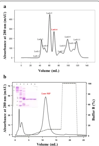
Lmr-MP isolation was performed through two chroma-tographic steps. Figure 1a shows the first step on a HiPrep Sephacryl® S-100 HR column, in which 11 frac-tions were obtained and named LmS-1 to LmS-11. LmS-6 showed previously (data not shown) a metallo-protease activity on azocasein and was subsequently sub-mitted to an ion exchange chromatography on HiTrap™ IEX SP XL column (Fig.1b). In this step, three fractions were obtained. The third fraction was responsible for metalloprotease activity on azocasein (as described below) and was named Lmr-MP.
SDS-page
Figure1c insert shows the analysis of the LmrV venom by SDS-PAGE. The crude venom, as well as fraction LmS-6 from the first chromatography step and Lmr-MP isolate in the second chromatographic step, were ana-lyzed. The analysis indicates that Lmr-MP was obtained with high purity and that its molecular mass is approxi-mately 23 kDa, being this mass consistent with metallo-proteases belonging to P-I class.
Mass spectrometry analysis of Lmr-MP
The MALDI-TOF mass spectrometry of Lmr-MP con-firmed its high purity level and the molecular mass deter-mined was 22.85 kDa (Fig.2a). The amino acid sequence analysis was performed on the gel band in lane 5 (Fig.1
200 300 400 500 600 700 800 900 1000 1100 1200 1300 1400 1500 1600 1700 1800 m/z
0 10 20 30 40 50 60 70 80 90
100 1117.5833
776.4142 428.1402
244.0924
904.4744
547.3083 618.3454
411.1140 1003.5417
1230.6669 476.2713
361.2448
330.1395 887.4465
689.3815
1100.5642
1458.7688 1343.7280 869.4245
1636.7911
y14
y13
y12
y11
y11–NH3
y10
y9
y8
y9–NH3
y7
y6
y5
b5
a5–NH3
y4
b4–NH3
b4–3NH3
y3
b3
b3–NH3
b2
QD internal ion
y2
b6
b8
a
c
b
22858.442
0 200 400 600 800 1000
In
te
n
s
.[
a
.u
.]
25000 30000 35000 40000 45000
m/z
N-glycosylation site in the 70th position in Lmr-MP in comparison to LHF-II (Fig.2b and c).
Lmr-MP activity with different inhibitors
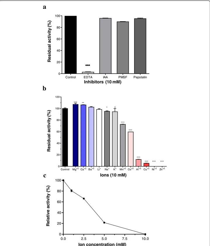
Lmr-MP proteolytic activity was performed with different inhibitors, as EDTA (an ionic chelator that can act as metal-loprotease inhibitor), IAA (cysteine protease inhibitor), PMSF (serine protease inhibitor) and pepstatin (aspartyl protease inhibitor). According to Fig. 3a, Lmr-MP activity was completely abolished when incubated with EDTA, sug-gesting that this enzyme is a metalloprotease, since EDTA is a zinc chelator.
Effect of different ions and ZnCl2on Lmr-MP activity
Lmr-MP activity was evaluated by enzyme incubation with different ions (CoCl2, LiCl, MgCl2, KCl, ZnCl2,
NiSO4, CuCl2, CaCl2, MnCl2, AlCl3, BaCl2and NaCl). It
was observed that Ca2+, Mg2+ and Ba2+ raised the en-zyme activity, whereas Al3+, Cu2+, Ni2+ and Zn2+ de-creased and inhibited the activity (Fig. 3b). Moreover,
Zn2+, in excess, negatively influences Lmr-MP activity as showed in Figure 3c. Increasing Zn2+concentration de-creases Lmr-MP activity.
LmS-6 activity with different substrate
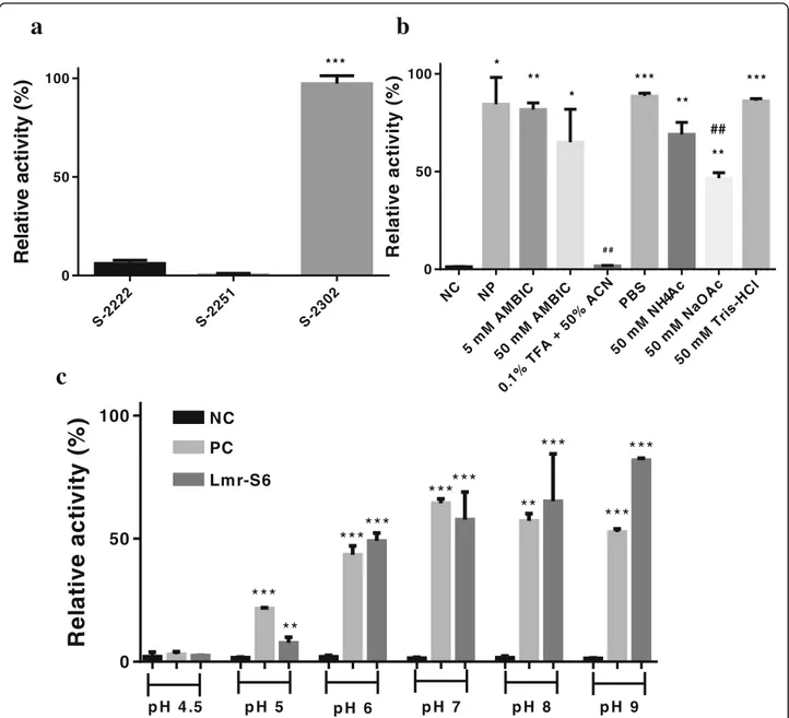
LmS-6 fraction was submitted to chromogenic substrates assay, and the substrate plasma kallikrein (S-2302), H-D-Pro-Phe-Arg-pNA sequence, was cleaved after the arginine residue. The S-2222 substrate with the sequence Bz-Ile-Glu-Gly-Arg-pNA was also cleaved, however with lower affinity. Moreover, the S-2251 substrate was not de-graded by LmS-6 (Fig.4a).
Lms-6 activity with different solutions and optimal pH
The metalloprotease activity in different solutions and the pH profile were evaluated by azocasein activity with LmS-6 fraction. LmS-6 had the highest activity when in-cubated with PBS, pH 7.4 and Tris-HCl, pH 8.0, while the enzyme activity was abolished in 0.1% TFA + 50% ACN (Fig. 4b). Moreover, the enzyme was optimally
(See figure on previous page.)
Fig. 2Spectrometry analysis and alignment.aMass spectrum of Lmr-MP. Molecular mass of Lmr-MP was obtained by MALDI-TOF (positive linear mode) using sinapinic acid (SA) matrix.bHCD MS/MS of the (M + 2H)+ 2ion of the tryptic peptide SNQDLINVQSAAADTLK acquired on an Orbitrap Elite™Mass Spectrometer with 15,000 resolution (at 400 m/z). N-terminal ions (aandb) are shown in red and indicated by˩whereas C-terminal ions (y) are shown in blue and indicated byГ. Internal ions are shown in green. Mass accuracy for all fragment ions is better than 20 ppm. The mass spectrometer used cannot differentiate between leucine and isoleucine residues, and the assignment is made here solely with homology matching.cSequence alignments of sv-MP hemorrhagic factor-2 (LHF-II) fromL. m. muta(UniProt ID P22796) and Lmr-MP fromL. m. rhombeata. The highly conserved residues are highlighted in black. Cys residues are shaded gray. Asn-aaX-Ser/Thr residue (star symbol) represents the N-glycosylation site. X = Leu/Ile. Alignment and figure were generated by MultAlin and ESPript servers, respectively
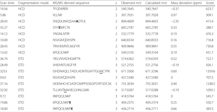
Table 1Tryptic peptides identified through MS/MS analysis
Scan time Fragmentation mode MS/MS derived sequence z Observed m/z Calculated m/z Mass deviation (ppm) Score
19.56 HCD TFGEWRER 2 540.7645 540.7647 −0.37 623.7
5.86 HCD VLLNR 2 307.7031 307.7028 0.97 309.1
28.43 HCD SNQDLINVQSAAADTLK 2 894.4609 894.4603 −2.35 415.4
35.27 HCD DYYEMFLTK 2 605.2787 605.2785 0.33 288.7
14.12 HCD YNGNLNTIR 2 532.7779 532.7778 0.19 476.3
10.00 HCD NSVGIVQDHSPK 2 640.8334 640.8333 0.16 716.8
28.43 HCD TRVHEIVNTLNGFYR 2 909.9846 909.9841 0.55 730.8
13.42 HCD KPQCILNKP 2 549.3105 549.3104 0.19 431.7
36.74 ETD YIELVVVADHGMFTK 3 574.6362 574.6359 0.52 722.1
28.49 ETD VHEIVNTLNGFYR 3 521.2755 521.2756 −0.19 926.1
3253 ETD ISHDNAQLLTAIDLADNTIGIAYTGGMCYPK 5 671.3300 671.3296 0.60 1359.6
9.63 ETD NSVGIVQDHSPK 3 427.5580 427.5580 0 707.5
37.16 ETD HDENHCHCSASFCIMPPSISEGPSYEFSDCSK 5 755.3034 755.3031 0.40 1248.5
32.92 ETD TLLIAVTMAHELGHNLGMK 4 517.0287 517.0288 −0.19 1093.1
9.72 ETD RKPQCILNKP 3 418.5764 418.5764 0 545.7
19.86 ETD KPQCILNKPX 3 404.2375 404.2374 0.25 550.7
18.00 ETD RKPQCILNKPX 3 456.2714 456.2711 0.66 383.5
Control EDTA IAA PMSF Pepstatin 0
20 40 60 80 100
***
a
Residual
activity
(%)
Inhibitors (10 mM)
Control Mg+2 Ca+2 Ba+2 Li+ Na+ K+ Mn+2 Co+2 Al+3 Cu+2 Ni+2 Zn+2 0
20 40 60 80 100 120
* ** **
***
***
***
***
*** *** ***
b
Residual activity
(%)
Ions (10 mM)
0.0 2.5 5.0 7.5 10.0
0 20 40 60 80 100
c
Relative activity (%)
Ion concentration (mM) ***
Fig. 3Enzymatic activity of Lmr-MP upon inhibitors and different ions.aAzocaseinolytic activity of Lmr-MP (10μg/100μL): in the absence (control) or presence of 10 mM different inhibitors (EDTA, IAA, PEPS and PMSF).bin the presence of 10 mM different ions (CoCl2, LiCl, MgCl2, KCl,
ZnCl2, NiSO4, CuCl2, CaCl2, MnCl2, AlCl3, BaCl2and NaCl).cin the presence of ZnCl2at different concentrations (2.5, 5.0, 7.5 and 10 mM). The
active in a pH range from 7.0 to 9.0 and the activity de-creased in acidic pHs (Fig.4c).
Discussion
Accidents caused byLachesisgenus are less common than
Crotalus and Bothrops envenomings in Brazil. However, large amount of venom is injected in victims bitten by bush-masters leading to severe symptoms such as hypotension, profuse diarrhoea, oedema and abnormal bleeding [3, 30].
These symptoms can be caused by different proteins and peptides present in Lmr venom.
Few components have been purified from L. muta rhombeata venom until now, including a L-amino acid oxidase, PLA2, PLB, hyaluronidase, serine protease and
BPPs [9, 10, 12, 13]. Although there are studies about these components, little is known about some mecha-nisms induced by these proteins and peptides, especially in Lachesisenvenomation, since its venom is difficult to obtain and is hard to keep bushmasters in captivity [5,31].
Re
la
ti
v
e
a
c
ti
v
it
y
(%)
0 50
100 NC
PC
p H 4 .5 p H 5 p H 6 p H 7 p H 8 p H 9
Lmr-S6
***
**
***
***
***
***
**
***
***
***
b
a
c
R
e
la
ti
ve
ac
ti
vi
ty
(%
)
S-2 222
S-22 51
S-2 302
0 50 100
* * *
R
el
at
ive
a
c
ti
vi
ty
(%
)
NC NP
5m MA
MB IC
50 mM
AM BIC
0.1% TFA
+50
%ACN PB S
50 mM
NH4 Ac
50 mM
NaO Ac
50 mM
Tri s-H
Cl
0 50
100 * **
*
* * *
**
** * * *
##
# #
Fig. 4Proteolytic activity of the fraction LmS-6.aupon 0.4 mM chromogenic substrates (Chromogenix®) for plasma kallikrein (S-2302), plasmin and plasminogen activated by streptokinase (S-2251) and Factor Xa (S-2222). ***p< 0.0001 compared among the tested substrates.bin the presence of different solutions. LmS-6 fraction (5μg) was incubated with 5 and 50 mM AMBIC, pH 7.8; 0.1% TFA + 50% ACN; PBS, pH 7.4; 50 mM NaOAc, pH 4; 50 mM NH4OAc, pH 4.5 and 50 mM Tris-HCl, pH 8.0 at 37 °C for 90 min. Each point represents the mean ± SD (n= 3), *p< 0.05, **p
In this paper we described the isolation of the first metal-loprotease from L. m. rhombeata subspecies through two chromatographic steps: a molecular exclusion followed by an ion exchange chromatography (Fig.1). The metallopro-tease activity was confirmed by EDTA inhibition on enzym-atic assay and no effect caused by other proteases inhibitors (PMSF, IAA and pepstatin) was observed (Fig. 3a). The metalloprotease was named as Lmr-MP and the SDS-PAGE and MALDI-TOF analysis showed that it was efficiently purified (Fig.1c insert and Fig.2a). Moreover, the molecular mass determined (22.85 kDa) is in accordance to the mass of other SVMPs and indicates that Lmr-MP is a metallopro-tease from P-I class [14,32,33].
P-I to P-IIId metalloproteases classification compre-hends different multidomains. In P-I case, there is only a signal peptide, a pro-domain and a metalloprotease do-main. The signal peptide is responsible for protein secre-tion, the pro-domain is related to catalytic activation and the metalloprotease domain encodes the enzyme sequence [16, 34]. In general, P-I class metalloproteases are less haemorrhagic than P-III classes possibly because of the multiple domains associated with P-III sequences [32].
Until now, two metalloproteases have been purified from
L. muta muta venom, LHF-I and LHF-II (also named Mutalysin-II) [35–37]. An alignment between only LHF-II and Lmr-MP was performed since both are P-I SVMPs [38]. The alignment revealed high similarity between these en-zymes despite differences in some amino acids (Fig. 2c). LHF-II has a N-glycosylation site in Asn70, while Lmr-MP has an Ala in the same position (Fig2b). The calculated mass of LHF-II, from its primary sequence, was 22,595.77 Da [39]. In comparison to the molecular mass of Lmr-MP deter-mined by MALDI-TOF, the difference between them is only 262.67 Da, consisted with mass variation regarding amino acid substitutions and not due to glycosylations [40]. Further-more, the change of Asn by Ala in Lmr-MP lead to the loss of a potential N-glycosylation site.
Pla et al. [41] described the proteome analysis of La-chesis muta rhombeatavenom. Around 29.5% of venom composition was SVMPs (10.3% of this value are related to metalloproteases from class P-III and 19.2% from P-I class). The authors found similarity of metalloprotease from P-I class with LHF-II described above. In addition, another study from dos Santos [42], also revealed the presence of one metalloprotease from P-I class similar to LHF-II. The metalloprotease found in both studies are probably the Lmr-MP or an isoform.
Although P-I metalloproteases class have no rele-vant haemorrhagic activity, they degrade other mem-brane components and appear to be related to pathogenic effects of local damage observed in enve-noming [43]. Because of their fibrinolytic and not haemorrhagic effects, the therapeutic potential of these metalloproteases for thrombolytic events has
been studied [37], suggesting a potential therapeutic effect for Lmr-MP.
We evaluated Lmr-MP activity in the presence of dif-ferent ions (Fig. 3b). The enzymatic activity increased with Ca2+, Mg2+ and Ba2+. In contrast, the activity was inhibited by Al3+, Cu2+, Ni2+ and Zn2+. Therefore, Lmr-MP is activated by divalent ions. However, zinc ions inhibited this activity. This inhibition by zinc was dose-dependent, as demonstrated in Fig.3c. Some previ-ous studies have shown that divalent ions, such as Ca2+, help to stabilize the protein. On the other hand, al-though Zn2+is present in metalloprotease structures and is fundamental to proteolytic activity, if in excess, it can inhibit enzyme activity by causing stereochemical distur-bances in the stabilization of the molecule [33,36,44].
The proteolytic activity of LmS-6 fraction upon chromogenic substrates showed the highest activity when the enzyme was incubated with S- 2302, the substrate for plasma kallikrein (H-D-Pro-Phe-Arg-pNA) (Fig. 4a). Plasma kallikrein is important in human physiology, spe-cifically in release of bradykinin (BK). It is activated by fac-tor XIIa and then cleaves high-molecular-mass kininogen to generate bradykinin [45]. Therefore, the metallopro-tease isolated in this study may act in important systems, bringing perspectives for the use of Lmr-MP as a thera-peutic agent in haemostatic disorders. Despite this, more studies are needed to prove the activity of the enzyme on the substrate and to infer its activity in envenoming and as therapeutic agent.
Additionally, Lmr-MP activity was evaluated in the presence of different solutions, such as 5 and 50 mM AMBIC (pH 7.8), 0.1% TFA + 50% ACN, PBS pH 7.4, 50 mM NaOAc pH 4, 50 mM NH4 OAc pH 4.5 and
50 mM Tris-HCl pH 8.0. The proteolytic activity on azo-casein was higher in 50 mM Tris-HCl buffer (pH 8.0) and PBS pH 7.4, which corroborated with optimal pH analysis (Fig. 4c) in which the optimal range was deter-mined between 7.0 and 9.0 (Fig. 4b and c). Moreover, it was observed the loss of metalloprotease activity in 0.1% TFA + 50% ACN solution (Fig. 4b) when compared to the negative control of the assay, probably due to the loss of its cofactor and because of acetonitrile, which may interrupt hydrophobic and ionic interactions [46]. These results also indicate that the metalloprotease loses its activity in acidic pHs. The MP LHF-II is also stable in the pH range from 8 to 10 [36].
Conclusion
that ion zinc inhibit its activity in a dose-dependent man-ner and this enzyme lose activity in acidic pHs. These re-sults open new perspectives for Lachesis venom and metalloproteases knowledge and, although more studies must be performed to provide a therapeutic activity, Lmr-MP showed preferentially inhibit the kallikrein plasma substrate, that plays a critical role in physiological processes, making it a favorable candidate for future pharmaceutical tools.
Abbreviations
ACN:Acetonitrile; AlCl3: Aluminium chloride; AMBIC: Ammonium
bicarbonate; ANOVA: Analysis of variance; BaCl2: Barium chloride;
BK: Bradykinin; CaCl2: Calcium chloride; CoCl2: Cobalt chloride; CuCl2: Cupric
chloride; EDTA: Ethylenediaminetetraacetic acid; ETD: Electron-transfer dissociation; FPLC:Fast protein liquid chromatography; HCD: High-energy collisional dissociation; HEPES: 4-(2-hydroxyethyl)-1-piperazineethanesulfonic acid; IAA: Iodoacetic acid; KCl: Potassium chloride; LAAO: L-aminoacid oxidase; LHF-I and II: Metalloproteases fromLachesis muta mutavenom; LiCl: Litium chloride; Lmr:Lachesis muta rhombeata; Lmr-MP: Metalloprotease fromLachesis muta rhombeatavenom; LmrV:Lachesis muta rhombeatavenom; LmS-6: Sixth fraction of theLachesis muta rhombeatavenom on Sephacryl chromatography; Magnesium chloride: Sodium chloride; MALDI-TOF: Matrix Assisted Laser Desorption Ionisation; MES: 2-(N-morpholino) ethanesulfonic acid; MetOH: Methanol; MgCl2: Manganese(II) chloride; MnCl2: Magnesium chloride;
NaOAc: Sodium acetate; NH4OAc: Ammonium acetate; NiSO4: Nickel sulfate;
PBS: Phosphatebufferedsaline; PEPS: Pepstatin; PLA2: Phospholipases A2;
PLB: Phospholipase B; PMSF: Phenylmethylsulfonyl fluoride; SA: Sinapinic acid; SD: Standard deviation SD; SDS-PAGE: Sodium dodecyl sulfate polyacrylamide gel electrophoresis; svMPs: Snake venom metalloproteases; TFA: Trifluoracetic acid; Tris-HCl: Tris hydrochloride buffer; ZnCl2: Zinc chloride
Funding
Financial support of Coordination for the Improvement of higher Education Personnel (CAPES Finance Code 001). This study was supported by the National Institute of Health (NIH, USA grant R41 GM103362); São Paulo Research Foundation (FAPESP, grants 2011/23236–4 and scholarship to GAW 2014/23285–3 and BMC 2016/09711–5); Coordination for the Improvement of Higher Education Personnel (CAPES, through Programa Editoração CAPES, edital 13/2016, grant 0722/2017, 88881.142062/2017–01) and National Council for Scientific and Technological Development (CNPq, through Programa Editorial CNPq/CAPES, chamada 26/2017, grant 440954/2017–7; grant 303689/2013–7 and scholarship to FAC 140949/2015–1).
Availability of data and materials
Not applicable.
Author’s contributions
FAC was responsible for project development, enzyme purification, major part of the assays, analysis of the results and writing of the manuscript. BMC performed the enzymatic experiments and participated in the Lmr-MP isolation. GCW did spectrometry analysis and participated of data interpretation and analysis. KCFB and CBS participated of purification and structural characterization of Lmr-MP. NGRG and HC participated of some enzymatic assays. BU contributed with mass spectrometry analysis and coordinated GCW analysis during her internship. ECA coordinated and designed the experiments and contributed in writing the manuscript. All authors read and approved the final manuscript.
Ethics approval and consent to participate
Not applicable.
Consent for publication
Not applicable.
Competing interests
The authors declare that they have no competing interests.
Publisher’s Note
Springer Nature remains neutral with regard to jurisdictional claims in published maps and institutional affiliations.
Author details
1Department of Physics and Chemistry, School of Pharmaceutical Sciences of
Ribeirão Preto, University of São Paulo (USP), Av. do Café s/n°, Monte Alegre, Ribeirão Preto, SP 14040-903, Brazil.2Universidad Latina de Costa Rica, San
José, Costa Rica.3Department of Pharmaceutical Sciences, School of
Pharmaceutical Sciences of Ribeirão Preto, University of São Paulo (USP), Ribeirão Preto, SP, Brazil.4Proteomics Resource Center, New York University
Langone Medical Center, 430 East 29th St, New York City 10016, USA.
Received: 29 June 2018 Accepted: 1 November 2018
References
1. Chippaux JP. Epidemiology of envenomations by terrestrial venomous animals in Brazil based on case reporting: from obvious facts to contingencies. J Venom Anim Toxins Incl Trop Dis. 2015;21:13.https://doi. org/10.1186/s40409-015-0011-1.
2. Brasil. Ministério da Saúde. Acidentes por animais peçonhentos -Notificações registradas no Sistema de Informação de Agravos de Notificação. 2018.
3. Jorge MT, Sano-Martins IS, Tomy SC, Castro SC, Ferrari RA, Ribeiro LA, et al. Snakebite by the bushmaster (Lachesis muta) in Brazil: case report and review of the literature. Toxicon. 1997;35(4):545–54.
4. Cremonez CM, Leite FP, Bordon KD, Cerni FA, Cardoso IA, Gregório ZM, et al. ExperimentalLachesis muta rhombeataenvenomation and effects of soursop (Annona muricata) as natural antivenom. J Venom Anim Toxins Incl Trop Dis. 2016;22:12.https://doi.org/10.1186/s40409-016-0067-6.
5. Duque AMH, Corrales G. First report of the reproduction in captivity of the Chocoan bushmaster,Lachesis acrochorda(García, 1896). Herpetol Notes. 2015;8:315–20.
6. Stephano MA, Guidolin R, Higashi HG, Tambourgi DV, Sant'Anna OA. The improvement of the therapeutic anti-Lachesis muta serum production in horses. Toxicon. 2005;45(4):467–73.
7. Pardal PPO, Bezerra IS, Rodrigues LS, Pardal JSO, Farias PHS. Acidente por Surucucu (Lachesis muta muta) em Belém-Pará: Relato de caso. Rev Para Med. 2007;21(1):37–42.
8. Bolaños R, Muñoz G, Cerdas L. Toxicity, neutralization and
immunoelectrophoresis of the venom ofLachesis mutafrom Costa Rica and Colombia (author's transl). Toxicon. 1978;16(3):295–300.
9. Wiezel GA, dos Santos PK, Cordeiro FA, Bordon KCF, Selistre-de-Araujo HS, Ueberheide B, et al. Identification of hyaluronidase and phospholipase B in Lachesis muta rhombeatavenom. Toxicon. 2015;107(Pt B):359–68. 10. Cordeiro FA, Perini TG, Bregge-Silva C, Cremonez CM, Rodrigues RS,
Boldrini-França J, et al. A new phospholipase A2 fromLachesis muta rhombeata: purification, biochemical and comparative characterization with crotoxin B. Protein Pept Lett. 2015;22(9):816–27.
11. Damico DC, Vassequi-Silva T, Torres-Huaco FD, Nery-Diez AC, de Souza RC, Da Silva SL, et al. LmrTX, a basic PLA2(D49) purified fromLachesis muta
rhombeatasnake venom with enzymatic-related antithrombotic and anticoagulant activity. Toxicon. 2012;60(5):773–81.
12. Bregge-Silva C, Nonato MC, de Albuquerque S, Ho PL, Junqueira de Azevedo IL, Vasconcelos Diniz MR, et al. isolation and biochemical, functional and structural characterization of a novel L-amino acid oxidase fromLachesis mutasnake venom. Toxicon. 2012;60(7):1263–76. 13. Pinheiro-Júnior EL, Boldrini-França J, de Campos Araújo LMP, Santos-Filho
NA, Bendhack LM, Cilli EM, et al. LmrBPP9: a synthetic bradykinin-potentiating peptide fromLachesis muta rhombeatavenom that inhibits the angiotensin-converting enzyme activityin vitroand reduces the blood pressure of hypertensive rats. Peptides. 2018;102:1–7.
14. Markland FS Jr, Swenson S. Snake venom metalloproteinases. Toxicon. 2013; 62:3–18.
15. Moura-da-Silva AM, Almeida MT, Portes-Junior JA, Nicolau CA, Gomes-Neto F, Valente RH. Processing of snake venom metalloproteinases: Generation of toxin diversity and enzyme inactivation. Toxins (Basel). 2016;8(6):pii: E183. 16. Fox JW, Serrano SM. Insights into and speculations about snake venom
17. Calderon LA, Sobrinho JC, Zaqueo KD, de Moura AA, Grabner AN, Mazzi MV, et al. Antitumoral activity of snake venom proteins: new trends in cancer therapy. Biomed Res Int. 2014;2014:203639.
18. Maria DA, da Silva MG, Correia Junior MC, Ruiz IR. Antiproliferative effect of the jararhagin toxin on B16F10 murine melanoma. BMC Complement Altern Med. 2014;14:446.
19. Guimarães DO, Lopes DS, Azevedo FV, Gimenes SN, Silva MA, Achê DC, et al.In vitroantitumor and antiangiogenic effects of Bothropoidin, a metalloproteinase fromBothrops pauloensissnake venom. Int J Biol Macromol. 2017;97:770–7.
20. Urra FA, Araya-Maturana R. Targeting metastasis with snake toxins: molecular mechanisms. Toxins (Basel). 2017;9(12):pii E390.
21. Rowan AD, Litherland GJ, Hui W, Milner JM. Metalloproteases as potential therapeutic targets in arthritis treatment. Expert Opin Ther Targets. 2008; 12(1):1–18.
22. Itoh Y. Metalloproteinases in rheumatoid arthritis: potential therapeutic targets to improve current therapies. Prog Mol Biol Transl Sci. 2017;148:327–38. 23. Kini RM, Koh CY. Metalloproteases affecting blood coagulation, fibrinolysis
and platelet aggregation from snake venoms: definition and nomenclature of interaction sites. Toxins (Basel). 2016;8(10):pii: E284.
24. Laemmli UK. Cleavage of structural proteins during the assembly of the head of bacteriophage T4. Nature. 1970;227(5259):680–5.
25. Bern M, Kil YJ, Becker C. Byonic: advanced peptide and protein identification software. Curr Protoc Bioinformatics. 2012;13(13):20.
26. Beynon RJ, Bond JS. Proteolytic enzymes: a practical approach. Oxford: IRL Press at Oxford University Press; 1989.
27. Morita Y, Hasan Q, Sakaguchi T, Murakami Y, Yokoyama K, Tamiya E. Properties of a cold-active protease from psychrotrophic Flavobacterium balustinum P104. Appl Microbiol Biotechnol. 1998;50(6):669–75.
28. Ducros E, Ferrari M, Pellegrino M, Raspanti C, Bogni C. Effect of aeration and agitation on the protease production byStaphylococcus aureusmutant RC128 in a stirred tank bioreactor. Bioprocess Biosyst Eng. 2009;32(1):143–8. 29. Wang WJ, Shih CH, Huang TF. A novel P-I class metalloproteinase with
broad substrate-cleaving activity, agkislysin, fromAgkistrodon acutusvenom. Biochem Biophys Res Commun. 2004;324(1):224–30.
30. Pardal PP, Souza SM, Monteiro MR, Fan HW, Cardoso JL, França FO, et al. Clinical trial of two antivenoms for the treatment ofBothropsandLachesis bites in the north eastern Amazon region of Brazil. Trans R Soc Trop Med Hyg. 2004;98(1):28–42.
31. Dias L, Rodrigues MA, Rennó AL, Stroka A, Inoue BR, Panunto PC, et al. Hemodynamic responses toLachesis muta(south American bushmaster) snake venom in anesthetized rats. Toxicon. 2016;123:1–14.
32. Gutiérrez JM, Escalante T, Rucavado A, Herrera C, Fox JW. A comprehensive view of the structural and functional alterations of extracellular matrix by snake venom metalloproteinases (SVMPs): novel perspectives on the pathophysiology of envenoming. Toxins (Basel). 2016;8(10):pii: E304. 33. Gutiérrez JM, Rucavado A, Escalante T, Díaz C. Hemorrhage induced by
snake venom metalloproteinases: biochemical and biophysical mechanisms involved in microvessel damage. Toxicon. 2005;45(8):997–1011.
34. Ramos OH, Selistre-de-Araujo HS. Snake venom metalloproteases--structure and function of catalytic and disintegrin domains. Comp Biochem Physiol C Toxicol Pharmacol. 2006;142(3–4):328–46.
35. Sánchez EF, Magalhães A, Diniz CR. Purification of a hemorrhagic factor (LHF-I) from the venom of the bushmaster snake,Lachesis muta muta. Toxicon. 1987;25(6):611–9.
36. Sánchez EF, Magalhës A, Mandelbaum FR, Diniz CR. Purification and characterization of the hemorrhagic factor II from the venom of the bushmaster snake (Lachesis muta muta). Biochim Biophys Acta. 1991; 1074(3):347–56.
37. Agero U, Arantes RM, Lacerda-Queiroz N, Mesquita ON, Magalhães A, Sanchez EF, et al. Effect of mutalysin II on vascular recanalization after thrombosis induction in the ear of the hairless mice model. Toxicon. 2007; 50(5):698–706.
38. Sanchez EF, Diniz CR, Richardson M. The complete amino acid sequence of the haemorrhagic factor LHFII, a metalloproteinase isolated from the venom of the bushmaster snake (Lachesis muta muta). FEBS Lett. 1991;282(1):178–82. 39. Gasteiger E, Hoogland C, Gattiker A, Duvaud S, Wilkins MR, Appel RD, et al.
Protein identification and analysis tools on the ExPASy Server. In: The Proteomics Protocols Handbook, Humana Press. Walker JM editor. 2005. pp. 571–607. 40. Parker CE, Mocanu V, Mocanu M, Dicheva N, Warren MR. Mass spectrometry
for post-translational modifications. Alzate O, editor. Neuroproteomics. Boca
Raton: CRC Press/Taylor & Francis; 2010. Available from:https://www.ncbi. nlm.nih.gov/books/NBK56018/
41. Pla D, Sanz L, Molina-Sánchez P, Zorita V, Madrigal M, Flores-Díaz M, et al. Snake venomics ofLachesis muta rhombeataand genus-wide antivenomics assessment of the paraspecific immunoreactivity of two antivenoms evidence the high compositional and immunological conservation across Lachesis. J Proteome. 2013;89:112–23.
42. dos Santos PK. Proteoma da peçonha deLachesis muta rhombeata. Universidade Federal de São Carlos, Departamento de Ciências Fisiológicas; 2013.https://repositorio.ufscar.br/handle/ufscar/5511. Acessed 20 Aug 18. 43. Rucavado A, Flores-Sánchez E, Franceschi A, Magalhaes A, Gutiérrez JM.
Characterization of the local tissue damage induced by LHF-II, a metalloproteinase with weak hemorrhagic activity isolated fromLachesis muta mutasnake venom. Toxicon. 1999;37(9):1297–312.
44. Gomez-Ortiz M, Gomis-Rüth FX, Huber R, Avilés FX. Inhibition of carboxypeptidase a by excess zinc: analysis of the structural determinants by X-ray crystallography. FEBS Lett. 1997;400(3):336–40.
45. Bryant JW, Shariat-Madar Z. Human plasma kallikrein-kinin system: physiological and biochemical parameters. Cardiovasc Hematol Agents Med Chem. 2009;7(3):234–50.