Structural insights into selectivity and cofactor binding in snake venom
L
-amino
acid oxidases
A. Ullah
a, T.A.C.B. Souza
b, J.R.B. Abrego
a, C. Betzel
c, M.T. Murakami
b, R.K. Arni
a,⇑aCentro Multiusuário de Inovação Biomolecular, Departamento de Física, Universidade Estadual Paulista (UNESP), 15054-000 São José do Rio Preto, SP, Brazil bLaboratório Nacional de Biociências, Centro Nacional de Pesquisa em Energia e Materiais, Campinas, 13083-970 SP, Brazil
cInstitute of Biochemistry and Molecular Biology, University of Hamburg, Laboratory of Structural Biology of Infection and Inflammation, c/o DESY, Notkestrasse 85, Build. 22a,
D-22603 Hamburg, Germany
a r t i c l e
i n f o
Article history: Received 21 March 2012 Available online 3 April 2012
Keywords:
L-Amino acid oxidase
Bothrops jararacussu Crystal structure Amino acid specificity FAD-binding mode
a b s t r a c t
L-Amino acid oxidases (LAAOs) are flavoenzymes that catalytically deaminateL-amino acids to
corre-spondinga-keto acids with the concomitant production of ammonia (NH3) and hydrogen peroxide
(H2O2). Particularly, snake venom LAAOs have been attracted much attention due to their diverse clinical
and biological effects, interfering on human coagulation factors and being cytotoxic against some path-ogenic bacteria andLeishmaniassp. In this work, a new LAAO fromBothrops jararacussuvenom (Bjsu-LAAO) was purified, functionally characterized and its structure determined by X-ray crystallography at 3.1 Å resolution. BjsuLAAO showed high catalytic specificity for aromatic and aliphatic large side-chain amino acids. Comparative structural analysis with prokaryotic LAAOs, which exhibit low specificity, indi-cates the importance of the active-site volume in modulating enzyme selectivity. Surprisingly, the flavin adenine dinucleotide (FAD) cofactor was found in a different orientation canonically described for both prokaryotic and eukaryotic LAAOs. In this new conformational state, the adenosyl group is flipped towards the 62–71 loop, being stabilized by several hydrogen-bond interactions, which is equally stable to the classical binding mode.
Ó2012 Elsevier Inc. All rights reserved.
1. Introduction
A number of proteins, enzymes and biologically active peptides that interfere with key physiological processes are present in snake venoms, triggering a wide spectrum of secondary effects such as blood clotting, myotoxicity, neurotoxicity, platelet aggregation and lipid digestion [1,2]. Proteins that act at specific points and interfere with the highly-regulated blood coagulation cascade and platelet aggregation have been recruited to serve as diagnostic and clinical tools[3,4].
L-Amino acid oxidases (LAAOs; E.C. 1.4.3.2) are flavoenzymes
that catalytically deaminate L-amino acids to the corresponding
a
-keto acids with the production of ammonia (NH3) and hydrogenperoxide (H2O2)[5,6]. LAAOs are cytotoxic proteins[7,8], which
in-hibit platelet aggregation[8,9]and are active againstLeishmania spp.[10], bacteria, and viral proteins[5–11]. Since the bactericidal activity of LAAO is inhibited by catalase, this suggests that hydro-gen peroxide is important in these processes[11].
LAAOs can be inactivated by decreasing the pH and can be reac-tivated by increasing pH[12]and inactivation by freezing is more pronounced between20 and30°C[13]. These results suggest
that inactivation of the enzyme is likely due to conformational changes in the protein structure, particularly around the flavin binding site[13].
Although their content varies between genera and species, most of the Viperidae venoms contain LAAOs. InBothropsspecies, LAAOs represent approximately 2% of the total weight of the desiccated
crude venom and leads to the typical yellow color [10,11].
Although LAAOs have been isolated from different organisms, snake venom LAAOs are the best characterized[7]; however, there is as yet no clear correlation between their structural and toxic properties. Here we present the results of the purification and structural characterization of a new L-amino acid oxidase from
the venom of Bothrops jararacussu (BjsuLAAO) to improve this
correlation.
2. Materials and methods
2.1. Two-step purification procedure
Desiccated crude venom (125 mg) was suspended in 1.5 ml of 0.02 M Tris–HCl buffer containing 0.15 M NaCl, pH 8.0 and centri-fuged at 10,000g for 10 min. The clear supernatant (1 ml) was ap-plied onto a 16/60 Sephacryl S-100 column previously equilibrated with the aforementioned buffer. The protein fractions were eluted
0006-291X/$ - see front matterÓ2012 Elsevier Inc. All rights reserved. http://dx.doi.org/10.1016/j.bbrc.2012.03.129
⇑Corresponding author. Fax: +55 17 3221 2247. E-mail address:[email protected](R.K. Arni).
Contents lists available atSciVerse ScienceDirect
Biochemical and Biophysical Research Communications
at a flow rate of 0.2 ml/min and fractions of 1 ml/tube were col-lected, the absorption was monitored at 280 nm and the fractions were analyzed by SDS–PAGE[14]. The fractions forming the second peak of size-exclusion chromatography (SEC) were pooled and were concentrated to 0.5 ml using a micro-concentrator (AMICON) with a 30 kDa membrane (Fig. S1) and applied onto a Mono Q 5/50 GL column. The column was washed with 0.02 M Tris–HCl buffer pH 8.0 (eluent A) and eluted with a nonlinear salt gradient (eluent A + 1 M NaCl). Protein concentration was determined according to a microbiuret method described by[15], using bovine serum albu-min as standard.
2.2. Substrate specificity
Substrate specificity was determined by dissolving 2 mM of the amino acidsL-Histidine,L-Glutamine,L-Threonine,L-Serine,L
-Ly-sine,L-Arginine,L-Phenylalanine,L-Tryptophan,L-Leucine,L
-Isoleu-cine,L-Methionine,L-Cystine,L-Cysteine,L-Valine andL-tyrosine in
500
l
l of 0.1 M Tris–HCl pH 7.2 with the addition of 5l
l of solution containingO-phenylenediamine (OPD) (10 mg/ml) and 1l
l of per-oxidase (1 mg/ml). The final solution was incubated with 3l
g ofenzyme for 30 min at 25°C. The reaction was blocked with
500
l
l of 10% citric acid (w/v) and the absorbance was determined at 490 nm.2.3. Crystallization
The purified LAAO fromB. jararacussu(BjsuLAAO) was concen-trated to 9 mg/ml in micro-concentrators (AMICON MWC 30 kDa) and stored in a 0.02 M Tris–HCl pH 8.0 buffer at 4°C. Crystalliza-tion was performed by the hanging-drop vapor-diffusion method using 24-well tissue-culture plates[16] and commercially avail-able crystallization screens such as crystal screen 1 and 2, polyeth-ylene glycol 6000, ammonium sulfate kits (Hampton research) and the PEG suite (Quiagen). Typically, 1
l
l of a protein solution was mixed with an equal volume of the screening solution and equili-brated over a reservoir containing 0.5 ml of the latter solution. Crystals suitable for diffraction were obtained with 0.1 M sodium acetate trihydrate pH 4.6 and 25% (w/v) polyethylene glycol 1000.2.4. Data collection and structure determination
A BjsuLAAO crystal was directly flash-cooled in a 100 K nitro-gen-gas stream and X-ray diffraction data were collected on the W01B-MX2 beamline at the Brazilian Synchrotron Light Laboratory (Campinas, Brazil). The wavelength of the radiation source was set to 1.458 Å and a MarMosaic 225 mm CCD detector was used to re-cord the X-ray diffraction intensities. The data were indexed, inte-grated and scaled using the DENZO and SCALEPACK programs from
the HKL-2000 package [17]. Molecular replacement was carried
out using the MOLREP program [18] and a model based on the
atomic coordinates of native L-amino acid oxidase from Vipera
amodytes amodytes(PDB code 3KVE)[2].
3. Results and discussion
BjsuLAAO was isolated from theB. jararacussuvenom by two chromatographic steps (SEC and AEC) yielding 5 mg of purified en-zyme from an initial amount of 250 mg of crude venom (Figs. S1– S4).
3.1. BjsuLAAO has higher specificity for hydrophobic residues
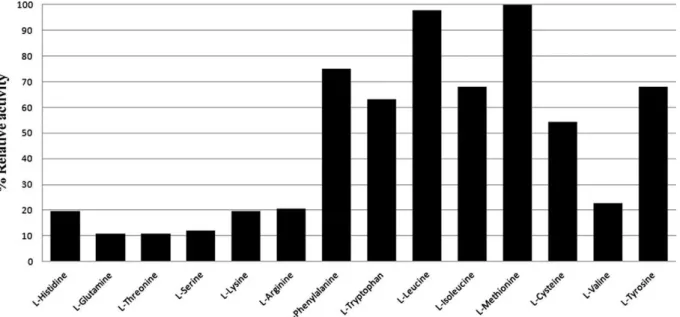
The BjsuLAAO activity was tested using different amino acids as substrate (Fig. 1). The enzyme showed high activity for aromatic and aliphatic amino acids with large side chain includingL
-Methi-onine,L-Leucine,L-Phenylalanine,L-Isoleucine,L-Tryptophan andL
-Tyrosine. A significant activity was also observed for L-Cysteine.
Over other amino acids, BjuLAAO had low catalytic activity (Fig. 1). This pattern shows a clear preference for hydrophobic res-idues with voluminous side chain and the affinity for resres-idues with polar and/or small side-chains is significantly reduced or absent (Fig. 1). The LAAO fromBothrops pauloensisalso showed preference for Met-, Leu-, Phe- and Ile- as substrates [7], suggesting that LAAOs encountered inBothropsgenus retain similar functions. In contrast, the LAAO fromBungarus fasciatusdisplayed higher speci-ficity towards Tyr- and Asp- [19], whereas the bacterial LAAO
(Rhodococcus opacus) has a very low substrate specificity hydrolyzing
aromatic, aliphatic and polar amino acids[20].
Fig. 1.Substrate specificity histogram. The LAAO activity was tested using different amino acids as substrate. We detected high activity for aromatic and hydrophobic amino acids,L-Methionine,L-Leucine,L-Phenylalanine,L-Isoleucine,L-Tryptophan andL-Tyrosine. The LAAO showed low catalytic activity forL-Histidine,L-Arginine,L-Valine,L -Threonine,L-Glutamine,L-Lysine andL-Serine.
3.2. The dimeric structure of BjsuLAAO
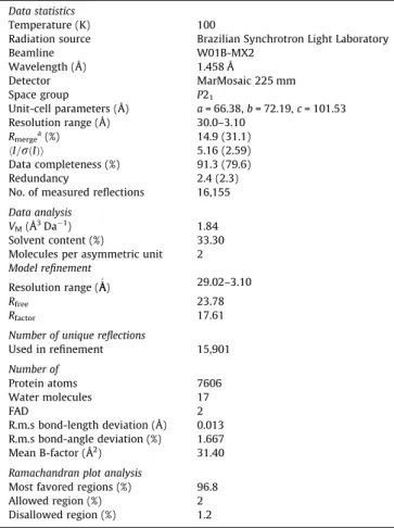
The BjsuLAAO structure was refined to a finalRfactorof 17.6% and
Rfreeof 23.8% at 3.1 Å resolution. Data-processing and refinement
statistics are presented in Table 1. Analogously to other LAAOs [2,19–21], BjsuLAAO is comprised of three domains: FAD-binding domain (residues 35–72, 240–318 and 446–483), substrate bind-ing domain (residues 5–25, 73–129, 231–239 and 319–445) and
helical domain (130–230) (Fig. 2A). Sequence alignment among
BjsuLAAO,Agkistrodon halys pallasandVipera ammodytes
ammo-dytesshowed an average identity of 85% (Fig. 2B) with relevant
amino acid substitutions in the FAD-binding domain and dimer interface (Fig. 2C). Other differences are limited to protein surface without any apparent contribution to protein function.
Electrostatic surface analysis of monomers of BjsuLAAO and
LAAO fromVipera ammodytes ammodytesindicates that
dimeriza-tion is mediated by Coulombic forces between substrate-binding domain (highly negatively charged) and FAD-binding domain (highly positively charged) (Fig. 3A and B). The crystallographic dimer that corresponds to the biological unit of BjsuLAAO was confirmed by in solution DLS analysis (Fig. S5) with a diameter of approximately 70 Å and an intermolecular interface of approx-imately 2081 Å2. The key residues involved in dimer stabilization
are K191, R317, H314, R317, S318, R300, R301, Y436, D376, D349, D210, D201, H320, T182, D205, D388, K186, R297 and H440.
In the structure of LAAO fromVipera ammodytes ammodytes, a zinc ion present in the tetrameric interface being coordinated by residues H75 and G279 was addressed as functionally and struc-turally important[2]. In the dimericB. jararacussuandCalloselasma
rhodostomaLAAO structures, no zinc ion was observed since the
corresponding interface is absent. Furthermore, H75 is not con-served in BjsuLAAO being substituted by a Tyrosine. Biochemical data showed that the presence or absence of zinc ion did not inter-fere with the catalytic activity (results not shown). These observa-tions strongly suggest that the zinc ion plays a role in tetramer stabilization, but not in LAAO activity.
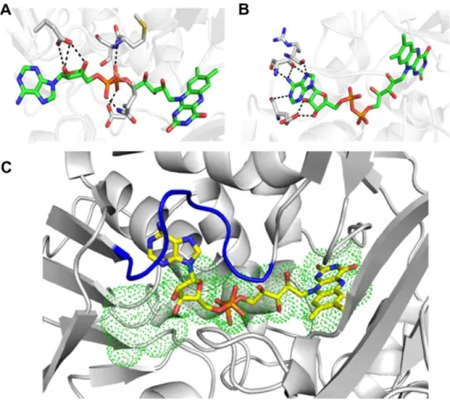
3.3. A new FAD-binding mode with adenosyl moiety buried in the 62– 71 loop
The FAD-binding domain is strategically located in the cleft formed by cofactor- and substrate-binding domains and is highly conserved in both prokaryotes and eukaryotes. The isoalloxazine ring of the FAD molecule is stabilized by extensive hydrogen bonds and Van der Waals contacts with residues G87, P88, M89, R90, L91, P92, G464, W465 and I466. Surprisingly, the adenosyl moiety was found in two different conformations in the asymmetric unit. In molecule B, this FAD portion presents a canonical binding mode mediated by Van der Waals contacts and being stabilized in this orientation by the ribose and di-phosphoryl groups that are tightly anchored to E63, Q71 and E457 side chains and backboneNatoms from M43 and S44 (Fig. 4A and C). In molecule A, the adenosyl group was found buried in the 62–71 loop representing a new
Table 1
Data collection and refinement statistics.
Data statistics
Temperature (K) 100
Radiation source Brazilian Synchrotron Light Laboratory
Beamline W01B-MX2
Wavelength (Å) 1.458 Å
Detector MarMosaic 225 mm
Space group P21
Unit-cell parameters (Å) a= 66.38,b= 72.19,c= 101.53
Resolution range (Å) 30.0–3.10
Rmergea(%) 14.9 (31.1)
hI=rðIÞi 5.16 (2.59)
Data completeness (%) 91.3 (79.6)
Redundancy 2.4 (2.3)
No. of measured reflections 16,155
Data analysis
VM(Å3Da1) 1.84
Solvent content (%) 33.30
Molecules per asymmetric unit 2 Model refinement
Resolution range (ÅA0) 29.02–3.10
Rfree 23.78
Rfactor 17.61
Number of unique reflections
Used in refinement 15,901
Number of
Protein atoms 7606
Water molecules 17
FAD 2
R.m.s bond-length deviation (Å) 0.013 R.m.s bond-angle deviation (%) 1.667
Mean B-factor (Å2) 31.40
Ramachandran plot analysis
Most favored regions (%) 96.8
Allowed region (%) 2
Disallowed region (%) 1.2
aR
merge¼Phkl
P
ijIiðhklÞ hIðhklÞij=Phkl
P
iIiðhklÞ, whereIiðhklÞis theith
obser-vation of reflectionhklandhðhklÞiis the weighted average intensity for all obser-vationsIof reflectionhkl.
binding mode (Fig. 4B and C). In this novel orientation, the adenosyl group is stabilized by hydrogen bonds formed with resi-dues E63, S65 and R67, and extensive hydrophobic contacts with most of the residues forming the 62–71 loop (Fig. 4B and C). This new conformation modifies the solvent accessibility to the
FAD-binding domain increasing the cleft volume, previously occupied by the adenosyl group in the canonical-binding mode. Structural and energetic analyses indicated that the FAD molecule in the new orientation is equally stable to the canonical-binding mode suggesting to be biologically relevant for the activity of LAAOs.
Fig. 3.Electrostatic surface analysis of BjsuLAAO (A) and LAAO fromVipera ammodytes ammodytes(B).
Fig. 4.FAD-binding mode in molecules B (A) and A (B) of BjsuLAAO crystal structure. (C) Superposition of FAD in molecule A (sticks in atom colors), and molecule B (green dots). The 62–71 loop is colored in blue. (For interpretation of the references to color in this figure legend, the reader is referred to the web version of this article.)
3.4. Active-site volume is related to LAAO substrate specificity
The BjsuLAAO activity is higher for aromatic and aliphatic ami-no acids with large side chains (Fig. 1). This result showed that BjsuLAAO is more specific thanC. rhodostomaLAAO[22]that binds to a wide range ofL-amino acids. Comparative structural analysis
suggests that the small side-chain residues cannot adopt a specific or stable orientation in the active site of BjsuLAAO mainly due to the large cavity volume. InC. rhodostomaLAAO, the substitution of isoleucine at position 430 to a tryptophan makes the cavity vol-ume smaller; implying in additional contacts of small side-chain residues with the active-site residues. Moreover, structural super-position of several snake venom LAAOs showed that the key resi-dues for amino acid recognition are fully conserved including Y372, R90, W465, I430 and R322. This conservation indicates a highly specific function of snake venom LAAOs under the enven-omation process.
In this work, a LAAO fromB. jararacussuvenom was purified and its functional and structural properties were determined. It was demonstrated that BjsuLAAO has higher specificity for hydropho-bic residues and structural analysis indicates that the volume of ac-tive site is important for substrate specificity. Remarkably, a new FAD-binding mode equally stable to the classical-binding mode was observed, which could be biologically relevant for LAAO activity.
Acknowledgments
This work was supported by grants from FAPESP, CNPq, CAPES and TWAS.
Appendix A. Supplementary data
Supplementary data associated with this article can be found, in the online version, athttp://dx.doi.org/10.1016/j.bbrc.2012.03.129.
References
[1] R. Doley, R.M. Kini, Protein complexes in snake venom, Cell Mol. Life Sci. 66 (2009) 2851–2871.
[2] D. Georgieva, M. Murakami, M. Perband, R.K. Arni, C. Betzel, The structure of a nativeL-amino acid oxidase, the major component of theVipera ammodytes ammodytes venomic, reveals dynamic active site and quaternary structure stabilization by divalent ions, Mol. BioSyst. 7 (2011) 379–384.
[3] R.J. Lewis, M.L. Garcia, Therapeutic potential of venom peptides, Nat. Rev. Drug Discovery 2 (2003) 790–802.
[4] R.L. Lewis, L. Gutmann, Snake venoms and the neuromuscular junction, Semin. Neurol. 24 (2004) 175–179.
[5] Y.J. Zhang, J.H. Wang, W.H. Lee, Q. Wang, H. Liu, Y.T. Zheng, Y. Zhang, Molecular characterization ofTrimeresurus stejnegerivenomL-amino acid oxidase with
potential anti-HIV activity, Biochem. Biophys. Res. Commun. 309 (2003) 598– 604.
[6] M.Z. Sun, C. Guo, Y. Tian, D. Chen, F.T. Greenaway, S. Liu, Biochemical, functional and structural characterization of Akbu-LAAO: a novel snake venom
L-amino acid oxidase from Agkistrodon blomhoffii ussurensis, Biochimie 92 (2010) 343–349.
[7] R.S. Rodrigues, J.F. da Silva, J. Boldrini Franca, F.P. Fonseca, A.R. Otaviano, F.H. Silva, A. Hamaguchi, A.J. Magro, A.S. Braz, J.I. dos Santos, M.I. Homsi-Brandeburgo, M.R. Fontes, A.L. Fuly, A.M. Soares, V.M. Rodrigues, Structural and functional properties of Bp-LAAO, a newL-amino acid oxidase isolated
fromBothrops pauloensissnake venom, Biochimie 91 (2009) 490–501. [8] R.M. Alves, G.A. Antonucci, H.H. Paiva, A.C.O. Cintra, J.J. Franco, E.P. Mendonca
Franqueiro, D.J. Dorta, J.R. Giglio, J.C. Rosa, A.L. Fuly, M. Dias-Baruffi, A.M. Soares, S.V. Sampaio, Evidence of caspase-mediated apoptosis induced by l-amino acid oxidase isolated fromBothrops atroxsnake venom, Comp. Biochem. Physiol. A: Mol. Integr. Physiol. 151 (2008) 542–550.
[9] Z.Y. Li, T.F. Yu, E.C.Y. Lian, Purification and characterization of l-amino acid oxidase from king cobra (Ophiophagus hannah) venom and its effects on human platelet aggregation, Toxicon 32 (1994) 1349–1358.
[10] A.G. Tempone, H.F. Andrade, P.J. Spencer, C.O. Lourenço, J.R. Rogero, N. Nascimento,Bothrops moojenivenom kills Leishmaniaspp. with hydrogen peroxide generated by its L-amino acid oxidase, Biochem. Biophys. Res.
Commun. 208 (2001) 620–624.
[11] R.G. Stábeli, C.D. Sant’Ana, P.H. Ribeiro, T.R. Costa, F.K. Ticli, M.G. Pires, A. Nomizo, S. Albuquerque, N.R. Malta-Neto, M. Marins, S.V. Sampaio, A.M. Soares, CytotoxicL-amino acid oxidase fromBothrops moojeni: biochemical and functional characterization, Int. J. Biol. Macromol. 41 (2007) 132–140. [12] D. Wellner, Evidence for conformational changes in l-amino acid oxidase
associated with reversible inactivation, Biochemistry 5 (1966) 1585–1591. [13] B. Curti, V. Massey, M. Zmudka, Inactivation of snake venom l-amino acid
oxidase by freezing, J. Biol. Chem. 243 (1968) 2306–2314.
[14] U.K. Laemmli, Cleavage of structural proteins during the assembly of the head of bacteriophage T4, Nature 227 (1970) 680–685.
[15] R.F. Itzhaki, D.M. Gill, A micro-biuret method for estimating proteins, Anal. Biochem. 9 (1964) 401–410.
[16] J. Jancarik, S.H. Kim, Sparse matrix sampling: a screening method for crystallization of proteins, J. Appl. Crystallogr. 24 (1991) 409–411.
[17] Z. Otwinowski, W. Minor, Processing of X-ray diffraction data collected in oscillation mode, in: Macromolecular Crystallography. Part A, Academic Press, 1997, pp. 307–326.
[18] A. Vagin, A. Teplyakov, MOLREP: an automated program for molecular replacement, J. Appl. Crystallogr. 30 (1997) 1022–1025.
[19] J.F. Wei, H.W. Yang, X.L. Wei, L.Y. Qiao, W.Y. Wang, S.H. He, Purification, characterization and biological activities of theL-amino acid oxidase from
Bungarus fasciatussnake venom, Toxicon 54 (2009) 262–271.
[20] A. Faust, K. Niefind, W. Hummel, D. Schomburg, The structure of a bacterial l-amino acid oxidase fromRhodococcus opacus gives new evidence for the hydride mechanism for dehydrogenation, J. Mol. Biol. 367 (2007) 234–248. [21] H. Zhang, M. Teng, L. Niu, Y. Wang, Y. Wang, Q. Liu, Q. Huang, Q. Hao, Y. Dong,
P. Liu, Purification, partial characterization, crystallization and structural determination of AHP-LAAO, a novelL-amino-acid oxidase with cell apoptosis-inducing activity fromAgkistrodon halys pallasvenom, Acta Crystallogr. D: Biol. Crystallogr. 60 (2004) 974–977.