Arq. Bras. Med. Vet. Zootec., v.69, n.2, p.285-292, 2017
Serum concentrations of acute phase proteins in goats and ewes with naturally acquired Staphylococcus aureus mastitis
[Concentração de proteínas de fase aguda em cabras e ovelhas com mastite de ocorrência natural causada por Staphylococcus aureus]
K.M.M.G. Simplício1, T.G. Rocha2; D.C.C. Sanchez², F.S. Cotrim², P.C. Silva²; J.J. Fagliari²
1Programa de pós-graduação FCAV-UNESP Jaboticabal, SP 2Faculdade de Ciências Agrárias e Veterinárias – FCAV-UNESP – Jaboticabal, SP
ABSTRACT
Serum protein concentrations, including acute phase proteins (APPs), of goats and ewes with naturally acquired Sthaphylococcus aureus mastitis were determined by means of SDS-PAGE electrophoresis to evaluate the relevance of these APPs as biomarkers of the disease in these species. Fifteen healthy goats and 5 goats with naturally acquired staphylococci mastitis, as well as fifteen healthy ewes and 5 ewes with staphylococci mastitis were submitted to daily blood sampling during 7 days. In goats, an increase of 570%, 125%, 621%, and 279% in serum concentrations of ceruloplasmin, fibrinogen, haptoglobin and α1
-acid glycoprotein, respectively, was observed. In sheep the increase in serum concentrations of ceruloplasmin, fibrinogen, haptoglobin and α1-acid glycoprotein was of 337%, 90%, 461%, and 225%,
respectively. Our results indicate that these APPs have considerable potencial as early and sensible biomarkers of mastitis caused by S. aureus in goats and sheep.
Keywords: ceruloplasmin, fibrinogen, haptoglobin, α1-acid glycoprotein
RESUMO
O proteinograma, incluindo proteínas de fase aguda (PFAs), de cabras e ovelhas com mastite de origem natural causada por Staphylococcus aureus, foi determinado por meio de eletroforese em gel de poliacrilamida contendo dodecil sulfato de sódio (SDS-PAGE) a fim de avaliar a importância destas PFAs como biomarcadores da enfermidade nestas espécies. Amostras de sangue foram colhidas diariamente de cinco cabras e cinco ovelhas com mastite estafilocócica naturalmente adquirida, bem como de quinze cabras e quinzes ovelhas saudáveis durante 6 dias consecutivos. Nas fêmeas caprinas, foi verificado aumento dos teores séricos de ceruloplasmina (570%), fibrinogênio (125%), haptoglobina (621%), e α1-glicoproteína ácida (279%). Nas fêmeas ovinas as concentrações de ceruloplasmina,
fibrinogênio, haptoglobina e α1-glicoproteína ácida elevaram-se em 337%, 90,9%, 461% e 225%,
respectivamente. Os resultados permitem inferir que estas PFAs são marcadores sensíveis e precoces de mastite causada por S. aureus em cabras e ovelhas.
Palavras-chave: Ceruloplasmina, fibriniogênio, haptoglobina, α1-glicoproteína ácida
INTRODUCTION
Clinical and subclinical mastitis are serious problems in dairy herds worldwide. Although there are several bacteria causing mastitis in goats and ewes, Staphylococcus aureus is by far the most important agent causing udder
Recebido em 3 de setembro de 2015 Aceito em 9 de agosto de 2016 E-mail: [email protected]
infection, especially since it can evolve to persistent infection with recurring clinical episodes (Aires-de-Sousa et al., 2007; Radostitis et al., 2007; Erskine, 2013).
identification of early biomarkers. Research along the last few decades showed that quantification of acute-phase proteins (APPs) either in blood plasma or serum is useful for diagnosis and prognosis of disease, as well as to evaluate the severity of inflammation; APPs quantification may also contribute with human food security by early disease detection at slaughterhouse (Eckersall, 2000; González et al., 2007). Proteins whose serum concentrations decrease as a response to inflammation are called negative acute-phase proteins and include albumin and transferrin. Contrarily, proteins whose serum concentrations increase during inflammation are defined as positive acute-phase proteins, e.g., ceruloplasmin, fibrinogen, haptoglobin, α1-acid glycoprotein.
Concentrations of the latter proteins are mostly low or non-detectable in healthy animals. Thus, increased serum levels of these proteins are used as auxiliary tools for diagnosing and monitoring disease. The type of APPs increasing as a result of inflammation, as well as the time that these APPs remain elevated varies among species and disease (Thomas, 2010; Ceciliani et al., 2012).
Sodium dodecyl sulfate-polyacrylamide gel electrophoresis (SDS-PAGE) allows the identification of up to 30 protein bands. It is an easy, low-cost technique which requires small samples, allowing detection of extremely low concentrations of protein. By using this technique, it is possible to evaluate the APPs of interest in veterinary medicine (Fagliari et al., 2007).
Despite the importance of determining APPs in ruminant clinic, there are only few reports available on this subject, particularly on goats and sheep. Thus, the aim of the present study was to quantify APPs in blood serum from goats and ewes with naturally acquired Staphylococcus aureus mastitis using SDS-PAGE, in order to assess the importance of such proteins as early biomarkers of the disease in these species.
MATERIALS AND METHODS
Fifteen healthy goats and 15 healthy ewes (control groups) as well as five goats and five ewes with naturally acquired Staphylococcal mastitis were used. The experimental protocol was approved by the Committee of Ethics and Animal Welfare of the School of Agrarian
Sciences and Veterinary Medicine, São Paulo State University (Unesp), Brazil, under protocol number 029033-08.
Selection of animals was performed by physical examination and microbiological isolation and identification of Staphylococcus aureus in milk samples by assessing the colony characteristics on blood agar, Gram staining and positivity to biochemical tests such as catalase, coagulase, mannitol, Voges-Proskauer and anaerobic fermentation.
Control animals that presented no clinical signs of mastitis and were negative for bacterial detection in milk culture belonged to the School of Agrarian Sciences and Veterinary Medicine (UNESP). Ill animals were selected from among patients with acute mastitis (i.e. fever, swelling and/or local pain in the mammary gland, inappetence, changes in the characteristics of milk, etc) admitted into the School Veterinary Hospital at the same institution. Animals were included in the study after acknowledgment (through anamnesis) that the manifestation of clinical signs begun no longer than 3 days before admittance.
Once included into the experiment, consecutive daily blood samples were taken for six days and moments identified as M1, M2, M3, M4, M5 and M6. During the study period (M1 - M6), animals remained in the Hospital and were treated (M2-M4), once daily for three days with M1 being the first sampling moment when the animal had no therapy influence on its immune response to disease. Treatment consisted of intramammary administration of 200 mg of gentamicin once daily. All females were only discharged from Hospital after microbiological cure of the infection.
Blood samples were collected in tubes without anticoagulant and centrifuged to obtain serum, which was frozen in aliquots at -80ºC for ulterior analysis.
Serum total protein was determined by the biuret method using a commercial kit (Labtest, Labtest, Labtest Diagnostica, Lagoa Santa, Minas Gerais, Brazil). Separation of protein fractions and determination of ceruloplasmin, haptoglobin, α1-acid glycoprotein concentrations were
sulfate-polyacrylamide gel electrophoresis (SDS-PAGE) as described by Laemmli (1970). Concentrations were obtained through computerized densitometer (Shimadzu CS-9301PC, Tokyo, Japan). A marker solution (Sigma, St. Louis, MO, USA) with molecular weights of 29,000, 45,000, 66,000, 97,400, 116,000 and 205,000 Daltons (Da) was used to identify protein fractions associated with the electrophoretic mobility of patterns of purified immunoglobulin A, immunoglobulin G, ceruloplasmin, transferrin, and haptoglobin (Sigma, St. Louis, MO, USA). Plasma fibrinogen levels were measured by the heat precipitation method, as described by Jain (1993).
Results were analyzed by ANOVA. The means were compared using the Tukey's test set at 5.0% significance value (Petrie and Watson, 2009).
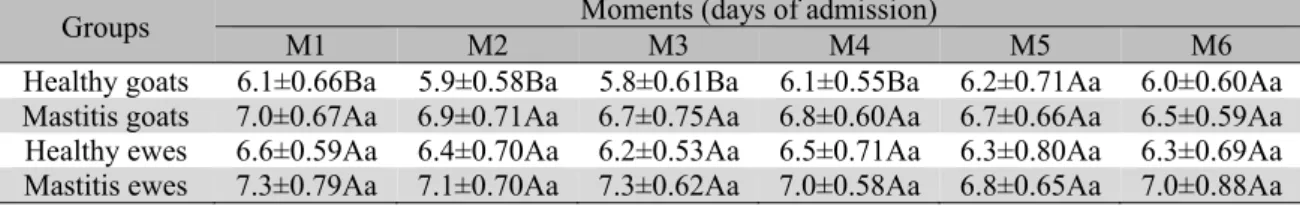
RESULTS AND DISCUSSION Goats with mastitis presented mild increase in concentrations of total protein in serum when compared with the control group. Total protein levels subsequently decreased five days after admission (M5). There was no difference in serum total protein in ewes between groups and moments (Table 1, Figure 1).
Table 1. Means and standard deviations of serum total protein concentrations (g/dL) in both healthy and ill (staphylococcal mastitis) goats and ewes from admission (M1) to the end of the experiment (M6)
Groups M1 M2 Moments (days of admission) M3 M4 M5 M6 Healthy goats 6.1±0.66Ba 5.9±0.58Ba 5.8±0.61Ba 6.1±0.55Ba 6.2±0.71Aa 6.0±0.60Aa Mastitis goats 7.0±0.67Aa 6.9±0.71Aa 6.7±0.75Aa 6.8±0.60Aa 6.7±0.66Aa 6.5±0.59Aa Healthy ewes 6.6±0.59Aa 6.4±0.70Aa 6.2±0.53Aa 6.5±0.71Aa 6.3±0.80Aa 6.3±0.69Aa Mastitis ewes 7.3±0.79Aa 7.1±0.70Aa 7.3±0.62Aa 7.0±0.58Aa 6.8±0.65Aa 7.0±0.88Aa
Means followed by different capital letters in the same column and different small letters in the same line indicate significant difference by the Tukey's test (P>0.05).
4 5 6 7 8
M1 M2 M3 M4 M5 M6
Moment (day)
To
ta
l P
ro
te
in (
g/d
L)
Healthy goats Goats w ith mastitis Healthy sheep Sheep w ith mastitis
Figure 1. Means of serum protein (g/dL) in both healthy and ill (staphylococcal mastitis) goats and ewes from admission (M1) to the end of the experimental period (M6).
Contrary to the present study, Patelli et al. (2008) did not report an increase in total protein concentrations in goats experimentally infected with Trypanossoma evansi when compared to the control group. Brum et al. (2007) reported similar results to the findings of the present study in sheep naturally infected with facial eczema. The mild increase in concentrations of serum total protein detected in the goats of this
experiment may have been caused by the significant increase of APPs, e.g., ceruloplasmin.
ceruloplasmin concentrations in ewes with mastitis were 337% higher than in the control group. Between moments, a gradual decrease in
ceruloplasmin levels it was evident along the experimental period in the mastitis groups.
Table 2. Means and standard deviations of serum ceruloplasmin (mg/dL) in both healthy and ill (staphylococcal mastitis) goats and ewes from admission (M1) to the end of the experiment (M6)
Groups M1 M2 Moments (days of admission) M3 M4 M5 M6 Healthy
goats 9.2±3.53Ba 11.3±3.77Ba 9.8±2.95Ba 12.3±3.11Ba 10.9±3.44Ba 13.0±3.16Ba Mastitis
goats 83.7±7.45Aa 90.7±8.33Aa 84.5±6.65Aa 73.1±7.32Ab 60.9±5.89Ac 52.7±6.12Ad Healthy
ewes 12.4±3.32Ba 10.8±2.92Ba 13.0±2.87Ba 11.7±3.06Ba 14.6±3.21Ba 16.1±2.21Ba Mastitis
ewes 77.1±9.1Aa 65.3±7.21Aab 60.3±7.03Ab 53.4±5.22Ab 46.5±4.66Ac 40.8±4.06Ad
Means followed by different capital letters in the same column and different small letters in the same line indicate significant difference by the Tukey's test (P>0.05).
0 10 20 30 40 50 60 70 80 90 100
M1 M2 M3 M4 M5 M6
Moment (day)
C
er
ulo
pl
asm
in
(m
g/d
L)
Healthy goats Goats w ith mastitis Healthy sheep Sheep w ith mastitis
Figure 2. Means of serum ceruloplasmin (mg/dL) in both healthy and ill (staphylococcal mastitis) goats and ewes from admission (M1) to the end of the experiment (M6).
Costa et al. (2010) reported an increase of 298% in serum levels of ceruloplasmin in ewes with experimentally-induced mastitis, which agrees with the results of the present study. There are few publications on ceruloplasmin levels in small ruminants presenting mastitis (Eckersall, 2006). Nevertheless, the increase of this protein´s concentration reaffirms its anti-inflammatory and biocatalytic effect reported by Patel et al. (2002). In cows with clinical or sub-clinical mastitis, serum ceruloplasmin levels drastically increased in both blood plasma and milk (Eckersall et al., 2001; Grönlund et al., 2005). Therefore, although using ceruloplasmin as inflammatory marker is less common than using other APPs, it seems to be a reliable signaler of bovine
infection (Segelmark et al., 1997; Murata et al., 2004). These results emphasize the clinical importance of this biomarker in goats and sheep.
Table 3. Means and standard deviations of serum fibrinogen (mg/dL) in both healthy and ill (staphylococcal mastitis) goats and ewes from admission (M1) to the end of the experiment (M6)
Groups M1 M2 Moments (days of admission) M3 M4 M5 M6 Healthy
goats 379±41Ba 382±44Ba 375±36Ba 402±49Ba 391±45Ba 432±51Ba Mastitis
goats 883±92Aa 907±98Aa 923±94Aa 886±91Aa 875±83Aa 845±90Aa Healthy
ewes 442±38.7Ba 439±40.1Ba 455±33.7Ba 471±39.9Ba 463±41.2Ba 456±37.5Ba Mastitis
ewes 912±90.3Aab 930±94.1Aa 919±87.3Aab 865±81.7Aab 821±75.6Ab 757±70.5Ab
Means followed by different capital letters in the same column and different small letters in the same line indicate significant difference by the Tukey's test (P>0.05).
300 400 500 600 700 800 900 1000
M1 M2 M3 M4 M5 M6
Moment (day)
Fibr
inoge
n (
m
g/dL)
Healthy goats Goats w ith mastitis Helathy sheep Sheep w ith mastitis
Figure 3. Means of serum fibrinogen (mg/dL) in both healthy and ill (staphylococcal mastitis) goats and ewes from admission (M1) to the end of the experiment (M6).
Costa et al. (2010) reported that the peak in plasma fibrinogen occurred 132 hours after the experimental intra-mammary infection with Staphylococcus aureus in ewes. This time lapse matches with M5-M6 in our experiment, when, contrary to the report of the previous authors, there was a significant decrease in serum fibrinogen concentrations. However, it is important to consider the more accurate control regarding the beginning of clinical signs in experimental works, as compared to naturally occurring disease, in which it is also difficult to control some variables, as in the present study.
In goats with mastitis there was an increase of 621% in levels of haptoglobin when compared with the control group. The highest concentration was observed at M3, whereupon levels decreased and were significantly lower at M6, although still
higher than in the control group. Serum levels of haptoglobin were also high in ewes with mastitis, with maximum value at the second day in hospital (M2). Despite serum concentrations of haptoglobin decreased throughout the experimental period in mastitis groups, levels of this parameter remained higher than in the control group until the end of the experimental period (M6).
this protein presented the higher responsiveness in ewes, with an increase of 461% in ill animals when compared with the control group. Costa et
al. (2010) reported concentrations that rose as high as 607% in experimental infection with S. aureus when compared to control group.
Table 4. Means and standard deviations of serum haptoglobin (mg/dL) in both healthy and ill (staphylococcal mastitis) goats and ewes from admission (M1) to the end of the experiment (M6)
Groups M1 M2 Moments (days of admission) M3 M4 M5 M6 Healthy
goats 20.7±3.09Ba 22.5±3.17Ba 19.8±3.01Ba 21.0±2.87Ba 23.1±3.18Ba 20.5±3.03Ba Mastitis
goats 160±25Aa 168±26Aa 175±30Aa 151±26Aa 139±32Aab 127±24Ab Healthy
ewes 27.0±2.21Ba 30.2±2.89Ba 28.2±2.33Ba 31.2±2.17Ba 28.6±2.33Ba 26.8±2.71Ba Mastitis
ewes 167±22.7Aa 174±25.1Aa 163±20.3Aa 166±24.3Aa 153±20.8Aab 142±17.5Ab
Means followed by different capital letters in the same column and different small letters in the same line indicate significant difference by the Tukey's test (P>0.05).
20 40 60 80 100 120 140 160 180
M1 M2 M3 M4 M5 M6
Moment (day)
H
apt
og
lo
bi
n
(m
g/
dL
)
Healthy goats Goats w ith mastitis Healthy sheep Sheep w ith mastitis
Figure 4.Means of serum haptoglobin (mg/dL) in both healthy and ill (staphylococcal mastitis) goats and ewes from admission (M1) to the end of the experiment (M6).
In general, haptoglobin is considered a sensitive and reliable inflammatory biomarker in goats and ewes suffering staphylococcal mastitis. The present study agrees with Eckersall et al. (2001) who stated that haptoglobin is highly sensitive in detecting inflammatory/infectious diseases in ruminants; and with Skinner and Roberts (1994) who reported that this protein may be used as a safe indicator of bacterial infection and inflammation in sheep.
Serum concentrations of α1-acid glycoprotein in
goats with mastitis were 279% higher when compared with the control group and decreased along the experimental period. Likewise, levels of α1-acid glycoprotein in ewes with mastitis
Table 5. Means and standard deviations of serum α1-acid glycoprotein (mg/dL) in both healthy and ill
(staphylococcal mastitis) goats and ewes from admission (M1) to the end of the experiment (M6) Groups M1 M2 Moments (days of admission) M3 M4 M5 M6 Healthy goats 7.4±2.7Ba 9.2±2.2Ba 9.0±2.03Ba 8.6±1.93Ba 9.5±2.09Ba 8.3±1.97Ba Mastitis goats 38.1±4.8Aa 33.9±5.3Aab 37.2±5.0Aa 31.8±4.7Ab 30.2±4.9Ab 25.6±4.0Ac Healthy ewes 8.7±1.2Ba 10.1±1.8Ba 9.6±0.95Ba 8.3±1.2Ba 10.0±1.4Ba 9.2±0.93Ba Mastitis ewes 33.4±5.2Aa 36.1±6.1Aa 35.3±5.4Aa 28.6±5.5Ab 26.3±4.3Ab 21.7±4.2Ac
Means followed by different capital letters in the same column and different small letters in the same line indicate significant difference by the Tukey's test (P>0.05).
5 10 15 20 25 30 35 40
M1 M2 M3 M4 M5 M6
Moment (day)
α1
-A
ci
d
G
ly
copr
ot
ei
n
(m
g/
dL)
Healthy goats Goats w ith mastitis Helathy sheep Sheep w ith mastitis
Figure 5. Means of serum α1-acid glycoprotein (mg/dL) in both healthy and ill (staphylococcal mastitis)
goats and ewes from admission (M1) to the end of the experiment (M6).
Results by Rinaldi et al. (2008), showed that α1
-acid glycoprotein had immunomodulatory properties acting on leucocytes and cytokines during inflammation, which justifies the increase of this APP along with the acute phase response. Recent studies in sheep and other species revealed that α1-acid glycoprotein may be an
indicator of chronic inflammation, due to its long plasma half-life compared with other APPs (Eckersall et al., 2007; González et al., 2008). However, the early detection and significant high level of this protein at the beginning of disease, associated with its decrease by the fourth day in hospital (M4) in both goats and ewes, confirm its characteristic of positive APP, acting early in the acute phase response in females with staphylococcal mastitis.
CONCLUSIONS
Detection and quantification of APPs by SDS-PAGE was useful and reliable as a complementary test in the diagnosis of mastitis caused by Staphylococcus aureus in goats and
ewes. Moreover, the APPs ceruloplasmin, haptoglobin, and α1–acid glycoprotein were early
and sensitive indicators of mammary inflammation in both species.
ACKNOWLEDGEMENT
To Fundação de Amparo e Pesquisa do Estado de São Paulo (FAPESP) for the financial support to this work.
REFERENCES
AIRES-DE-SOUSA, M.; PARENTE, C.E.S.R.; MOTTA, O.V. et al. Characterization of Staphylococcus aureus isolated from buffalo, bovine,
ovine, and caprine milk samples from Rio de Janeiro state, Brazil. Appl. Environ. Microbiol., v.73,
p.3845-3849, 2007.
BRUM, K.B.; HARAGUCHI, M.; LEMOS, R.A.A. et al. Crystal-associated cholangiopathy in sheep grazing Brachiaria decumbens containing the saponin
CECILIANI, F.; CERON, J.J.; ECKERSALL, P.D. et al. Acute phase proteins in ruminants. J. Proteomic’s,
v.75, p.4207-4231, 2012.
COORAY, R.; WALLER, K.P.; VENGE, P. Haptoglobin comprises about 10% of granule protein extracted from bovine granulocytes isolated from healthy cattle. Vet. Immunol. Immunopathol., v.119,
p.310-315, 2007.
COSTA, N.A.; SIMÃO, L.C.V.; FAGLIARI, J.J. et al.
Alterações na concentração do fibrinogênio e da haptoglobina em ovelhas com mastite infectadas experimentalmente com Staphylococcus aureus. Arch. Vet. Sci., v.12, p.119-121, 2010.
ECKERSALL, P.D. Acute phase proteins as biomarkers of disease in production animals. In: PROCEEDINGS OF THE ANNUAL MEETING OF THE AMERICAN COLLEGE OF VETERINARY PATHOLOGISTS AND AMERICAN SOCIETY FOR VETERINARY CLINICAL PATHOLOGY, 57.,
2006, Tucson. Proceedings… Tucson,
Arizona:[ACVP], 2006. p.57. (Resumo)
ECKERSALL, P.D. Recent advances and future prospects for the use of acute phase proteins as markers of disease in animals. Rev. Méd. Vét., v.151,
p.577-584, 2000.
ECKERSALL, P.D.; LAWSON, F.P.; BENCE, L. et al. Acute phase protein response in an experimental
model of ovine caseous lymphadenitis. BCM Vet. Res.,
v.3, p.35, 2007.
ECKERSALL, P.D.; YOUNG, F.J.; MCCOMB, C. et al. Acute phase proteins in serum and milk from dairy
cows with clinical mastitis. Vet. Rec., v.14, p.35-41,
2001.
ERSKINE, R.J. Mastite em grandes animais. In: KAHN, C.M.; LINE, S. (Eds.). Manual merck de veterinária. São Paulo: Roca, 2013. 3420p.
FAGLIARI, J.J.; PASSIPIERI, M.; OKUDA, H.T. et al. Serum protein concentrations, including acute
phase proteins, in calves with hepatogenous photosensitization. Arq. Bras. Med. Vet. Zootec., v.59,
p.1355-1358, 2007.
GONZÁLEZ, F.H.D.; MARTÍNEZ-SUBIELA, S.; CERÓN, J.J. Haptoglobina en rumiantes: generalidades y posibles aplicaciones clínicas. Ann. Vet. Murcia, v.23, p.5-17, 2007.
GONZÁLEZ, F.H.D.; TECLES, F.; MARTÍNEZ-SUBIELA, S. et al. Acute phase proteins response in
goats. J. Vet. Diagn. Investig., v.20, p.580-584, 2008.
GRÖNLUND, U.; SANDGREN, C.H.; WALLER, K.P. Haptoglobin and serum amyloid A in milk from dairy cows with chronic sub-clinical mastitis. Vet. Res., v.36, p.191-198, 2005.
JAIN, N.C. Essentials of veterinary hematology.
Philadelphia: Lea & Febinger, 1993. 417p.
JAIN, S.; GAUTAM, V.; NASEEM, S. Acute phase proteins: as diagnostic tool. J. Pharm. Bioallied Sci.,
v.3, p.118-127, 2011.
LAEMMLI, U.K. Cleavage of structural proteins during the assembly of the head of bacteriophage T4.
Nature, v.227, p.680-685, 1970.
MURATA, H.; SHIMADA, N.; YOSHIOKA, M. Current research on acute phase proteins in veterinary diagnosis: an overview. Vet. J., v.168, p.28-40, 2004.
PATEL, B.N.; DUNN, R.J.; JEONG, S.Y. et al.
Ceruloplasmin regulates iron levels in the CNS and prevents free radical injury. J. Neuroscienc., v.22,
p.6578-6586, 2002.
PATELLI, T.H.C.; MARQUES, L.C.; FAGLIARI, J.J.
et al. Perfil eletroforético das proteínas de fase aguda
em caprinos experimentalmente infectados com
Trypanosoma evansi. Braz. J. Vet. Res. Anim. Sci.,
v.45, p.481-187, 2008.
PETRIE, A.; WATSON, P. Estatística em ciência animal e veterinária. 2.ed. São Paulo: Roca, 2009.
248p.
RADOSTITIS, O.M.; GAY, C.C.; BLOOD, D.C. et al. Veterinary medicine. 10.ed. London: W.B.
Saunders, 2007. 2156p.
RINALDI, M.; CECILIANI, F.; LECCHI, C. et al.
Differential effects of α1-acid glycoprotein on bovine neutrophil respiratory burst activity and IL-8 production. Vet. Immunol. Immunopathol., v.126,
p.199-210, 2008.
SEGELMARK, M.; PERSSON, B.; HELLMARK, T.
et al. Binding and inhibition of myeloperoxidase: a
major function of ceruloplasmin? Clin. Exp. Immunol.,
v.108, p.167-174, 1997.
SKINNER, J.G.; ROBERTS, L. Haptoglobin as an indicator of infection in sheep. Vet. Rec., v.134,
p.33-36, 1994.
THOMAS, J.S. Overview of plasma proteins. In: FELDMAN, B.F.; ZINKL, J.G.; JAIN, N.C. (Eds.).
Schalm´s veterinary hematology. Philadelphia: