(Annals of the Brazilian Academy of Sciences) ISSN 0001-3765
www.scielo.br/aabc
Superoxide dismutase and lipid hydroperoxides in blood and endometrial
tissue of patients with benign, hyperplastic and malignant endometrium
SNEŽANA PEJI ´C1, ANA TODOROVI ´C1, VESNA STOJILJKOVI ´C1, DRAGANA CVETKOVI ´C3, NENAD LU ˇCI ´C2, RATKO M. RADOJI ˇCI ´C4, ZORICA S. SAI ˇCI ´C5 and SNEŽANA B. PAJOVI ´C1
1Laboratory of Molecular Biology and Endocrinology, Vinˇca Institute of Nuclear Sciences
P.O. Box 522, 11001 Belgrade, Serbia
2Clinic for Gynecology and Maternity, Clinical Centre, Ulica 12 beba bb, 51000 Banja Luka, Bosnia and Herzegovina 3Institute of Zoology, Faculty of Biology, University of Belgrade, Studentski trg 3, 11060 Belgrade, Serbia
4Institute for Biochemistry and Physiology, Faculty of Biology, University of Belgrade
Studentski trg 3, 11060 Belgrade, Serbia
5Institute for Biological Research, Siniša Stankovi´c, Department of Physiology, Bulevar despota Stefana 142
11060 Belgrade, Serbia
Manuscript received on August 10, 2007; accepted for publication on April 14, 2008; presented byALEXANDERW.A. KELLNER
ABSTRACT
Epidemiological and experimental data point to involvement of oxygen derived radicals in the pathogenesis of gyne-cological disorders, as well as in cancer development. The objective of the present study was to examine changes in activities and levels of copper/zinc superoxide dismutase (CuZnSOD) and lipid hydroperoxides (LOOH) in blood and endometrial tissue of patients diagnosed with uterine myoma, endometrial polypus, hyperplasia simplex, hyperplasia complex and adenocarcinoma endometrii. The results of our study have shown decreased SOD activities and unchanged SOD protein level in blood of all examined patients in comparison to healthy subjects. Decrease of both SOD activity and level was found in endometrium of patients with hyperplasia simplex, hyperplasia complex and adenocarcinoma in comparison to women with polypus or myoma. LOOH level was elevated in both tissues of patients with hyperplasia or adenocarcinoma in comparison to healthy subjects or patients with benign diagnosis. Our findings suggest that the decrease in SOD activity and level, as well as the increase in LOOH level, in patients with gynecological disorders, render these patients more susceptible to oxidative damage caused by reactive oxygen species (ROS). An imbalance in ROS formation and SOD level may be important in the pathogenesis and/or perpetuation of tissue damage in gyneco-logical patients. Since evidence suggests that SOD may be a therapy target for cancer treatment, our findings provide a basis for further research and options for clinical applications.
Key words: copper/zinc superoxide dismutase, lipid hydroperoxides, uterine myoma, endometrial polypus, endome-trial hyperplasia, endomeendome-trial adenocarcinoma.
INTRODUCTION
Reactive oxygen species (ROS), such as superoxide
(O∙−
2 ) and hydrogen peroxide (H2O2), are produced in
metabolic processes. A balance between O∙−
2 production
and elimination is important for maintenance of a proper
Correspondence to: Dr. Snežana Peji´c E-mail: [email protected]
ROS participate in the pathogenesis of various diseases including carcinogenesis (Valko et al. 2007).
Endometrial carcinoma is the most frequently diag-nosed malignancy of the female genital tract. It is often preceded by histopathologic lesions known as endome-trial hyperplasia (Mutter 2002), and it is generally con-sidered a precursor of endometrial cancer (Münstedt et al. 2004). According to the current World Health Orga-nization (WHO) nomenclature, endometrial hyperplasia is classified in four categories, simple hyperplasia (SH), complex hyperplasia (CH), simple atypical hyperplasia (SAH) and complex atypical hyperplasia (CAH).
In recent years, there has been a growing interest in the investigation on the role played by ROS and anti-oxidants in gynecological patients. Chiou and Hu (1999) reported on the elevated LPO and decreased plasma and erythrocytes SOD activity in both uterine cervicitis and myoma patients, while activities of CAT and GPx were elevated in cervicitis patients and lowered in myoma patients. Similar observations were made on erythro-cytes SOD, CAT and GPx activities of cervicitis patients, whereas activities of examined enzymes decreased in cervical cancer patients (Manoharan et al. 2004). LPO was found to be increased in both groups of patients (Manoharan et al. 2004). In endometrial cancer tis-sue of both Finnish and Japanese women, the activity of SOD was found to be significantly lower than in normal endometrium (Punnonen et al. 1993). Our recent re-sults showed that SOD activity was lower in the blood of patients with uterine polypus or myoma, hyperplasia endometrii and adenocarcinoma endometrii than in that of healthy subjects (Peji´c et al. 2006). There is a limited amount of information regarding the relative levels of SOD expression in gynecological patients. It was shown that levels of both CuZnSOD and MnSOD in endome-triosis and adenomyosis were persistently higher than their respective levels throughout the menstrual cycle in fertile controls (Ota et al. 1999). Increase in SOD level has also been observed in ovarian cancer patients (Hu et al. 2005). Other studies showed that SOD levels in various cancer tissues were unchanged or lower than in normal tissue (Bostwick et al. 2000, Monari et al. 2006). The aim of this study was to investigate changes in CuZnSOD activities and protein levels, as well as LPO level in blood and endometrial tissue of patients
diagnosed with uterine myoma, endometrial polypus, hyperplasia simplex, hyperplasia complex and adeno-carcinoma endometrii.
MATERIALS AND METHODS SUBJECTS
The material used in this study comprised 103 blood samples and 88 tissue specimens of women admitted to the Department of Gynecology and Obstetrics for gy-necological evaluation within routine checkups or for abnormal uterine bleeding. The specimens were taken after obtaining informed consent and the study was con-ducted prospectively. The protocol followed is consis-tent with the World Medical Association Declaration of Helsinki (Ethical Principles for Medical Research In-volving Human Subjects).
On the basis of diagnosis and histological exami-nation, subjects were divided into the following groups:
healthy control patients (C, n=15, mean ± SEM: 49±
3 yr); patients with benign uterine changes: polypus
en-dometrii (PE, n= 18, 45±3 yr) or uterus
myomato-sus (UM, n= 12, 47±2 yr); patients with abnormal
bleeding: hyperplasia simplex endometrii (SH, n=31,
48±1 yr), hyperplasia complex endometrii (CH, n=22,
48±2 yr) or adenocarcinoma endometrii, stage I (ACE,
n=5, 59±3 yr).
SAMPLES
Venous blood samples were collected into heparinized tubes on the same day of endometrial biopsy and
ali-quoted immediately. For SOD assay (OxisResearchTM),
blood was centrifuged at 2500 g for 5 min. Plasma was discarded and the pellet resuspended in 4 packed-cell vol-ume of ice-cold demineralized ultrapure water (MilliQ reagent grade water system, Millipore Corp., Bedford, MA, USA). After the addition of ethanol/chloroform extraction reagent (62.5/37.5 vol/vol) to remove hemo-globin, samples were centrifuged at 3000 g for 10 min (Eppendorf centrifuge 5417, Eppendorf AG, Hamburg, Germany). The upper aqueous layer was collected and
stored at−70◦C until assay.
Endometrial tissue samples were washed in saline solution and homogenized in phosphate buffer
contain-ing 0.05M KH2PO4and 1 mM EDTA, pH 7.8 (1 g tissue
& Hoyer, Göttingen, Germany) and frozen at −70◦C
for 20 h to disrupt cell membranes. For SOD assay
(OxisResearchTM), thawed homogenates were vortexed
for 1 min and centrifuged at 8600 g, for 20 min at 4◦C
(Eppendorf centrifuge 5417, Eppendorf AG, Hamburg, Germany). According to the manufacturer’s recommen-dation, after addition of ethanol/chloroform extraction reagent (62.5/37.5 vol/vol) to completely remove hemo-globin, samples were centrifuged at 6000 g for 20 min (Beckman centrifuge J2-21, Beckman Instruments Inc., Palo Alto, CA, USA). The upper aqueous layer was
col-lected and stored at−70◦C until assay. SOD assay was
performed spectrophotometrically (Perkin Elmer Spec-trophotometer, Lambda 25, Perkin Elmer Instruments, Norwalk, CT, USA). The specific enzyme activity was expressed as Units (U) per milligram of total protein (U/mg protein). Determination of protein concentration was performed in crude hemolysates using the method of Lowry et al. (1951) and expressed as mg/ml.
SODACTIVITY ASSAY
Determination of SOD activity was performed using
Oxis Bioxytechr SOD-525TM Assay (Oxis
Interna-tional, Inc., Portland, OR, USA). The method is based on SOD-mediated increase of autoxidation of 5,6,6a11b-tetrahydro-3,9,10-tryhydroxybenzo[c]fluorene in aque-ous alkaline solution to yield a chromophore with max-imum absorbance at 525 nm. The SOD activity is de-termined from the ratio of the autoxidation rates in the presence (Vs) and absence (Vc) of SOD. One SOD-525 activity unit is defined as the activity that doubles the autoxidation rate of the blank control.
LIPIDHYDROPEROXIDES
LOOH was measured by Oxis BioxytechrLPO-560TM
Assay (Oxis International, Inc., Portland, OR, USA),
which is based on the oxidation of ferrous (Fe2+) ions
to ferric (Fe3+) ions by hydroperoxides, in acidic
condi-tions. Ferric ions then bind to the indicator dye, xylenol orange, yielding a colored complex. The absorbance of the complex was measured at 560 nm. Since hydrogen peroxide content in many biological samples is much higher than that of other hydroperoxides, samples were pretreated with catalase to decompose the existing H2O2 and eliminate the interference. LOOH concentration was expressed as nmol/mg protein.
WESTERNBLOTTING
Equal amounts of protein were dissolved in SDS-PAGE sample loading buffer and electrophoresed in polyacry-lamide gel (10%), according to Laemmli (1970). For Western blotting, the proteins were transferred to ni-trocellulose membranes. Non-specific binding sites on membranes were blocked with TBST (10 mM Tris, 150 mM NaCl, 0.1% Tween 20) containing 1% BSA and then probed with rabbit anti-CuZnSOD polyclonal an-tibody (SOD-100, Stressgen Biotechnologies; 1:7500). After the blots were washed, alkaline phosphatase-con-jugated goat anti-rabbit IgG (SAB-301, Stressgen Bio-technologies; 1:7500) was added. Each blot was trip-licated and scanned. The density of bands was deter-mined by ImageJ processing software and normalized to the loading control.
STATISTICALANALYSIS
Statistical analysis was carried out using the Kruskal-Wallis method, whereas significances were evaluated by Mann-Whitney test (values marked with asterisk(s)
are significantly different from control: *p<0.05, **p<
0.01, ***p<0.001) and Dunn’s test (values with
dif-ferent letter designations are significantly difdif-ferent,
p<0.05). Spearman’s rank correlation coefficient was
used to investigate associations between lipid
peroxida-tion and SOD activities. Two-tailed pvalues are given
throughout. All data were analyzed using GraphPad Prism 4 software.
RESULTS
Superoxide dismutase activity in blood is shown in Fig-ure 1. The obtained values showed significant
varia-tion among the examined groups (H= 46.83, df = 5,
p<0.001). The activity was 26% (p<0.05) and 20%
(p> 0.05) lower in polypus and myoma patients,
re-spectively, than in controls. It was also significantly decreased in patients suffering from hyperplasia
sim-plex (42%, p<0.001), hyperplasia complex (45%, p<
0.001) and adenocarcinoma (53%, p<0.01). A
signifi-cant difference between hyperplasia or adenocarcinoma patients and those suffering from myoma was also ob-served (p<0.05).
C PE UM SH CH ACE 0
1 2 3 4 5 6 a
ac
*
a bc
***
bc***
bc**
U
/m
g
p
ro
te
in
Fig. 1 – CuZnSOD activity in the blood of control patients (C) and patients diagnosed with: polypus endometrii (PE), uterus myomatosus (UM), simple hyperplasia (SH), complex hyperplasia (CH) and ade-nocarcinoma endometrii (ACE). Mean SOD activities (±SD) are
rep-resented by the box; medians are plotted inside the box; the whiskers extend to the 5thand 95thpercentiles. Values which are not desig-nated by the same letter (a, b, c) are significantly different (p<0.05).
Values marked with asterisk(s) are significantly different (*p<0.05,
**p<0.01, ***p<0.001) than the control (Peji´c et al. 2006).
in all examined patients (p>0.05). Also, there were no
significant differences of SOD protein levels among the patient groups (p>0.05).
In endometrium of the same patients (Fig. 3), SOD activity was similar in polypus and myoma patients. However, compared to subjects with benign endome-trial changes (polypus and myoma), SOD activity was significantly lower in patients suffering from
hyperpla-sia simplex (∼41%), hyperplasia complex (∼53%) and
adenocarcinoma (∼72%) (p<0.05).
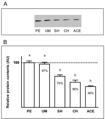
Similarly, SOD protein levels were found to be
sig-nificantly decreased (p<0.05) in the endometrium of
pa-tients diagnosed with hyperplasia simplex (∼30%),
hy-perplasia complex (∼44%) and adenocarcinoma (∼54%),
compared to polypus or myoma subjects (Fig. 4). LOOH level (Table I) showed significant variation
among the examined groups in blood plasma (H=23.38,
df=5, p<0.001) and endometrial tissue (H=37.11, df
=4, p<0.001). Compared to controls, moderate
eleva-tion of lipid hydroperoxides in plasma was observed in patients diagnosed with polypus endometrii or uterine
myoma (4% and 27%, respectively, p>0.05) whereas
it was significantly higher in both types of hyperplasia
(SH: 41%, CH: 52%, p <0.001) and adenocarcinoma
(57%, p<0.01). Also, in patients with simple
hyperpla-100
C PE UM SH CH ACE
100
102% 102%
89% 90% 90%
C UM CH PE SH ACE
A
B
a a a
a a a
R
e
la
ti
v
e
p
ro
te
in
c
o
n
te
n
ts
(
A
U
)
Fig. 2 –A.Relative protein contents. Example of Western blot im-munoassay for CuZnSOD in blood of control patients (C) and patients diagnosed with polypus endometrii (PE), uterus myomatosus (UM), simple hyperplasia (SH), complex hyperplasia (CH) and adenocarci-noma endometrii (ACE).B.Comparison of relative SOD protein con-tents in the blood of control patients (C) and patients diagnosed with polypus endometrii (PE), uterus myomatosus (UM), simple hyperpla-sia (SH), complex hyperplahyperpla-sia (CH) and adenocarcinoma endometrii (ACE). Values are expressed in arbitrary units (AU), as a percent of the protein content in controls which is considered as 100% (mean±SEM).
sia, levels of LOOH were higher than in patients with polypus (p<0.05).
LOOH level in endometrial tissue was significantly increased in patients with simple or complex
hyperpla-sia (∼50%) and adenocarcinoma (∼100%), compared to
polypus or myoma subjects.
We found a negative correlation between the level
of lipid hydroperoxides and SOD activity in blood (r=
−0.33, p<0.001) and endometrial tissue (r = −0.21,
p<0.05) of the examined patients.
DISCUSSION
TABLE I
LOOH concentration in plasma and endometrial tissue of control patients (C) and patients diagnosed with: polypus endometrii (PE), uterus myomatosus (UM), simple hyperplasia (SH), complex hyperplasia (CH)
and adenocarcinoma endometrii (ACE), (mean±SD).
Parameters/Patients C PE UM SH CH ACE
Plasma LOOH
0.19±0.02a 0.20±0.09ac 0.24±0.04ad* 0.27±0.07bd*** 0.27±0.07bcd*** 0.29±0.04bcd**
(nmol/mg protein) Endometrial
tissue LOOH 0.22±0.06a 0.24±0.09a 0.33±0.09b 0.34±0.07b 0.47±0.08b
(nmol/mg protein)
Values with different letter designations are significantly different (p<0.05). Values marked with asterisks are significantly different (*p<0.05,
**p<0.01, ***p<0.001) from the control.
PE UM SH CH ACE 0
1 2 3 4 5 6
a a
b b
b
U
/m
g
p
ro
te
in
Fig. 3 – CuZnSOD activity in the endometrium of patients diagnosed with: polypus endometrii (PE), uterus myomatosus (UM), simple hy-perplasia (SH), complex hyhy-perplasia (CH) and adenocarcinoma en-dometrii (ACE). Mean SOD activities (±SD) are represented by the
box; medians are plotted inside the box; the whiskers extend to the 5th and 95thpercentiles. Values not designated by the same letter (a, b) are significantly different (p<0.05).
superoxide dismutases are the main enzymes responsible for the elimination of superoxide radicals and are con-sidered to be key antioxidants in aerobic cells (Hileman et al. 2001).
In the blood of the examined patients, SOD activi-ties were decreased while SOD protein levels remained stable in comparison to healthy subjects. Also, the de-crease in SOD activity was more pronounced in women with hyperplasia or adenocarcinoma than in those suf-fering from polypus or myoma, while higher levels of LOOH were recorded in all examined groups, in com-parison to controls, except for patients with polypus en-dometrii. These results are in accordance with findings of other authors. Patients with cervicitis or myoma had lower plasma SOD activity and elevated lipid
peroxida-a
100
PE UM SH CH ACE
100
97%
70%
56% 46% a
b b
b
PE UM SH CH ACE
B
A
R
e
la
ti
v
e
p
ro
te
in
c
o
n
te
n
ts
(
A
U
)
Fig. 4 –A.Relative protein contents. Example of Western blot im-munoassay for CuZnSOD in the endometrium of patients diagnosed with polypus endometrii (PE), uterus myomatosus (UM), simple hy-perplasia (SH), complex hyhy-perplasia (CH) and adenocarcinoma en-dometrii (ACE).B.Comparison of relative SOD protein contents in endometrium patients diagnosed with polypus endometrii (PE), uterus myomatosus (UM), simple hyperplasia (SH), complex hyperplasia (CH) and adenocarcinoma endometrii (ACE). Values are expressed in arbitrary units (AU), as a percent of the protein content in PE patients which is considered as 100% (mean±SEM). Values not designated by
tion, compared to healthy women, whereas erythrocyte SOD activity was significantly lower only in myoma pa-tients (Chiou and Hu 1999). Decreased SOD activity and enhanced lipid peroxidation were found in erythrocytes of cervical cancer patients as well, compared to healthy subjects and cervicitis patients (Manoharan et al. 2004). A profound decrease in SOD activity and an in-crease in lipid peroxidation were also found in endo-metrium of patients with hyperplasia simplex, hyper-plasia complex and adenocarcinoma, in comparison to women with polypus or myoma. In adenocarcinoma patients, we observed a more pronounced decrease in SOD activity than in hyperplastic subjects. Levels of SOD protein were found to be 30-45% lower in endo-metrial tissue with hyperplasia or adenocarcinoma, com-pared to polypus and myoma. Previous analysis of en-dometrial samples from two different human popula-tions (Japanese and Finnish women) showed signific-antly decreased SOD activity and higher LPO in en-dometrium of cancer patients than in the normal tis-sue, which pointed to an impairment in the enzymic AO defense system (Punnonen et al. 1993). Lower SOD concentrations were recorded in the peritoneal fluid of infertile women with endometriosis (Liu et al. 2001, Szczepanska et al. 2003). Recent evidence also indi-cates decreased SOD activity and expression in many disease states or pathological conditions, including can-cer (Bostwick et al. 2000, Cullen et al. 2003). Other studies, however, showed that SOD levels or activities in various tissues with neoplastic changes appeared un-changed or elevated compared to normal tissues (Bala-subramaniyan et al. 1994, Chung-man Ho et al. 2001, Hu et al. 2005). Such observations are likely because of the different assays and various cell types used in those studies.
Decrease of enzyme activity could be a consequen-ce of elevated endogenous ROS production, as indicated by several studies. Kong et al. (2000) showed that, in response to oxidative stress, SOD enzyme may be con-sumed to prevent oxidative damage since it was shown that the overproduction of ROS exhausts the SOD ca-pacity. Similar observation was recorded by Onodera et al. (2003) regarding the antioxidant defense system in general. It was also shown that oxygen radical produc-tion, which elevates lipid peroxidaproduc-tion, increases with
clinical progression of diseases (Bagchi and Puri 1998, Skrzydlewska et al. 2005). In support of these observa-tions, we found negative correlation between LOOH and SOD activity in blood and endometrial tissue.
Evidence suggests that transformed tissues produce high levels of ROS and are constantly under oxidative stress (Hileman et al. 2001). The increase in ROS, such
as O−
2, is able to stimulate cell cycle progression and
promote cell proliferation. Although the precise mecha-nisms responsible for such elevation of ROS are not yet defined, several potential mechanisms have been
sug-gested. They include the oncogenic signals, such asc
-mycand Ras (Hu et al. 2005) or mitochondrial
mu-tations and respiratory chain malfunction, which may lead to increased superoxide production (Pelicano et al. 2004). In addition, molecular mechanisms by which the expression of cytosolic SOD responds to ROS stress, have not been well characterized (Hu et al. 2005). De-creased cytosolic SOD level in gynecological disorders, observed in our study, may be due to a reduced expres-sion of the SOD gene. Other possibilities include a di-minished translation rate for SOD mRNA in endometrial hyperplastic and carcinoma cells. Such a discrepancy in mRNA levels and the levels of corresponding proteins, suggesting possible translational regulation, has been re-ported for many gene products in normal and malignant cells (Klausner et al. 1993, Bommer et al. 2004, Lauer et al. 1999). In addition, Brown et al. (2004) demonstrated
an essential role of O2or O∙−2 in the posttranslational
ac-tivation of CuZnSOD and in the ratio of active to inactive CuZnSOD, which may be relevant to various diseases.
Most studies have shown bothin vivoandin vitro
Recent findings show that SOD might be a promis-ing therapeutic target for cancer treatment. Dependpromis-ing on the cell type and the level of SOD activity, inhibition or enhancement of SOD using molecular biology tech-niques reflects two possible approaches to cancer ther-apy. For the cells with lower SOD activity, enhancement of SOD expression may be used to reverse the premalig-nant and maligpremalig-nant phenotype (Hileman et al. 2001).
Our study shows that gynecological patients have lower SOD activity in both blood and endometrium, while the SOD protein level is decreased only in the en-dometrial tissue. There is a possibility that women with hyperplasia or adenocarcinoma are more susceptible to oxidative stress since a more pronounced decrease of SOD level was observed in those patients.
Further investigation is necessary to reveal if al-terations of SOD may contribute to the understanding of molecular mechanisms of carcinogenesis and to the development of new therapeutic approaches in clinical practice.
ACKNOWLEDGMENTS
The work was financially supported by the Ministry of Science and Technology of the Republic of Serbia (Grants 143044B and 143035B).
RESUMO
Resultados epidemiológicos e experimentais apontam para o envolvimento dos radicais derivados do oxigênio na patogê-nese das moléstias ginecológicas, assim como no desenvolvi-mento do câncer. O objetivo do presente estudo foi o de exa-minar as alterações nas atividades e níveis de Cu/Zn superóxi-do dismutase (CuZnSOD) e hidroperóxisuperóxi-dos lipídicos (LOOH) no sangue e tecido endometrial de pacientes diagnosticados com mioma uterino, pólipo endometrial, hiperplasia simplex, hiperplasia complex e adenocarcinoma do endométrio. Os re-sultados de nosso estudo mostraram atividades de SOD dimi-nuídas e nível de SOD proteína inalterado no sangue de todos os pacientes examinados em comparação a indivíduos saudá-veis. Diminuição de ambos, atividade de SOD e nível protéi-co, foram encontrados no endométrio de pacientes com hiper-plasia simplex, hiperhiper-plasia complex e adenocarcinoma em comparação às mulheres com pólipos e/ou mioma. O nível de LOOH estava elevado em ambos os tecidos de pacientes com hyperplasia e adenocarcinoma em comparação a
indiví-duos saudáveis ou pacientes com diagnóstico benigno. Nossos resultados sugerem que um decréscimo na atividade e nível pro-téico de SOD, assim como um incremento no nível de LOOH, em pacientes com desordens ginecológicas, tornam esses pa-cientes mais susceptíveis ao dano oxidativo causado pelas espé-cies reativas de oxigênio (ROS). Um desequilíbrio na formação de ROS e no nível de SOD pode ser importante na patogênese e/ou perpetuação do dano tecidual em pacientes ginecológicos. Desde que existe evidência de que SOD pode ser um alvo para terapia de câncer, nossos resultados fornecem uma base para futura pesquisa e opções para aplicações clínicas.
Palavras-chave: Cu/Zn superóxido dismutase, hidroperóxi-dos lipídicos, mioma uterino, pólipo endometrial, hiperplasia endometrial, adenocarcinoma endometrial.
REFERENCES
BAGCHIKANDPURIS. 1998. Free radicals and antioxidants in health and disease. East Mediterr Health J 4: 350–360. BALASUBRAMANIYANN, SUBRAMANIANSANDGOVIN -DASAMYS. 1994. Status of antioxidant systems in human
carcinoma of uterine cervix. Cancer Lett 87: 187–192. BOMMERUA, LAZARIS-KARATZASA, DEBENEDETTIA,
NURNBERGP, BENNDORFR, BIELKAHANDSONEN
-BERG N. 1994. Translational regulation of the mam-malian growth-related protein P23: involvement of eIF-4E. Cell Mol Biol Res 40: 633–641.
BOSTWICKDGET AL. 2000. Antioxidant enzyme expres-sion and reactive oxygen species damage in prostatic in-traepithelial neoplasia and cancer. Cancer 89: 123–134. BRIGELIUS-FLOHÉR. 2006. Glutathione peroxidases and
redox-regulated transcription factors. Biol Chem 387: 1329–1335.
BROWNNM, TORRESAS, DOANPE ANDO’HALLORAN
TV. 2004. Oxygen and the copper chaperone CCS regulate posttranslational activation of Cu, Zn superoxide dismutase. Proc Natl Acad Sci 101: 5518–5523. CHIOUJFANDHUML. 1999. Elevated lipid peroxidation
and disturbed antioxidant enzyme activities in plasma and erythrocytes of patients with uterine cervicitis and myoma. Clin Biochem 32: 189–192.
CHUNG-MAN HOJ, ZHENGS, COMHAIRSA, FARVERC
AND ERZURUM SC. 2001. Differential expression of manganese superoxide dismutase and catalase in lung cancer. Cancer Res 61: 8578–8585.
pancreas: Another link between chronic pancreatitis and pancreatic cancer. Pancreas 26: 23–27.
DRÖGEW. 2002. Free radicals in the physiological control of
cell function. Physiol Rev 82: 47–95.
HILEMANEA, ACHANTAGANDHUANGP. 2001. Super-oxide dismutase: An emerging target for cancer therapeu-tics. Expert Opin Ther Targets 5: 697–710.
HUY, ROSENDG, ZHOUY, FENGL, YANGG, LIUJAND
HUANGP. 2005. Mitochondrial manganese-superoxide dismutase expression in ovarian cancer: role in cell pro-liferation and response to oxidative stress. J Biol Chem 280: 39485–39492.
KLAUSNER RD, ROUAULT TA ANDHARFORD JB. 1993. Regulating the fate of mRNA: the control of cellular iron metabolism. Cell 72: 19–28.
KONGQ, BEELJAANDLILLEHEIKO. 2000. A threshold
concept for cancer therapy. Med Hypotheses 55: 29–35. LAEMMLIUK. 1970. Cleavage of structural proteins during
the assembly of the head of bacteriophage T4. Nature 227: 680–685.
LAUERC, VÖLKLA, RIEDLS, FAHIMIHDANDBEIERK. 1999. Impairment of peroxisomal biogenesis in human colon carcinoma. Carcinogenesis 20: 985–989.
LIUY, LUOLANDZHAOH. 2001. Levels of lipid peroxides and superoxide dismutase in peritoneal fluid of patients with endometriosis. J Tongji Med Univ 21: 166–167. LOWRYOH, ROSEBROUGHNJ, FARRALANDRANDALL
RJ. 1951. Protein measurement with the Folin phenol reagent. J Biol Chem 193: 265–275.
MANOHARANS, KOLANJIAPPANKANDKAYALVIZNIM.
2004. Enhanced lipid peroxidation and impaired enzymic antioxidant activities in the erythrocytes of patients with cervical carcinoma. Cell Mol Biol Lett 9: 699–707. MONARIMET AL. 2006. Superoxide dismutase in gastric
adenocarcinoma: is it a clinical biomarker in the develop-ment of cancer? Biomarkers 11: 574–584.
MÜNSTEDTK, GRANTP, WOENCKHAUSJ, ROTHGAND
TINNEBERGHR. 2004. Cancer of the endometrium: cur-rent aspects of diagnostics and treatment. World J Surg Oncol 2: 24.
MUTTERGL. 2002. Diagnosis of premalignant endometrial
disease. J Clin Pathol 55: 326–331.
OBERLEY TD. 2002. Oxidative damage and cancer. Am J
Pathol 160: 403–408.
OBERLEYTDANDOBERLEYLW. 1997. Antioxidant en-zyme levels in cancer. Histol Histopathol 12: 525–535. ONODERAK, OMOIN-O, FUKUIK, HAYASAKAT, SHIN
-KAI T, SUZUKIS, ABE KANDURANO S. 2003.
Ox-idative damage of rat cerebral cortex and hippocampus, and changes in antioxidative defense systems caused by hyperoxia. Free Radic Res 37: 367–372.
OTAH, IGARASHIS, HATAZAWAMANDTANAKAT. 1999. Immunohistochemical assessment of superoxide dismu-tase expression in the endometrium in endometriosis and adenomyosis. Fertil Steril 72: 129–134.
PEJIC´ S, KASAPOVIC´ J, TODOROVIC´ A, STOJILJKOVIC´ V
AND PAJOVIC´ SB. 2006. Lipid peroxidation and anti-oxidant status in blood of patients with uterine myoma, endometrial polypus, hyperplastic and malignant endo-metrium. Biol Res 39: 71–82.
PELICANOH, CARNEYDANDHUANGP. 2004. ROS stress in cancer cells and therapeutic implications. Drug Resist Updat 7: 97–110.
PUNNONEN R, KUDO R, PUNNONEN K, HIETANEN E,
KUOPPALAT, KAINULAINENH, SATO KANDAHO -TUPA M. 1993. Activities of antioxidant enzymes and
lipid peroxidation in endometrial cancer. Eur J Cancer 29A: 266–269.
SKRZYDLEWSKAE, SULKOWSKIS, KODAM, ZALEWSKI
B, KANCZUGA-KODA L AND SULKOWSKA M. 2005. Lipid peroxidation and antioxidant status in colorectal cancer. World J Gastroenterol 11: 403–406.
SZCZEPANSKAM, KOZLIKJ, SKRZYPCZAKJANDMIKO
-LAJCZYK M. 2003. Oxidative stress may be a piece in the endometriosis puzzle. Fertil Steril 79: 1288–1293. TOYOKUNI S. 2006. Novel aspects of oxidative
stress-as-sociated carcinogenesis. Antioxid Redox Signal 8: 1373– 1377.
VALKO M, LEIBFRITZ D, MONCOL J, CRONIN MT,
MAZURMANDTELSERJ. 2007. Free radicals and
anti-oxidants in normal physiological functions and human disease. Int J Biochem Cell Biol 39: 44–84.