This Accepted Author Manuscript is copyrighted and published by Elsevier. It is posted here by agreement between Elsevier and University of Brasilia. Changes resulting from the publishing process - such as editing, corrections, structural formatting, and other quality control
mechanisms - may not be reflected in this version of the text. The definitive version of the text was subsequently published in [Research in Microbiology, Volume 155, Issue 8, October 2004, Pages 681–687, doi:10.1016/j.resmic.2004.04.012].You may download, copy and otherwise use the AAM for non-commercial purposes provided that your license is limited by the following restrictions:
(1) You may use this AAM for non-commercial purposes only under the terms of the CC-BY-NC-ND license.
(2) The integrity of the work and identification of the author, copyright owner, and publisher must be preserved in any copy.
(3) You must attribute this AAM in the following format: [agreed attribution language, including link to CC BY-NC-ND license + Digital Object Identifier link to the published journal article on Elsevier’s ScienceDirect® platform].
________________________________________________________________________
Este Manuscrito do Autor Aceito para Publicação (AAM) é protegido por direitos autorais e publicado pela Elsevier. Ele esta disponível neste Repositório, por acordo entre a Elsevier e a Universidade de Brasília. As alterações decorrentes do processo de publicação - como a edição, correção, formatação estrutural, e outros mecanismos de controle de qualidade - não estão refletidas nesta versão do texto. A versão definitiva do texto foi posteriormente publicado em [Research in Microbiology, Volume 155, Número 8, Outobro de 2004, Páginas 681–687, doi:10.1016/j.resmic.2004.04.012]. Você pode baixar, copiar e utilizar de outra forma o AAM para fins não comerciais , desde que sua licença seja limitada pelas seguintes restrições:
(1) Você pode usar este AAM para fins não comerciais apenas sob os termos da licença CC- BY- NC-ND.
(2) A integridade do trabalho e identificação do autor, detentor dos direitos autorais e editor deve ser preservado em qualquer cópia.
Purification and ultrastructural localization of a copper
–
zinc superoxide
dismutase (CuZnSOD) from the entomopathogenic and acaricide fungus
Metarhizium anisopliae
Luiza Amaral de Castro Sandra Estrazulas Farias Sônia Nair Bao
Augusto Schrank
Marilene Henning Vainstein
Abstract
The entomopathogenic fungus Metarhizium anisopliae contains three superoxide dismutases. One of these enzymes was purified and partially characterized as a CuZnSOD. The enzyme has an estimated molecular mass o 30 690 D d sp y 3838.89 U mg−1. D -PAGE and 2D gels show a single band of protein in the fractions eluted from the gel filtration column with a molecular mass of 20 000 and ∼15 000 Da, respectively, and a pI of 6.0. These results suggest that the native enzyme is a dimer consisting of two subunits. Polyclonal antiserum were raised against purified CuZnSOD and used to determine its subcellular localization by immunoelectron microscopy. M. anisopliae CuZnSOD is present in the cell wall.
Keywords: Metarhizium anisopliae; Superoxide dismutase (SOD); Antioxidant enzyme; Active oxygen species (AOS); Oxygen free radicals (OFR); Biological control
1. Introduction
Metarhizium anisopliae is a broad host range entomopathogenic fungus first
recognized as a potential candidate for biological control of agriculture pests in the 1880s. The
fungus actively invades the hosts through the cuticle by mechanical pressure and synergistic
action of hydrolytic enzymes [18,47,53]. The whole process of host infection occurs in
successive phases of adhesion of spores, hyphal germination, differentiation (apressorium
formation), penetration, colonization, reproduction and dissemination for the start of a new
cycle [9]. During host colonization, fungi usually encounter a host resistance response, the
generation of an environment around the pathogen rich in active oxygen species (AOS), and
death of host cells surrounding the infection site. The dead host cells become a defensive layer
that deprives the biotroph of a food supply. This layer also acts as a mechanical barrier, which
the pathogen finds diffi- cult to breach [22,34].
Superoxide dismutases (SODs) are metalloenzymes with a redox metal at their active
sites that rapidly convert two molecules of superoxide radical to hydrogen peroxide and
cofactors: MnSOD [19], CuZnSOD [40], FeSOD [58] and NiSOD [31,59]. The CuZnSODs are
typically found in the cytosol of eukaryotes, while FeSODs are mainly found in prokaryotes and
chloroplasts, and MnSODs are found in prokaryotes and in mitochondria. There are, however,
some exceptions to this simplified evolutionary scheme [23]. MnSOD from filamentous fungi
[13–15,45] does not contain a typical mitochondrial signal peptide, characteristically rich in
basic and hydrophilic amino acids [46]. FeSOD and MnSOD share a high degree of amino acid
sequence homology [46], while CuZnSOD represents a distinct class. A subclass of SOD exists
which is capable of utilizing either Mn or Fe as the catalytic metal ion, and it is referred to as
combialistic SOD [56].
In 1993, Schrank et al. [51] described a FeSOD in M. anisopliae. The discovery of a
eukaryotic FeSOD directly contradicted the prevailing scheme of SOD distribution, in which the
iron type of SOD was found only in prokaryotes. Loss of infectivity has been observed in highly
pathogenic strains of M. anisopliae; however, the implicated causes are still not clear. Fungal
virulence is a function of many attributes and it is difficult to assess their individual
contribution to host mortality [9]. One of the factors responsible for virulence variation may be
the presence of superoxide dismutases (SODs) [51]. In addition to their essential role in
protecting living organisms against the toxicity of free oxygen radicals generated by
metabolism, SODs play a significant role in the pathogenicity of several organisms, including
Salmonella typhimurium [16], Mycobacterium tuberculosis [57] and Cryptococcus neoformans
[8,12,42]. The deletion of the SOD1 gene in C. neoformans resulted in defects in the expression
of a number of virulence factors, i.e., laccase, urease and phospholipase. Recovery of virulence
factor expression and restoration of virulence was achieved by sod1 mutant complementation
[42]. Candida albicans cells lacking CuZnSOD showed increased susceptibility to macrophage
attack and had attenuated virulence in mice [29].
A wide variety of fungi have been shown to contain at least two forms of SOD,
CuZnSOD and MnSOD [10,28, 32,35,36,44]. Thirty-two complete deduced amino acid
sequences have been reported in the SwissProt, EMBL and GenBank data libraries. These
include sod1, encoding a major CuZnSOD in Neurospora crassa [7]; SOD2 genes encoding
MnSOD from Penicillium chrysogenum [14], Aspergillus fumigatus[13], Ganoderma
microsporum [45], and Colletotrichum graminicola [15]; and sod1 and sod2, encoding a
CuZnSOD and an MnSOD, respectively, in Saccharomyces cerevisiae [5,6,37], and a gene
encoding FeSOD from Rhodotorula glutinis [49]. Fungal parasites have the ability to secrete
enzymes in order to protect them from a hostile chemical in the environment. SODs may also
s bs (s p x d s, O− 2 ) and product (hydrogen peroxide, H2O2) have been implicated in these processes in animal and plant cell systems [35,48,50].
The description of the purification of the Metarhizium SODs provides the framework
for ascertaining the potential role of these enzymes in protection against externally generated
superoxide.
2. Materials and methods
2.1. Organisms and culture conditions
M. anisopliae strain E6 was obtained from infected insects (Escola Superior de
Agronomia Luiz de Queiroz, USP, Brazil, Microbial Genetics Group Collection). Mycelia were
g w q d C ’s m d m (MC ) [11] d s y 106 sp s m −1 rotator-shaking platform (180 pm) 28 ◦C [47]. After 48 h of incubation, mycelia were
harvested by filtration onto a Whatman 1 filter paper and washed twice with sterile distilled
water.
2.2. Analytical procedures
SOD activity was determined by a riboflavin-nitroblue tetrazolium (NBT) assay adapted
from Beauchamp and Fridovich [1]. The photosensitive assay reagent contained 2.6 µM
riboflavin, 35 µM NBT, 14 mM N ,N ,N ,N -tetramethylethylethylenediamine (TEMED) in 0.05
M phosphate buffer, pH 7.8. The reaction mixture containing the photosensitive assay reagent
(0.5 ml) and the appropriately diluted enzyme preparation (up to 0.5 ml) was made up to a
total volume of 1 ml. The inhibition of the NBT reduction was measured at 650 nm and a liner
calibration plot was adopted. One unit of SOD represents the amount of enzyme that inhibits
50% of the rate of auto-oxidation of NBT under the defined assay conditions.
2.3. Purification of CuZnSOD
A p p d s w d 4 ◦C. h mycelia biomass (700 g) obtained from a 48-h shaking culture was collected on a sieve (160 µM), washed thoroughly
with deionized water, frozen by addition of liquid nitrogen, grounded to a fine powder and
suspended in lysis buffer (50 mM Tris–HCl, 1 mM EDTA, pH 7.5) containing protease inhibitors
(5 mM iodoacetamide, 50 µM Nα-p-tosylL-lysine chloromethyl ketone hydrochloride TLCK, 50
µM L-1-4 -tosylamino-2-phenylethylchloromethylketone TPCK, 1 mM phenylmethanesulfonyl
000 g, 15 m , 4 ◦C). h s g s p was brought to 50% saturation by gradually adding solid (NH4)2 O4 d w s s d 2 h 4 ◦C. h p p was removed by
centrifugation (10 000 g, 15 min, 4 ◦C) d h s pernatant was brought to 95% saturation
with (NH4)2SO4. The precipitate was collected by centrifugation (10 000 g, 30 m , 4 ◦C),
dissolved in 10 mM Tris–HCl/EDTA 1 mM, pH 7.5, and dialyzed against the same buffer for 24 h
4 ◦C. h s h s w re denominated TE (total extract), P50% and P95%, respectively. The dialyzed P95% fraction was directly applied to a DEAE-Sephacel column (25 ×
2.5 cm) equilibrated with 10 mM Tris–HCl pH 7.5. The column was washed thoroughly with
200 ml of equilibration buffer until all un- bound proteins were removed. The remaining bound
proteins were eluted by a continuous linear gradient of 500 ml of the same buffer containing 0
to 1.2 M NaCl at a flow 30 m h−1. F s 6 m w ected and those
containing SOD activity were pooled, concentrated by lyophilization and loaded onto a
Sephadex G-150 column (100 × 2.5 cm) equilibrated with 50 mM Tris–HCl containing 0.1 M
NaCl. Elution was continued with 200 ml of the same buffer at a flow 12 m h−1.
Fractions of 6 ml were collected and analyzed. BSA (67 000 Da), ovalbumin (43 000 Da),
chymotrypsinogen (25 000 Da) and ribonuclease A (13 700 Da) were used as molecular mass
markers, and blue dextran was used for determining the void volume. During the purification
steps, protein was determined spectrophotometrically at 280 nm or according to the Bradford
method, by using bovine serum albumin (BSA) as standard [4].
2.4. Electrophoresis
Sodium dodecyl sulfate–polyacrylamide gel electrophoresis (SDS-PAGE, 15%) was
according to Laemmli [33]. The purified enzyme was also analyzed by 2D gel electrophoresis
[43] stained with silver nitrate [3].
For the detection of SOD activity in gels, proteins were separated on an 8.5%
non-denaturing PAGE and stained using NBT, riboflavin and TEMED as previously described [17].
Potassium cyanide (KCN), an inhibitor of the CuZnSOD and not of the manganese and iron
forms, and hydrogen peroxide (H2O2), an inhibitor of the CuZnSOD and FeSOD, were used at
10 mM to ascertain the SOD type [41]. Total extracts of E. coli XL1-blue containing MnSOD and
FeSOD activity and purified CuZnSOD bovine (Sigma), and MnSOD and FeSOD from E. coli
2.5. Preparation of antisera and immunoblotting
Polyclonal antisera against purified SOD were raised in mice. Six-week-old BALB/c mice
were injected intraperitoneally w h 12.5 µg p d OD m s d w h F d’s complete
adjuvant followed by booster injection of 12.5 µg F d’s mp dj 25
days. Sera were obtained 8 days after the second injection and titrated by indirect ELISA.
Proteins resolved by SDS-PAGE were electroblotted onto nitrocellulose membranes. The
blotted membranes were blocked with 5% (w/v) milk powder in PBS (Na2HPO4 10 mM,
KH2PO4 1.7 mM, NaCl 137 mM, KCl 2.7 mM). After blocking, the membranes were incubated
with anti-SOD antiserum in blocked solution (1:2000). The membranes were washed three
times in PBS for 10 min, incubated with a secondary antibody (anti-mouse IgG peroxidase
conjugate), washed and revealed according to the manufacturers.
2.6. Immunoelectron microscopy
Spores and mycelia of M. anisopliae harvested from C ’s mp s d m d m (MC ) [11]
were collected for immunogold localization, fixed following the protocol previously described
and embedded in LR-gold [2]. The cured blocks were cut into ultrathin sections and mounted
on copper grids, followed by immunogold labeling with 15 nm gold protein A for 1 h. Finally,
specimens were stained with y 20 m d R y d’s d 5 m
prior to examination in a transmission electron microscope JEOL 100 C.
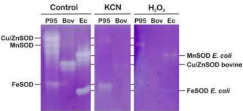
Fig. 1. Electrophoretic analysis of SODs from M. anisopliae. The non-denaturing gels were incubated with 10 mM KCN or 20 mM H2O2. (P95) P95 fraction (10 µl) containing 4.4 U of SOD; (Bov) Three units of purified bovine CuZnSOD (Sigma); (Ec) E. coli extract (10 µl) containing FeSOD and MnSOD activity (6 U).
3. Results and discussion
Three different SOD isozymes were resolved from total protein extracts of M.
anisopliae in native PAGE and NBT staining and identified by their cyanide and H2O2
inhibitors and was classi- fied as a CuZnSOD; the fastest moving band was resistant to cyanide
and sensitive to H2O2 as characteristic of FeSOD; the intermediary moving band was the least
prominent isozyme in a total protein extract when compared to CuZnSOD and FeSOD. This
enzyme was resistant to both H2O2 and low concentrations of cyanide (10 mM) resembling
the characteristics of MnSOD; however, the sensitivity to higher concentrations of cyanide
suggests that a different structure or different metals may be present in the active site (Fig. 1).
The presence of a cyanide-insensitive and H2O2-sensitive FeSOD was reported as being specifi-
cally and concomitantly expressed during differentiation of hypha/bud cell into chlamidospore
in Fusarium oxysporum f. sp. Raphani [32]. These results suggest that active intermediates of
oxygen and/or those detoxifying enzymes participate in the differentiation of the fungus upon
response to starvation [32]. However, direct evidence confirming the presence of a FeSOD in F.
oxysporum, was not provided [39]. Functional analysis of the SOD isozyme profile from C.
graminicola identified two major SOD activities, representing FeSOD and MnSOD differentiated
by their sensitivity to H2O2 [15]. More recently Raychaudhuri et al. (2003) reported for the
first time the presence of cytosolic FeSOD in a yeast system, Rhodotorula glutinis, confirmed
by atomic absorption spectroscopy and assay with different inhibitors. In silico analysis of the
sequence data suggested that it was not a classical cytosolic SOD, as it showed similarity
towards mitochondrial FeSOD/MnSOD [49].
Table 1
Purification steps of Metarhizium anisopliae SOD
Fig. 2. Anion-exchange chromatography profile of M. anisopliae SOD on DEAE-Sephacel. Concentrated
sample (173 mg of protein) was pumped m (25 × 2.5 m) w 30 m h−1. h
linear 0–1.0 M NaCl gradient (500 ml). Fractions of 6 ml were collected. (Ο) Protein absorption at 280
nm, (•) SOD activity and (- -) NaCl gradient
The isolation and purification of CuZnSOD was performed from mycelia of M.
anisopliae. Although the enzyme has been purified from numerous sources, only a few reports
exist concerning SOD purification from fungal sources [26–28,41], especially from
entomopathogenic fungi. The purification to homogeneity of CuZnSOD from M. anisopliae
involved four steps: homogenization of mycelia with removal of cell debris and dialysis;
precipitation with (NH4)2SO4; ion exchange and gel filtration chromatography (Table 1).
Fractional precipitation of the proteins was performed first at 50% saturation of (NH4)2SO4.
The extensive protein removal during this precipitation step was an advantage since all three
types of SOD remained present in the upper phase, increasing the specific activity 1.05-fold
when they were further precipitated with a 95% saturation of (NH4)2SO4. The P95% fraction
was submitted to DEAESephacel and three peaks with SOD activity were eluted (Fig. 2). The
pooled fractions containing CuZnSOD were concentrated and further purified by gel filtration
chromatography on Sephadex G-150. The enzyme was eluted with an estimated molecular
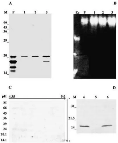
mass of 30 690 Da and a specific ac y 3838.89 U mg−1 ( b 1). D -PAGE (Fig. 3A),
non-denaturing PAGE (Fig. 3B) and 2D-gels (Fig. 3C) show a single band of protein in the
fractions eluted from the gel filtration column with an apparent molecular mass of 20 000 and
∼15 000 Da, respectively, and a pI of 6.0 (Figs. 3A and 3C). This value is approximately half of the molecular mass of the whole protein determined by gel filtration (30 690 Da) and indicates
that the enzyme is composed of dimers of equal size, whose association depends upon
noncovalent interactions. On the basis of inhibitory action of the known CuZnSOD inhibitor
potassium cyanide [20] the newly described SOD is clearly a member of the CuZnSOD group.
To date, most of the eukaryotic CuZnSOD have all been shown to be homodimers with each
subunit being approximately 15 000 Da in size [21,25,30]. One exception is the CuZnSOD of
Phanerochaete crysosporium, a homodimer with identical molecular mass of 22 000 Da [44].
For SOD from Aspergillus spp., a different pattern has been described in which the
corresponding enzyme has revealed tetrameric or pentameric structures [26,28]. The pI 6.0
encountered for CuZnSOD purified from M. anisopliae was almost identical to that of the SODs
Fig. 3. Electrophoretic and immunoblot analysis of purified CuZnSOD from M. anisopliae. (A) SDS-PAGE (15%) stained with silver nitrate; (B) Non-denaturing PAGE stained with nitroblue tetrazolium; (C) 2D SDS-PAGE stained with silver nitrate; (D) Western blot. Lanes: (M) Mw markers (panels A and C); (P) Pool of fractions eluted from the first peak of the DEAE-Sephacel chromatography (panels A and B); (1–3), fractions 58, 59, and 60 (10 µl each), respectively eluted from the Sephadex G-150 chromatography, (panels A and B); (4) Proteins extracted from mycelium of M. anisopliae (30 µg); (5) Concentrated supernatant of M. anisopliae culture (30 µg), (6) CuZnSOD purified from M. anisopliae (10 µg) (panel D).
M. anisopliae SOD was inhibited by 2 mM KCN. This was not surprising, since
CuZnSODs are generally known to be CN−-sensitive enzymes [30]. The activity of the purified
SOD was susceptible to inactivation by hydrogen peroxide. The inactivation of CuZnSOD has
been attributed to the reduction of the enzyme-bound Cu2+ to Cu1+ by H2O2, followed by a
Fenton-type reaction of Cu2+ with additional H2O2 to form CU2+–OH. This could oxidatively
attack an adjacent histidine residue of the enzyme [38].
The specificity of polyclonal antisera raised against puri- fied M. anisopliae CuZnSOD
was verified by western blot. These antisera showed specificity for a protein with apparent
molecular mass of 15 000 Da from mycelia extracts and not for proteins present in the culture
supernatant, indicating that CuZnSOD in M. anisopliae might not be secreted in the
investigated conditions. MnSOD and FeSOD did not share major antigenic determinants with
Fig. 4. Ultrastructural localization of CuZnSOD of M. anisopliae. (A) Spores; (B) mycelium from 24-h culture; (C) mycelium from 48-h culture. Lower case letters are: (c) cytoplasm; (cw) cell wall; (n) nucleus. Arrows heads show the depositions of gold particles.
Immunolocalization with CuZnSOD resulted in extensive gold labeling in the cell walls
and peripheral cytoplasm of mycelia, while control sections incubated with pre-immune serum
showed only a very low level of non-specific binding of gold particles (Fig. 4). Labeling of
sections from 24- and 48-h-old mycelia with antiserum to CuZnSOD indicated that the enzyme
accumulated within the cell walls. Different cells exhibited different labeling intensities (Figs.
4D, 4E, 4F). Intense labeling within and around the circumference of the fungal cell wall and, to
a lesser extent, within the peripheral cytoplasm-cell membrane regions, was observed,
suggesting a location for a rapid secretion. A. fumigatus CuZnSOD is present in substantial
quantities in the cell wall and it is secreted into culture supernatants, making it available to
exogenous sources of superoxide [24]. Usually, in eukaryotic cells CuZnSOD is found in the
superoxide production. In prokaryotes like Caulobacter crescentus, SODs are located in
different cellular compartments, CuZnSOD in the periplasm and FeSOD in the cytoplasm
[54,55].
The search for thigmotropically controlled proteins identified a CuZnSOD that was
posttranslationally modified upon appressorium induction in Uromyces appendiculatus, an
important structure for host penetration [35].
The cell wall location of CuZnSOD in M. anisopliae may have important implications for
further understanding its function. As an entomopathogenic fungus M. anisopliae lives in an
ecological habitat that creates extracellular or extracytoplasmic oxidative stress against which
a cytoplasmic SOD might not be effective. This CuZnSOD might be essential for tolerance to
that oxidative stress, while the other cytoplasmic SODs produced by the fungus detoxify
superoxide derived from cytoplasmic metabolism. Presumably, in filamentous fungi the
membrane and the thick cell wall are impermeable to superoxide. If CuZnSOD functions as
proposed, then the CuZnSOD form might more likely be found in fungi that are tolerant of
extracellular oxidative stress, like the entomopathogenic ones
Acknowledgements
This work was supported by grants and fellowships from: Programa de Apoio ao
Desenvolvimento Científico e Tecnológico (PADCT), Conselho Nacional de Desenvolvimento
Científico e Tecnológico (CNPq) and Fundação de Apoio a Pesquisa do Estado do Rio Grande do
Sul (FAPERGS). L.A.C. is the recipient of a scholarship from CNPq
References
[1] C. Beauchamp, I. Fridovich, Superoxide dismutase: Improved assays and an assay applicable to acrylamide gels, Anal. Biochem. 44 (1971) 276–287.
[2] M.A. Berryman, R.D. Rodewald, An enhanced method for postembedding immunocytochemical staining which preserves cell membranes, J. Histochem. Cytochem. 38 (1990) 159–170.
[3] H. Blum, H. Beier, H.J. Gross, Improved silver staining of plant proteins, RNA and DNA in polyacrylamide gels, Electrophoresis 8 (1987) 93–99.
[4] M.M. Bradford, A rapid and sensitive method for quantitation of microgram quantities of protein utilizing the principle of protein–dyebinding, Anal. Biochem. 58 (1976) 79–110.
[6] E.C. Chang, B. Crawford, Z. Hong, T. Bilinski, D.J. Kosman, Genetic and biochemical characterization of Cu,Zn superoxide dismutase mutants in Saccharomyces cerevisiae, J. Biol. Chem. 266 (1991) 4417–4424.
[7] P. Chary, D. Dillon, A.L. Schroeder, D.O. Natvig, Superoxide dismutase (sod-1) null mutants of Neurospora crassa: Oxidative stress sensitivity, spontaneous mutation rate and response to mutagens, Genetics 137 (1994) 723–730.
[8] S. Chaturvedi, A.J. Hamilton, P. Hobby, G. Zhu, C.V. Lowry, V. Chaturvedi, Molecular cloning, phylogenetic analysis and threedimensional modeling of Cu,Zn superoxide dismutase (CnSOD1) from three varieties of Cryptococcus neoformans, Gene 268 (2001) 41–51.
[9] J.M. Clarkson, A.K. Charnley, New insights into the mechanisms of fungal pathogenesis in insects, Trends Microbiol. 5 (1996) 197–203.
[10] G.J. Choi, H.J. Lee, K.Y. Cho, Involvement of catalase and superoxide dismutase in resistance of Botrytis cinerea to dicarboximide fungicide Vinclozolin, Pesticide Biochem. Physiol. 59 (1997) 1–10.
[11] D.J. Cove, The induction and repression of nitrate reductase in the fungus Aspergillus nidulans, Biochem. Biophys. Acta 113 (1966) 51–56.
[12] G.M. Cox, T.S. Harrison, H.C. McDade, C.P. Taborda, G. Heinrich, A. Casadevall, J.R. Perfect, Superoxide dismutase influences the virulence of Cryptococcus neoformans by affecting growth within macrophages, Infect. Immun. 71 (2003) 173–180.
[13] R. Crameri, A. Faith, S. Hemmann, R. Juassi, C. Ismail, G. Menz, K. Blaser, Humoral and cell-mediated autoimmunity in allergy to Aspergillus fumigatus, J. Exp. Méd. 184 (1996) 265–270.
[14] B. Díez, C. Schleissner, M.A. Moreno, M. Rodríguez, A. Collados, J.L. Barredo, The manganese superoxide dismutase from the penicillin producer Penicillium chrysogenum, Curr. Genet. 33 (1998) 387–394.
[15] G.-C. Fang, R.M. Hanau, L.J. Vaillancourt, The SOD2 gene, encoding a manganese-type superoxide dismutase, is up-regulated during conidiogenesis in the plant-pathogenic fungus Colletotrichum graminicola, Fungal Genet. Biol. 36 (2002) 155–165.
[16] J.L. Farrant, A. Sansone, J.R. Canvin, M.J. Pallen, P.R. Langford, T.S. Wallis, G. Dougan, J.S. Kroll, Bacterial copper- and zinc-cofactored superoxide dismutase contributes to the pathogenesis of systemic salmonellosis, Mol. Microbiol. 25 (1997) 785–796.
[17] L. Flohe, F. Otting, Superoxide dismutase assays, Methods Enzymol. 105 (1984) 93–104.
[18] A.P. Frazzon, I.S. Vaz Junior, A. Masuda, A. Schrank, M.H. Vainstein, In vitro assessment of Metarhizium anisopliae isolates to control the cattle tick Boophilus microplus, Vet. Parasitol. 94 (2000) 117–125.
[19] I. Fridovich, Superoxide dismutases, Annu. Rev. Biochem. 44 (1975) 147–159.
[21] I. F d h, p x d d (O.−2 ), superoxide dismutases, and related matters, J. Biol. Chem. 272 (1997) 18515–18517.
[22] D.G. Gilchrist, Programmed cell death in plant disease: The purpose and promise of cellular suicide, Ann. Rev. Phytopathol. 36 (1998) 393–414.
[23] B. Halliwell, J.M.C. Gutteridge, Free Radicals in Biology and Medicine, Clarendon, Oxford, 1999.
[24] A.J. Hamilton, M.D. Holdom, L. Jeavons, Expression of the Cu,Zn superoxide dismutase of Aspergillus fumigatus as determined by immunochemistry and immunoelectron microscopy, FEMS Immunol. Med. Microbiol. 14 (1996) 95–102.
[25] H.N. Hassan, Microbial superoxide dismutase, Adv. Genet. 26 (1989) 65–97.
[26] D.G. Hatzinikolaou, C. Tsoukia, D. Kekos, B. Macris, An efficient and optimized purification procedure for the superoxide dismutase from Aspergillus niger: Partial characterization of the purified enzyme, Bioseparation 7 (1997) 39–46.
[27] N.D. Holdom, R.J. Hay, A.J. Hamilton, Purification N-terminal amino acid sequence and partial characterization of Cu,Zn superoxide dismutase from the pathogenic fungus Aspergillus fumigatus, Free Rad. Res. 22 (1995) 519–531.
[28] N.D. Holdom, R.J. Hay, A.J. Hamilton, The Cu,Zn superoxide dismutases of Aspergillus flavus, Aspergillus niger, Aspergillus nidulans, and Aspergillus terreus: Purification and biochemical comparison with the Aspergillus fumigatus Cu,Zn superoxide dismutase, Infect. Immun. 64 (1996) 3326–3332.
[29] C. Hwang, S.G. Rhie, S.T. Kim, Y.R. Kim, W.K. Huh, Y.U. Baek, S.O. Kang, Copper- and zinc-containing superoxide dismutase and its gene from Cândida albicans, Biochim. Biophys. Acta 1427 (2002) 245–255.
[30] E.R. James, Superoxide dismutase, Parasitol. Today 10 (1994) 481–484.
[31] E.J. Kim, H.J. Chung, B. Suh, Y.C. Hah, J.H. Roe, Expression and regulation of the sodF gene encoding iron- and zinc-containing superoxide dismutase in Streptomyces coelicolor Muller, J. Bacteriol. 180 (1998) 2014–2020.
[32] Y. Kono, H. Yamamoto, M. Takeuchi, H. Komada, Alterations in superoxide dismutase and catalase in Fusarium oxysporum during starvation-induced differentiation, Biochim. Biophys. Acta 1268 (1995) 35–40.
[33] U.K. Laemmli, Cleavage of structure proteins during the assembly of the head of bacteriophage T4, Nature 227 (1970) 680–685.
[34] C. Lamb, R.A. Dixon, The oxidative burst in plant disease resistance, Ann. Rev. Plant. Physiol. Plant. Mol. Biol. 48 (1997) 251–275.
[36] C.T. Lin, M.T. Lin, D.C. Sheu, K.J. Duan, Cloning and characterization of a cooper/zinc-superoxide dismutase gene from Aspergillus japonicus, Taiwania 39 (1994) 73–79.
[37] X. Liu, F.I. Elashvill, E.B. Gralla, J. Valentine, P. Lapinskas, V. Culotta, Yeast lacking superoxide dismutase: Isolation of a genetic suppressors, J. Biol. Chem. 267 (1992) 18298– 18303.
[38] M.R. Mavelli, G. Ciriolo, G. Rotilio, Multiple electrophoretic variants of Cu, Zn superoxide dismutase as expression of the enzyme aging: Effects of H2O2, ascorbate and metal ions, Biochem. Biophys. Res. Commun. 117 (1983) 677–681.
[39] A.M. Mayer, R.C. Staples, N.L. Gil-Ad, Mechanisms of survival of necrotrophic fungal plant pathogens in hosts expressing the hypersensitive response, Phytochemistry 58 (2001) 33–41.
[40] J.M. McCord, I. Fridovich, Superoxide dismutase, an enzymatic function for erythrocuprein, J. Biol. Chem. 244 (1969) 6049–6055.
[41] H.P. Misra, I. Fridovich, Inhibition of superoxide dismutase by azide, Arch. Biochem. Biophys. 189 (1978) 317–322.
[42] S.D. Narasipura, J.G. Ault, M.J. Behr, V. Chaturvedi, S. Chaturvedi, Characterization of Cu,Zn superoxide dismutase (SOD!) gene knockout mutant of Cryptococcus neoformans var. gattii: Role in biology and virulence, Mol. Microbiol. 47 (2003) 1681–1694.
[43] P.H. O’F , H gh-resolution two-dimensional electrophoresis of proteins, J. Biol. Chem. 250 (1975) 4007–4021.
[44] R. Öztürk, L.A. Bozkaya, E. Atav, N. Saglam, L. Tarhan, Purification and characterization of superoxide dismutase from Phanerochaete chrysosporium, Enzyme Microbial. Technol. 25 (1999) 392–399.
[45] S.M. Pan, J.S. Ye, R.S. Hseu, Purification and characterization of the manganese superoxide dismutase from Ganoderma microsporum, Biochem. Mol. Biol. Int. 42 (1997) 1035–1043.
[46] M.W. Parker, C.C. Blake, Iron- and manganese-containing superoxide dismutases can be distinguished by analysis of their primary structures, FEBS Lett. 229 (1988) 337–382.
[47] A.S. Pinto, C.C. Barreto, A. Schrank, C.J. Ulhoa, M.H. Vainstein, Purification and characterization of an extracellular chitinase from the entomopathogen Metarhizium anisopliae, Can. J. Microbiol. 43 (1997) 322–327.
[48] A.H. Price, A. Taylor, S.J. Ripley, A. Griffiths, A.J. Trewavas, M.R. Knight, Oxidative signals in tobacco increase cytosolic calcium, Plant Cell 6 (1994) 1301–1310.
[49] S. Raychaudhuri, M.M. Reddy, N.R. Rajkumar, R. Rajasekharan, Cytosolic iron superoxide dismutase is a part of the triacylglycerol biosynthetic complex in oleaginous yeast, Biochem. J. 372 (2003) 587–594.
[50] D. Salvemini, G. De Nucci, J.M. Sneddon, J.R. Vane, Superoxide anions enhance platelet adhesion and aggregation, Br. J. Pharmacol. 97 (1989) 1145–1150.
M.S.N. Silva, Superoxide dismutases in the entomopathogenic fungus Metarhizium anisopliae, Ciência e Cultura 45 (1993) 200–205.
[52] O. Spiegelhalder, B. Gerstenecker, A. Kersten, E. Schiltz, M. Kist, Purification of Helicobacter pylori SOD and cloning and sequencing of the gene, Infect. Immun. 61 (1993) 5315–5325.
[53] R.J. St Leger, L. Joshi, M.J. Bidochka, D.W. Roberts, Construction of an improved mycoinsecticide overexpressing a toxic protease, Proc. Natl. Acad. Sci. USA 93 (1996) 6349– 6354.
[54] H.M. Steinman, Function of periplasmic copper–zinc superoxide dismutase in Caulobacter crescentus, J. Bacteriol. 175 (1993) 1198–1202.
[55] H.M. Steinman, B. Ely, Copper–zinc superoxide dismutase of Caulobacter crescentus: Cloning, sequencing, and mapping of the gene and periplasmic location of the enzyme, J. Bacteriol. 172 (1990) 2901–2910.
[56] S. Sugio, B.Y. Hiraoka, R. Yamakura, Crystal structure of combialistic superoxide dismutase from Porphyromonas gingivalis, Eur. J. Biochem. 267 (2000) 3487–3495.
[57] H.C.H. Wu, J. Tsai-Wu, Y. Huang, C. Lin, G. Lioua, F. Lee, Identification and subcellular localization of a novel Cu,Zn superoxide dismutase of Mycobacterium tuberculosis, FEBS Lett. 439 (1998) 192–196.
[58] F.J. Yost, I. Fridovich, An iron-containing superoxide dismutase from Escherichia coli, J. Biol. Chem. 248 (1973) 4905–4908.