BJRS
RADIATION SCIENCES
06-03 (2018) 01-12Risk management in radiation therapy with SEVRRA
G. S. Joana
a,b; M. Gonçalves
b; C. Salata
b; F. C. Teixeira
b; E. Sandrini
d; G. Bittencourt
e;
H. Salmon
e; L. Fairbanks
d; S. S. Fortes
f,g; M. S. Nogueira
caPós-Graduação em Ciências e Técnicas Nucleares - DEN/UFMG, 31270-901, Belo Horizonte, MG, Brazil b Comissão Nacional de Energia Nuclear - CNEN, 22290-901, Rio de Janeiro, RJ, Brazil
c Centro de Desenvolvimento de Tecnologia Nuclear - CDTN/CNEN, 31270-901, Belo Horizonte, MG, Brazil d Grupo COI, Clínicas Oncológicas Integradas, 22290-030, Rio de Janeiro, Brazil
e Oncoclínicas, 04536-132, São Paulo, Brazil
f Oncologia Rede D’OR - Unidade Caxias, 25075-025, Rio de Janeiro, Brazil
g Instituto Nacional do Câncer José Alencar Gomes da Silva - INCA, 20230-130, Rio de Janeiro, Brazil
ABSTRACT
Radiation therapy is a complex treatment modality involving several technological and professional resources as well as a large number of attributions of these professionals with a high degree of interdependence. Thus there are many possibilities of failure in the treatment process that may lead to an accidental exposure. Regulatory agencies have established guidelines in order to prevent accidental exposures through security analysis planning and risk management. The System of EValuation of Risk in RAdiotherapy, also known as SEVRRA, is based on the risk matrix methodology and allows the risk assessment of radiation therapy facilities in a prescriptive way, highlighting events leading to high-risk levels. With SEVRRA it is also possible to establish an analysis of importance of the barriers proposed to avoid or mitigate such events. This work aimed to demonstrate the application of SEVRRA for the risk management of radiation therapy facilities. The risk profile was determined by identifying the highest risk level events, and the barriers of greater impact for the overall risk level. After implementing some missing barriers in the original facility profile, it was achieved a reduction from 31% to 8% in the number of events leading to high-risk level, showing the effectiveness of SEVRRA for the risk management in radiation therapy facilities.
Keywords: Linear Accelerator, Radiation Therapy, Risk Management, Risk Matrix, SEVRRA.
1. INTRODUCTION
Radiation therapy is one of the primary treatment options in cancer management [1]. It is a complex therapeutic modality involving several professionals and a large number of activities developed with a high degree of interdependence. A radiation therapy facility encompasses specialized staff and advanced technology therapeutic modalities. Together with all alternatives for the choice of a treatment modality or advanced technological paths to achieve better results for the treatment, there are several possibilities of failure in the treatment process. The increase in the number of cancer types and the increasing number of patients requiring treatment further contribute to an accident-prone scenario concerning non-planned patient exposure. Thus, there is a need for some kind of preventive approach to risk management to increase safety in this practice [2].
The risk assessment consists in identifying possible errors or failures, evaluating their probability of occurrence and the severity of their consequences. It highlights the weaknesses of a process based on the risk rating and the robustness of the security mechanisms. The accidental sequence begins with the occurrence of an initiating event, followed by the actuation or failure of the safety mechanisms. The safety mechanisms are defined according to the moment they act in the accidental sequence. Frequency reducers are measures to avoid or prevent an initiating event; as such, they take effect before the initiating event occurs. Direct barriers are measures to detect an initiating event and prevent its consequences, such as accidental exposure of patients. Consequence reducers are measures to detect the initiating event and mitigate the consequences of accidental exposure; they take effect once the event has occurred and its consequences have started to become apparent [2].
Due to a number of accidents in the world in the last twenty years [3-9], the need for a systematic way of investigating and foreseeing non-planned dose events associated with patients, workers and public became fundamental for the radiation therapy good practices. In this regard, regulatory agencies have been establishing guidelines in their regulations to prevent accidental exposures through safety analysis planning and risk management [10-13]. The Ibero-American Forum of Radiological and Nuclear Regulatory Agencies (FORO) [14] recently developed in the context of a risk analysis project the System of EValuation of Risk in RAdiotherapy, also known as
SEVRRA [1, 15-18]. SEVRRA is an online tool that implements the risk matrix method applied to radiotherapy [2, 17, 19] also developed under the FORO, dealing semi-quantitatively with both retrospective and prospective risk analysis approaches. It is ready to perform a prescriptive risk assessment for the modalities of 3D conformational radiation teletherapy with linear accelerator and Cobalt 60 unit, High Dose [7] and Low Dose Rate Brachytherapy [2, 17, 19, 20]. Treatments with advanced technologies are beyond the scope of the current SEVRRA version. There is some intent to expand the SEVRRA and risk analysis methodology to take into account new modalities of radiation therapy such as intensity modulated radiation therapy (IMRT) and volumetric modulated arc therapy (VMAT) [21].
This work aims to demonstrate the feasibility of the application of SEVRRA for the risk management by establishing the risk profile of a Brazilian “average” radiation therapy facility, i.e., a facility in which the presence or absence of each barrier was defined from the average characteristic of most Brazilian facilities, identifying the highest risk accidental sequences and the related and most relevant direct barriers, frequency and consequence reducers to assess how the missing barriers could affect the facility risk profile. Then, it was evaluated the impact of implementing some barriers demanded by Brazilian regulatory guidelines on the overall risk of the average facility and, finally, these results were compared with risk profile of the reference facility built-in in SEVRRA.
2.
MATERIALS AND METHODS
The average Brazilian radiotherapy facility has been defined considering technologies, personnel and processes, among many other characteristics commonly observed in regulatory inspections carried out by the National Commission of Nuclear Energy on country's radiotherapy facilities. This typical installation features a linear electron accelerator of double photon energy and electron beam. It does not have a multileaf collimator (it uses protection blocks). It has no record and verifying system. It has a dedicated tomograph for simulation and has two radiotherapy technicians working per shift. Despite the fact that they have a radiation oncologist and a radiation protection supervisor, holders and substitutes, the latters only act eventually, not having a defined
weekly workload. Thus, the facility has only one radiation oncologist and one medical physicist during its period of operation. The facility has a quality assurance program based on national and international protocols and recommendations performed regularly, however, there is no quality assurance program for the planning system. There is a preventive maintenance program for the linear accelerator, but no established procedure for recording and communicating the necessity of beam dosimetry after maintenance followed by acceptance and release of the machine for clinical use.
The facility's human resources are qualified for their functions and undergo retraining annually. There is effectively only a radiation oncologist and a medical physicist responsible for treatment prescribing and planning. The processes are well standardized and the staff is aware of all the treatments performed in the service. They do not maintain a routine with weekly meetings to discuss the cases in course, this would be done whenever necessary. However, due to the high workload, there is no established routine for radiation oncologist and medical physicist participation on the first treatment session and the portal image of the first session is often verified with the treatment already being started. For the same reason, the patient clinical review with the radiation oncologist does not occur weekly. The facility does not go through an external audit. There is no treatment planning and dosimetry double check by a second medical physicist. The treatment sheet does not show patient's photograph nor a picture of its positioning on the treatment table.
The risk analysis has been performed with SEVRRA for each stage of the radiotherapy with linear accelerator, from equipment initial installation, acceptance, commissioning and maintenance to the treatment execution, going through all the intermediate steps such as prescription, acquisition of anatomical patient data and planning (Figure 1).
By definition, risk is the probability of occurrence of some kind of damage taking into account its severity. To reduce the risk of some accident-initiating event there are some safety measures that constitute barriers to avoid, prevent, detect, control and mitigate the consequences of the accident. In a redundancy system, two or more barriers must be established so that one acts in the case of failure of the other(s) [2].
Figure 1: SEVRRA's screen for the risk analysis of radiotherapy with a linear accelerator.
In SEVRRA, each initiating event is classified as having low, RL, medium, RM, high, RH, or very high-risk, RVH. Once the risk analysis has been carried out, the initiating events risks are rated and a risk acceptability criterion is applied as a way to focusing on those ones resulting in highest risk level and highest consequences. Any sequences that do not meet these criteria must have their safety measures enhanced. Additional measures should be adopted for those initiating event classified as very high or high-risk (in case of high consequences) by introducing some direct barriers, frequency reducers or consequence reducers, thus concluding one cycle of the process of risk assessment by reducing the overall risk level of the facility.
This study was accomplished in three steps: (1) At the first phase, it has been considered that the facility had no barriers or reducers in order to establish the baseline risk level for the radiotherapy with linear accelerator (Figure 2-a). (2) The second phase has been carried out considering the direct barriers, frequency and consequence reducers existing at the facility. Once the facility overall risk level has been obtained, the highest risk accidental sequences have been identified together with the direct barriers, frequency and consequence reducers representing the greatest impact on this risk level (Figure 2-b). (3) In the third phase, it has been analyzed which barriers could be
implemented on the facility to produce the greatest impact in reducing the risk level. The analysis has been performed again considering these modifications (Figure 2-c). The impact of these changes has been evaluated. Once the most important missing barriers have been identified, the impact of their implementation on the overall risk level of the facility has been analyzed.
Figure 2 - Distribution of initiating events by risk level (a) 1st phase: without direct barriers and
reducers, (b) 2nd phase: with facility's existing direct barriers and reducers, (c) 3rd phase: adding/ modifying direct barriers and reducers (d) for SEVRRA’s reference facility.
3.
RESULTS AND DISCUSSION
The risk matrix model implemented on SEVRRA has identified 148 accident-initiating events for the practice of radiotherapy with linear accelerator [2]. For the fictitious facility of this study, since it does not have multileaf collimator, only 141 events are indeed applicable. In the 1st phase, when it was considered that there were no direct barriers and frequency and consequence reducers, the facility presented one initiating event with very high risk, RVH, 120 with high risk, RH, and 20 with medium risk, RM, which corresponds to 0.7%, 85% and 14% of the initiating events applicable, respectively (Figure 2-a).
In the 2nd phase, when introducing in the analysis the barriers and reducers already existing in the “average” facility the risk profile becomes: 44 initiating events with RH, 69 with RM and 28 with
RL, corresponding to 31%, 49% and 20% of the initiating events, respectively (Figure 2-b), no longer presenting the very high-risk. At this phase, the acceptance and commissioning and treatment planning stages presented the highest number of accidental sequence with high-risk: a total of 27, representing 61% of the total of initiating events for the practice under analysis (Figure 3-a).
Figure 3 - Stages of highest risk (a) in the 2nd phase of the risk analysis and (b) in the 3rd phase of
the analysis, after modification/inclusion of additional barriers.
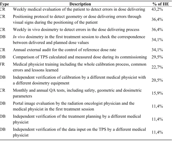
Table 1 presents direct barriers and reducers with highest impact at initiating events with high-risk level. The main missing barriers identified are related to the lack of radiation oncologist physician and medical physicist professionals in a number enough to comply the tasks related to these barriers on the facility. So the majority of these barriers can be implemented simply by establishing a minimum 40h / week workload (full time) for a second radiation oncologist physician and medical physicist.
Table 1: Direct Barriers (DB), frequency reducers (FR) and consequence reducers (CR) of
most significant impact. IIE summarizes “impacted initiating event”.
Type Description % of IIE
CR Weekly medical evaluation of the patient to detect errors in dose delivering 43,2% CR Positioning protocol to detect geometry or dose delivering errors through
visual signs during the positioning of the patient 36,4%
CR Weekly in vivo dosimetry to detect errors in the dose delivering process 36,4%
DB In vivo dosimetry in the first treatment session to check the correspondence
between delivered and planned dose values 34,1%
CR Annual external audit for the control of reference dose rate 34,1% DB Comparison of TPS calculated and measured dose during its commissioning 29,5% FR Medical physicist training including the whole calibration process, common
errors and lessons learned 22,7%
DB Independent verification of calibration by a different medical physicist with
a different dosimetry equipment 20,5%
CR Monthly and annual QA tests, including safety, geometric and dosimetric
parameters 15,9%
DB Portal image evaluation by the radiation oncologist physician and the
medical physicist in the first treatment session 11,4%
DB Independent verification of the treatment planning by a different medical
physicist 11,4%
DB Independent verification of the data input on the TPS by a different medical
physicist 11,4%
In the 3rd phase, repeating the analysis taking into account the hiring of one more of each of these two professionals, and consequently the implementation of the barriers related to the participation of these new professionals in the facility’s routine, it was obtained a reduction from 31% to 8% in the amount of high-risk initiating events. The number of RM and RL rose from 49% and 20% to 54% and 38% respectively (Figure 2-c). With these new barriers (the abundant number of professionals in the facility resulting in moderate workload, double check of treatment planning, participation in the first treatment session, periodical medical patient revision, among others), the risk level of the 16 initiating events from the stage of acceptance and commissioning was turned
from RH to RM or RL, also lowering the risk level of 9 of the 11 initiating events from the treatment planning stage from RH to RL (Figure 3).
The percentage of medium-risk initiating events for the “average” facility became higher than that for the SEVRRA's reference facility (Figure 2-d). According to acceptance criteria [2], the medium-risk events are tolerable depending on the cost-benefit analysis. Improvements should be made or measures should be taken to reduce the risk as much as possible, taking into account cost-benefit criteria.
4. CONCLUSION
This paper presented a risk analysis of an average Brazilian facility using SEVRRA. The objective was to provide the results obtained and show the efficiency of this system for the risk management.
SEVRRA makes easier the application of the risk matrix method and the evaluation of the risk analysis results for risk management. It is able to point out priorities and specific actions that must be implemented to reduce the overall facility risk level. With SEVRRA is possible to identify and concentrate efforts on the initiating events leading to the highest risk levels. It also allows highlighting the importance of some direct barriers and reducers that most significantly could impact on the facility risk profile.
The overall risk profile of an average Brazilian facility was established through the SEVRRA identifying the highest risk events as well as the highest impact barriers to this risk level. After considering the implementation of the missing and with highest impact barriers indicated by SEVRRA, it was possible to reduce the number of high-risk initiating events from 31% to 8%, demonstrating the effectiveness of this tool for risk management in radiotherapy. The SEVRRA is a tool that has the potential to contribute to the continuous improvement of the radiation therapy practice, increasing the radiotherapy safety to a higher level.
5. ACKNOWLEDGMENT
Authors would like to thank SEVRRA’s Mexico developers team and the Ibero-American Forum of Radiological and Nuclear Regulatory Agencies (FORO) working group, represented especially in the kind figures of Ramon Lopes Morones, Cruz Domenigo and M. L. Ramırez.
REFERENCES
1. EC – European Commission. General Guidelines on Risk Management in External Beam Radiotherapy. Radiation Protection N° 181, Luxembourg: European Union, 2015. 70p. 2. IAEA – International Atomic Energy Agency. Application of the Risk Matrix Method to
Radiotherapy. IAEA-TECDOC-1685, Vienna: IAEA, 2016. 74p.
3. COHEN L.; SCHULTHEISS T. E.; KENNAUGH R. C. A radiation overdose incident: Initial
data. International Journal of Radiation Oncology, Biology and Physics, v. 33, p. 217–224, 1995.
4. ASH, D.; BATES, T. Report on the clinical effects of inadvertent radiation underdosage in
1045 patients. Clinical Oncology, v. 6, p. 214-226, 1994.
5. LEVESON, N.; TURNER, C. S. An investigation of the Therac-25 accidents. IEEEComputer,
v. 26, p. 18-41, 1993.
6. IAEA – International Atomic Energy Agency. The accidental overexposure of radiotherapy patients in Bialystok. PUB-1180, Vienna: IAEA, 2002. 103p.
7. IAEA – International Atomic Energy Agency. Investigation of an accidental exposure of radiotherapy patients in Panama. PUB-1114, Vienna: IAEA, 2001. 115p.
8. IAEA – International Atomic Energy Agency. The overexposure of radiotherapy patients in San Jose. PUB-1027, Vienna: IAEA, 1998. 176p.
9. DERREUMAUX S.; ETARD C.; HUET C.; TROMPIER F.; CLAIRAND I.;
BOTTOLLIER-DEPOIS J. F.; AUBERT B.; GOURMELON P. Lessons from recent accidents in radiation therapy in France. Radiation Protection Dosimetry, v.131, p. 130-135, 2008.
10. IAEA – International Atomic Energy Agency. Programa Nacional de Protección Radiológica en las Exposiciones Médicas. IAEA-TECDOC-1710/S, Vienna: IAEA, 2013.
101p.
11. IAEA – International Atomic Energy Agency. Análisis Probabilista de Seguridad de Tratamientos de Radioterapia con Acelerador Lineal. IAEA-TECDOC-1670/S, Vienna:
IAEA, 2012. 74p.
12. CNEN – Comissão Nacional de Energia Nuclear. Requisitos de Segurança e Proteção Radiológica para Serviços de Radioterapia. Norma CNEN-NN-6.10, Rio de Janeiro:
CNEN, 2014.
13. ANVISA – Agência Nacional de Vigilância Sanitária. Documento de referência para o Programa Nacional de Segurança do Paciente, Brasília: ANVISA, 2013. 39p.
14. FORO – Ibero-American Forum of Radiological and Nuclear Regulatory Agencies. Available
at: <http://foroiberam.org/>. Last accessed: 05 Mar. 2018.
15. FORO – Ibero-American Forum of Radiological and Nuclear Regulatory Agencies. SEVRRA.
Available at: <http://foroiberam.org/sevrra>. Last accessed: 05 Mar. 2018.
16. CNSNS – Comisión Nacional de Seguridad Nuclear y Salvaguardias. Sistema de Evaluación del Riesgo en Radioterapia. SEVRRA. Available at:
<https://www.gob.mx/cnsns/acciones-y-programas/sistema-de-evaluacion-del-riesgo-en-radioterapia-sevrra >. Last accessed: 05 Mar. 2018.
17. CSN – Consejo de Seguridad Nuclear. Proyecto MARR (Matrices De Riesgo En Radioterapia). COE-05.01, Madrid: CSN, 2017. 62p.
18. CNEN – Comissão Nacional de Energia Nuclear. Sistema de Avaliação de Risco em Radioterapia. SEVRRA-BR. Available at: <http://www.cnen.gov.br/2015-07-16-18-05-60 >. Last accessed: 05 Mar. 2018.
19. VILARAGUT, J.J et al. Prevention of Accidental Exposure in Radiotherapy: The risk matrix
approach. Health Physics, v. 104(2), p. 139-150, 2013.
20. MC DONNELL, D. J.; PAPADOPULOS, S.; PAZ, A. B.; MORONES, R. L.; PEREZ, F. R.
Aplicacion de SEVRRA para la Evaluacion de Condiciones de Riesgo en Braquiterapia HDR, In: IRPA Regional Congress, 2013, Rio de Janeiro. Annals. Rio de Janeiro: Sociedade Brasileira de Proteção Radiológica, 2013.
21. FORO – Ibero-American Forum of Radiological and Nuclear Regulatory Agencies. Extensión de la Aplicación de la Metodología de Matrices de Riesgo Y SEVRRA a Nuevas Técnicas de Radioterapia. Available at: <http://foroiberam.org/web/guest/fondo-documental?