UNIVERSIDADE DA BEIRA INTERIOR
Ciências da Saúde
Evaluation of a-beta scavengers expression in rat
choroid plexus: effects of circadian rhythm and
a-beta levels
Mariya Hrynchak
Dissertação para obtenção do Grau de Mestre em
Ciências Biomédicas
(2º ciclo de estudos)
Orientador: Prof. Doutora Cecília Santos
Co-orientador: Prof. Doutora Telma Quintela
Acknowledgments
I must thank all those who directly or indirectly contributed to the realization of this dissertation.
Firstly, I would like to thank PhD professor Cecília Santos for the opportunity to work in his research group, for scientific guidance and knowledge transmitted over this year.
To Portuguese Foundation for Science and Technology (FCT) (PTDC/SAU-NEU/114800/2009) and COMPETE (Pest-C/SAU/UI0709/2011) for supporting this work.
To PhD professor Telma Quintela for all support, for scientific knowledge transmitted and for the important advices, for the confidence in me and for the important advices that enabled the execution of this work.
To the laboratory colleagues, especially a thank to Ana Catarina Duarte, Ana Raquel e Maria Maltez for their friendship, help, knowledge and patience provided throughout this year. To my friends of the Viseu, who, while away always supported me and motivated.
To my friends of colleges for their help and friendship who provided me during these two years.
To my boyfriend for all the support, friendship, encouragement, affection and dedication. And finally, but very important, to my parents, sister and brother for all the effort they did to get me here, for their understanding and patience. A great thank for always having believed in me and for education and love they gave me.
Abstract
The choroid plexus, a highly specialized tissue, is involved in a variety of functions such as production and secretion of the cerebrospinal fluid, synthesis and secretion of several bioactive substances and constituting the blood-cerebrospinal fluid barrier. Among others, the choroid plexus has the ability to produce molecules involved in the metabolism/clearance of amyloid β peptide such as transthyretin, gelsolin, apolipoprotein J, metallothionein 2, angiotensin-converting enzyme and insulin-degrading enzyme. Some of these amyloid β scavengers prevent the formation and deposition of amyloid fibrils.
The circadian clock, present in all mammal tissues, is synchronized by zeitgebers (e.g., light/dark) over a period of approximately 24 h. In mammals, the brain is involved in the control and coordination of the circadian rhythm, which in turn, is generated by the suprachiasmatic nucleus. Peripheral and extra-suprachiasmatic nucleus clocks exist in other brain areas which are under the control of the suprachiasmatic nucleus. Among these, stands out the choroid plexus by expressing the core clock genes that show a rhythmic expression in both females and males.
The aim of this project is to analyse whether the amyloid β scavengers are influenced by circadian rhythmicity in both sexes in rat choroid plexus explants by Real-time PCR. Moreover, it was also investigated if the expression of these genes/proteins undergoes changes using different concentrations of amyloid β (1 µg/ml and 3 µg/ml) in rats of different ages. Also, amyloid β scavengers expression was evaluated at different ages. For that, Real-time PCR and Western blot techniques were used.
The results herein presented indicate differential expression of amyloid β scavengers with age, sex and amyloid β concentration. However, more studies are needed to investigate, for instance, the circadian oscillation of these genes in vitro with different amyloid β concentrations and in vivo in Alzheimer´s Disease patients.
Keywords
Choroid plexus, circadian rhythm, amyloid β, transthyretin, gelsolin, apolipoprotein J, metallothionein 2, angiotensin-converting enzyme, insulin-degrading enzyme
Resumo alargado
O plexo coróide, localizado em cada um dos ventrículos cerebrais, é um tecido altamente especializado que se encontra envolvido numa variedade de funções importantes para a homeostasia do sistema nervoso central. Entre estas destacam-se a produção e secreção do líquido cefalorraquidiano, a síntese e secreção de inúmeras substâncias bioativas como fatores neurotróficos, citocinas, vitaminas e uma variedade de proteínas. Para além destas, as células epiteliais do plexo coróide constituem a barreira sangue-líquido cefalorraquidiano com o objetivo de regular a passagem de moléculas entre a corrente sanguínea e o líquido cefalorraquidiano. Quanto à função de síntese do plexo coróide, este possui a capacidade de produzir moléculas com função de metabolismo/clearance do péptido β amiloide. Entre outras, a transtirretina, gelsolina, apolipoproteína J, metalotioneína 2, enzima conversora de angiotensina e enzima de degradação da insulina, fazem parte deste grupo de proteínas. Depois de sintetizadas, as mesmas são secretadas para o líquido cefalorraquidiano onde formam um complexo estável com o péptido β amiloide e hidrolisam-no em fragmentos menos neurotóxicos. Desta forma, retardam a formação e deposição de fibrilas amiloides (principal característica no desenvolvimento da Doença de Alzheimer e, por sua vez, inibem os efeitos citotóxicos induzidos pelo péptido β amiloide.
A alteração dos ciclos luz/escuridão regula atividades metabólicas, comportamentais e fisiológicas dos organismos vivos e, estas atividades variam com a altura do dia. Estes ciclos diários são uma resposta da atividade de relógios endógenos, também denominados de relógios circadianos. Em mamíferos, o cérebro encontra-se envolvido no controlo e coordenação do ritmo circadiano. O ritmo circadiano está presente em todos os tecidos de mamíferos com uma periocidade de aproximadamente 24h sendo esta gerada pelo núcleo supraquiasmático. Para além do núcleo supraquiasmático, existem também osciladores circadianos noutras áreas do cérebro e órgãos designados de relógios periféricos os quais estão sob o controlo do núcleo supraquiasmático. De entre os osciladores extra - núcleo supraquiasmático está incluído o plexo coróide, que expressa genes do relógio regulados ritmicamente tanto nas fêmeas como nos machos.
Neste sentido, o principal objetivo deste trabalho foi analisar se os genes envolvidos no metabolismo/clerance (amyloid β scavengers) mencionados anteriormente são influenciados pelo ritmo circadiano em ambos os sexos em explantes de plexo coróide de ratos Wistar Han. Também se investigou se a expressão destes genes/proteínas sofre modificações em resposta a diferentes concentrações de β amiloide (1 µg/mL e 3 µg/mL) em ratos de diferentes idades. A expressão e localização de transtirretina, gelsolina, apolipoproteína J, metalotioneína 2, enzima conversora de angiotensina e enzima de degradação da insulina foram averiguadas em
explantes de plexos coróides recorrendo ao PCR convencional e imunohistoquímica por fluorescência (whole-mount). Após a confirmação, a expressão rítmica de cada um deles foi analisada em plexos de machos e fêmeas com 2 meses de idade recolhidos a diferentes horas. Os efeitos de β amiloide na expressão dos mesmos foram estudados nos explantes de plexo coróide de animais recém-nascidos, jovens e machos e fêmeas adultos. Para esse efeito, a técnica de Real-time PCR permitiu a quantificação da expressão génica a diferentes horas do dia e com diferentes concentrações de β amiloide. Além disso, o nível de expressão foi também avaliado em ratos de diferentes idades. A técnica de Western blot possibilitou verificar alterações de expressão provocadas pelo β amiloide ao nível proteico.
Na experiência do ritmo circadiano, apenas a apolipoproteína J, metalotioneína 2 e enzima de degradação da insulina apresentaram alterações significativas ao longo do dia nas fêmeas. Tanto a apolipoproteína J como a metalotioneína 2 atingiram o seu pico máximo de expressão às 14h (ZT7) e 2h (ZT19). Ao contrário destes, a expressão da enzima de degradação da insulina diminuiu estatisticamente às 2h (ZT19). Ao comparar a expressão destes genes ao longo da idade, estes mostraram ter perfis característicos: a transtirretina diminuiu com a idade, enquanto a gelsolina tem um comportamento contrário; a apolipoproteína J diminuíu nos animais jovens em comparação com os recém-nascidos e aumentou nos machos adultos em relação às duas primeiras idades; a expressão da enzima de degradação da insulina diminuiu em todas as idades relativamente aos animais recém-nascidos. Para além disso, a expressão de cada um deles, com exceção da enzima degradadora, varia entre géneros (machos e fêmeas adultos). Quando o plexo coróide retirado de animais de diferentes idades foi sujeito a diferentes concentrações de β amiloide, verificou-se que este estímulo aumentou a expressão de transtirretina, gelsolina e apolipoproteína J. Os níveis de transtirretina e gelsolina aumentaram com o aumento da concentração de β amiloide enquanto a expressão de apolipoproteína J é significativa apenas com 3 µg/mL de β amiloide. Nos animais jovens, o plexo coróide responde ao estímulo de β amiloide aumentando a expressão de transtirretina apenas com 3 µg/mL e diminuíndo a expressão de apolipoproteína J na presença de 1 µg/mL de β amiloide e da enzima de degradação da insulina com 1 e 3 µg/mL de β amiloide. Os machos adultos não apresentaram quaisquer alterações na expressão de genes, com exceção da enzima de degradação da insulina. Por último, as fêmeas adultas expressaram menor quantidade de transtirretina e apolipoproteína J na presença de ambas as concentrações. O contrário verifica-se quando analisados os níveis de enzima de degradação da insulina em que a sua expressão aumentou com 1 µg/mL de β amiloide. Em conclusão, os Aβ scavengers mostram uma expressão diferencial entre idades, géneros e concentração de β amiloide. No entanto, mais estudos são necessários para investigar, por exemplo, a oscilação circadiana desses genes in vitro com diferentes concentrações de β amiloide e in vivo em pacientes com doença de Alzheimer.
Palavras-chave
Plexo coróide, ritmo circadiano, β amiloide, transtirretina, gelsolina, apolipoproteína J, metalotioneína 2, enzima conversora de angiotensina, enzima de degradação de insulina
Index
I.
INTRODUCTION
1
1. Choroid Plexus 2
1.1. Anatomical location of the choroid plexus 2 1.2. Morphology of choroidal tissue 3 1.3. Biological functions of the choroid plexus 4 1.3.1. Production and secretion of polypeptides 5 1.4. Age-related changes of choroid plexus and Alzheimer’s Disease 6 1.5. Metabolism/clearance mechanisms of Aβ through the choroid plexus 7 1.6. Aβ scavengers and choroid plexus 9
1.6.1. TTR 9 1.6.2. GLS 10 1.6.3. ApoJ 11 1.6.4. MT2 12 1.6.5. ACE 13 1.6.6. IDE 14 1.7. Regulation of CP functions 14 1.7.1. Sex hormones 15
1.7.1.1. The choroid plexus as a target for sex hormones 15 1.7.1.1.1. Regulation of Aβ scavengers by sex hormones 16 1.7.2. Circadian rhythm 17 1.7.2.1. Concept of circadian rhythm: location and components 17 1.7.2.2. Molecular basis 19 1.8. Hormonal control of circadian rhythm 20 1.9. Circadian rhythm & Alzheimer Disease 22
II.
AIM
23
Aim 24
III.
MATERIALS AND METHODS
25
1. Animals experiments and tissue collection 26
1.1. Circadian rhythm 27
1.2. Aβ stimulus 24h 27
2. Total RNA extraction 27
3. cDNA synthesis 28
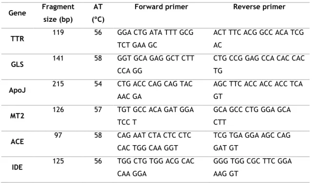
4. Conventional PCR 29
5. TTR, GLS and ApoJ Whole-mount Immunohistochemistry staining 30
6. Real time PCR 30
8. TTR & GLS Western blot 32
9. Statistical analysis 33
IV.
RESULTS
34
1. Expression of Aβ scavengers genes in CP 35
2. Cellular distribution of Aβ scavengers in rat CP 36 3. Regulation of Aβ scavengers by the circadian rhythm 37
3.1. Real time PCR 37
4. Regulation of Aβ scavengers through Aβ42 stimulus during 24h 39
4.1. Real time PCR 39
4.2. Western blot 43
V.
DISCUSSION
45
VI.
CONCLUSIONS AND FUTURE PERSPECTIVE
49
Figures list
Figure 1 - Anatomical location of the choroid plexus (CP) in brain and cerebrospinal fluid (CSF) flow.
Figure 2 - Morphological illustration of CP and its cellular structure.
Figure 3 - Illustrative pathway of Aβ formation and metabolism in the brain. Figure 4 - Illustration of the circadian system.
Figure 5 - Molecular organization of the circadian system. Figure 6 - Differences in action mechanisms of ER and AR in SCN.
Figure 7 - Schematic illustration of experimental studies performed in order to analyse Aβ scavengers regulation by circadian rhythm and Aβ42 stimulus for 24h in rat choroid plexus. Figure 8 - Aβ scavenger’s expression in rat CP.
Figure 9 - Cellular localization of Aβ scavenger’s in rat CP by confocal microscopy (A1-D3). Figure 10 - Relative Aβ scavenger’s expression in rat CP at different hours of the day.
Figure 11 - Comparison of Aβ scavenger’s expression between young (YG) and adult male (AM) and female (AF) rats considering as controls (100% expression) the newborn (NB) explants. Figure 12 - Relative Aβ scavenger’s expression in newborn, young (1 month) and adult male and female (3 months) rats CP in presence of Aβ42 stimulus for 24h.
Figure 13 - Evaluation of Aβ42 levels effect in TTR secretion by Western blot. Figure 14 - Evaluation of Aβ42 levels effect in GLS secretion by Western blot.
Tables list
Table 1 - Polypeptides and hormone receptors synthesized by CP Table 2 - Aβ regulation by SH
Abbreviations and acronyms list
ACE Angiotensin-Converting Enzyme AD Alzheimer’s Disease
AF Adult Female Ag Silver
AJs Adherens Junctions AM Adult Male
ApoJ Apolipoprotein J
APP Amyloid Precursor Protein AR Androgen Receptors ArKO Aromatase Knockout
ARNTL Aryl Hydrocarbon Receptor Nuclear Translocator-Like AT Annealing Temperature
Aβ Amyloid β
BBB Blood-Brain Barrier
BCSFB Blood– Cerebrospinal Fluid Barrier BSA Bovine Serum Albumin
Cd Cadmium
cDNA Complementary Deoxyribonucleic Acid CLOCK Circadian Locomotor Output Cycles Kaput CNS Central Nervous System
CP Choroid Plexus
CPEC Choroid Plexus Epithelial Cells CPs Choroid Plexuses CRY1 Cryptochrome 1 CRY2 Cryptochrome 2 CSF Cerebrospinal Fluid Cu Copper DEPC Diethylpyrocarbonate DHEA Dehydroepiandrosterone DHT Dihydrotestosterone
DMEM Dulbecco’s Modified Eagle Medium DR Dorsal Raphe
E2 17β-Estradiol
ECL Enhanced Chemiluminescent ERα Estrogen Receptor α
FBS Fetal Bovine Serum GDX Gonadectomise GLS Gelsolin
H2O2 Hydrogen Peroxide
Hg Mercury
IDE Insulin-Degrading Enzyme IGL Intergeniculate Leaflet ISF Interstitial Fluid MeOH Methanol MR Median Raphe MT Metallothionein MT2 Metallothionein 2 NB Newborn NC Negative Control NO Nitric Oxide OVX Ovariectomy
PBS Phosphate Buffered Saline PBS-T Phosphate Buffered Saline-Tween PER1 Period 1 PER2 Period 2 PER3 Period 3 PFA Paraformaldehyde PMSF Phenylmethanesulfonylfuoride PR Progesterone Receptor PVDF Polyvinylidene Difluoride qPCR Real Time PCR
RBP Retinol Binding Protein RHT Retinohypothalamic Tract RT Room Temperature SCN Suprachiasmatic Nucleus SHs Sex Hormones T4 Thyroxine TAE Tris-Acetate-EDTA TBS Tris Buffered Saline TBS-T Tris Buffered Saline-Tween TJs Tight Junctions
tRNA Total Ribonucleic Acid TTR Transthyretin
WR Work Reagent YG Young
Zn Zinc
1. Choroid Plexus
The choroid plexus (CP) is a highly specialized tissue that may provide a readout of the brain’s overall status and supply a variety of biological factors essential for normal brain function of the Central Nervous System (CNS). Therefore, it is involved in the establishment and maintenance of the baseline extracellular milieu throughout the CNS under both normal and pathological conditions (reviewed by Ransohoff (2014) and Emerich et al. (2004)).
1.1. Anatomical location of the choroid plexus
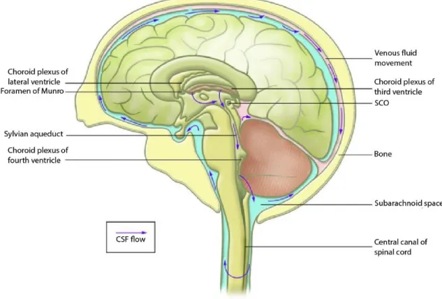
There are four interconnected channels within the brain, called ventricles, which are lined by the ependyma. Four choroid plexuses (CPs) reside inside these ventricular system of the brain: two in the lateral, one in the third and another in the fourth ventricles (Figure 1), where they form an interface that connects the peripheral blood to the cerebrospinal fluid (CSF) of the CNS (reviewed by Emerich et al. (2005)).
Figure 1 – Anatomical location of the choroid plexus (CP) in brain and cerebrospinal fluid (CSF) flow.
The CPs are inserted into the ventricular system and their main function is CSF production and secretion. The CSF is secreted from the CP and travels through the ventricular system in a rostrocaudal direction from the lateral ventricles to the third ventricle via the foramen of Munro, then through the Sylvian aqueduct to the fourth ventricle, and finally into the cisterna magna of the subarachnoid space and the central canal of the spinal cord(adapted from Picketts (2006)).
1.2. Morphology of choroidal tissue
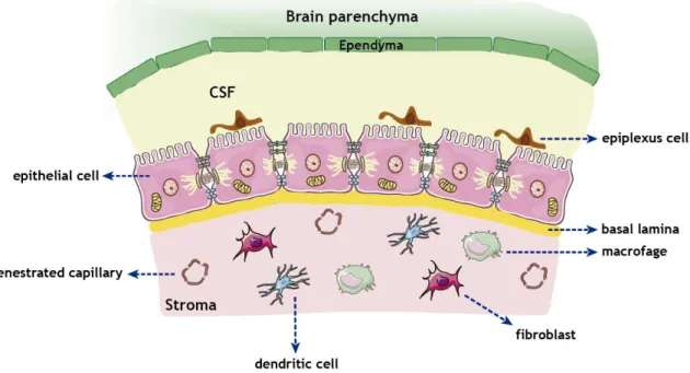
The CP, a highly irrigated structure, consists in epithelial–endothelial vascular convolutions developed from the dorsal roof of the ventricular system. The CP have a relatively simple structure, similar in the lateral, third and fourth ventricles (Figure 2). It is composed of three cellular layers: (i) the apical epithelial cells facing the CSF, (ii) the underlying supporting connective tissue, and (iii) the inner layer of endothelial cells with immediate contact with the blood. Despite its ependymal origin, these cells present characteristics of epithelial cells, being therefore designated by CP epithelial cells (CPEC). Beneath the epithelial layer there is connective tissue, which may contain fibroblasts, mast cells, macrophages, granulocytes or other infiltrates, a rich extracellular matrix, and a vascular network (reviewed by Wolburg and Paulus (2010), Damkier et al. (2013), Emerich et al. (2005) and Mortazavi et al. (2014)).
Figure 2 - Morphological illustration of CP and its cellular structure. The CP extends from the
ependymal cell layer of the ventricular wall forming a continuous strand of cuboidal epithelial cells resting upon a basal lamina and inner core of connective and highly vascularized tissue. The apical membrane of the epithelial cells faces the CSF where the cells contain numerous infoldings and scattered villi. Adjacent epithelial cells are bound together with the aid of tight junctions (TJs), adherent junctions (AJs) and desmosomes. Globular macrophages, dendritic cells and fibroblasts are found throughout the stroma. The epiplexus cells are located on the apical surface of the epithelial cells.
Tight junctions (TJs), adherens junctions (AJs) and desmosomes in the lateral surface of the CPEC are involved in the linkage of cells. TJs are concentrated in a short length of several complex strands close to the CSF (apical side) and play an important role to physically restrict
the movement of hydrophilic molecules to and from the CSF via the paracellular route between the cells, contributing to the blood–CSF barrier (BCSFB). Thus, the movement of polar molecules is limited by the lipid nature of cell walls of the CP and by the TJs. On the other hand, fenestrated capillaries derived from choroidal arteries are surrounded by connective tissue composed by fibroblasts and immune cells. Unlike the capillaries of the cerebral circulation, the capillaries in CP are large and sealed with thin diaphragms, thus providing a little resistance to the movement of small hydrophilic molecules and ions, and allowing the easy passage of other small molecules (e.g., nutrients, vitamins, etc.) into the interstitial fluid (ISF) of the CP (reviewed by Mortazavi et al. (2014), Damkier et al. (2013), Redzic and Segal (2004)). This branched structure is formed by numerous microvilli projected into the ventricles of the brain, providing a large contact surface area between CP epithelium and the CSF on one side, and epithelium and the CP ISF on the other, allowing the exchange of substances between the blood and the CSF (reviewed by Skipor and Thiery (2008) and Redzic (2013)).
CPEC has a large central spherical nucleus with abundant cytoplasm. In addition, there is a large mitochondrial content throughout the cytoplasm, needed to maintain the high cellular respiratory metabolism and energy requirements, a Golgi apparatus located laterally and toward the ventricular lumen, and smooth endoplasmic reticulum and clear vesicles, which are typical features of secretory cells. It was also demonstrated the presence of adrenergic, cholinergic, peptidergic and serotoninergic nerve fibres innervating the blood vessels and the epithelium (reviewed by Emerich et al. (2005), Damkier et al. (2013)).
1.3. Biological functions of the choroid plexus
The CP plays crucial roles in processes that establish, survey and maintain the biochemical and cellular status of the CNS under both normal and pathological conditions. Among these functions, stands out the CSF production and secretion (Wright, 1978), brain defence (Ghersi-Egea and Strazielle, 2001, Engelhardt et al., 2001, Zheng et al., 1991, Gonzalez et al., 2011), and production and secretion of polypeptides (reviewed by Chodobski and Szmydynger-Chodobska (2001)). Regarding the defence function, the BCSFB located at the CP epithelium regulates the paracellular diffusion of hydrophilic solutes and transcellular diffusion of lipophilic compounds from circulating plasma into extracellular fluids of the brain, and exclude xenobiotics. Thus, it provides the controlled environment required for optimal CNS function (reviewed by Redzic (2011)). Besides the BCSFB, CP is capable to monitor the CSF for the presence of noxious compounds, absorb and remove xenobiotics and endogenous waste products from the CSF to the circulating blood (Breen et al., 2002). Additionally, the CP is also involved in the neuro-immune system (Engelhardt et al., 2001), where it promotes a
surveillance mechanism defending against blood-borne pathogens. It also appears to assist in recovery processes by secreting neuroprotective compounds (reviewed by Chodobski and Szmydynger-Chodobska (2001)). In addition, there are several transport systems in CP that control the entry of nutrients (Spector and Lorenzo, 1975, Segal, 2001) and also facilitate the elimination of xenobiotics and endogenous waste products from the CSF to the circulating blood (Crossgrove et al., 2005). It has also been described its function in neurogenesis, suggesting an important role in cellular repair and replacement in the CNS (Li et al., 2002).
1.3.1. Production and secretion of polypeptides
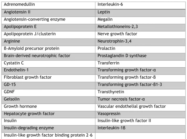
Choroidal epithelium is not only a target but also a major source of biologically active compounds in the CNS. mRNA and/or protein for a number of neuropeptides, growth factors, hormones and its receptors, and cytokines (Table 1) have been identified in the CP. Different hormone receptors mediate the action of hormones on the CP, affecting the local metabolism of the tissue and CSF secretion/composition (reviewed by Santos et al. (2011), Chodobski and Szmydynger-Chodobska (2001)).
In addition to synthesize the mentioned polypeptides and other molecules, the CP also secretes them into the CSF fluid to establish and maintain baseline levels of the extracellular milieu throughout the CNS (Johanson et al., 1999). Since the receptors for some of these polypeptides are localized in CP, this organ plays an integral role in autocrine/paracrine signaling. On the other hand, the release of these substances into the CSF allows CP to exert distal/endocrine-like effects on target cells in the brain and spinal cord due to bulk flow of this fluid (reviewed by Chodobski and Szmydynger-Chodobska (2001)).
The CP plays a key role in enzymatic processing and/or degradation of CSF-derived or CP-born polypeptides, through peptidases expressed on the apical surface of epithelial cells, and is also strategically positioned to ensure the CSF clearance of peptide degradation products and presumably noxious polypeptides, such as β-amyloid protein (Aβ). Thus, CP may play an important role in the pathogenesis of Alzheimer’s disease (AD) (reviewed by Krzyzanowska and Carro (2012)).
Table 1 - Polypeptides and hormone receptors synthesized by CP (reviewed by Chodobski and Szmydynger-Chodobska (2001))
1.4. Age-related changes of choroid plexus and Alzheimer’s
Disease
The CP tissue is subjected to various external factors and alters through structural and physiological changes during aging and pathological processes such AD. Aging of the CP is characterized by: (i) CPEC flattening, (ii) thicken of the basement membranes of both endothelial and epithelial cells and of the underlying connective tissue, and (iii) increasing of intracellular inclusions of both lipid byproducts and protein tangles. All of these age-related CP abnormalities are also observed in AD, although greatly enhanced (Serot et al., 2000). According to Serot et al., these morphological modifications imply problematic effects on brain functions, in part due to compromised CSF depuration capacities (Serot et al., 2003). Furthermore, the decreased rate of CSF secretion by CP and reduced drainage, leads to age-dependent increase in CSF/plasma concentration ratios of many compounds normally removed from the brain by CSF drainage (Garton et al., 1991). In literature reviewed by Serot
Adrenomedullin Interleukin-6 Angiotensin II Leptin Angiotensin-converting enzyme Megalin
Apolipoprotein E Metallothioneins-2,3 Apolipoprotein J/clusterin Nerve growth factor Arginine Neurotrophin-3,4 β-Amyloid precursor protein Prolactin
Brain-derived neurotrophic factor Prostaglandin D synthase Cystatin C Transferrin
Endothelin-1 Transforming growth factor-α Fibroblast growth factor Transforming growth factor-β GD-15 Transforming growth factor-β1-3 GDNF Transthyretin
Gelsolin Tumor necrosis factor-α
Growth hormone Vascular endothelial growth factor Hepatocyte growth factor Vasopressin
Insulin Insulin-like growth factor II Insulin-degrading enzyme Interleukin-1β
et al., and Kryzanowska et al., these modifications will probably impair CP functions
including synthesis, secretion and transport of proteins and other molecules. Therefore, CP dysfunction might be linked to an ineffective metabolism and clearance of AB, which is one of the principal late-onset AD features, since CP synthesizes and secretes several proteins involved in the regulation of AB brain levels, as will be discussed forward (reviewed by Serot et al. (2003), Krzyzanowska and Carro (2012)).
1.5. Metabolism/clearance mechanisms of Aβ through the
choroid plexus
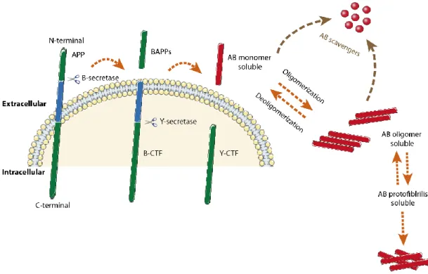
Amyloid precursor protein (APP) is processed by the sequential action of β- and ƴ-secretase, generating the Aβ peptide (~4kDa). Depending on the site of y-secretase cleavage, Aβ have 38 to 43 amino acids (Figure 3) (Haass et al., 1992). There are two major species: Aβ40 (sAβ – soluble Aβ) and Aβ42 (insoluble Aβ). Aβ40 is the major species produced and corresponds to 90% of the total Aβ peptide; Aβ42 is the minor species produced but is more prone to aggregation due to two additional hydrophobic amino acids, and it is also the predominant species accumulated in AD brain plaques. Aβ is normally produced by cells under physiological conditions (Shirwany et al., 2007) and is present in the CSF and ISF of both normal and AD brains (Tapiola et al., 2000). In soluble form, Aβ is known to bind several circulatory proteins like transthyretin (TTR), apolipoprotein J (ApoJ), among others, which have the ability to degrade Aβ peptide (Figure 3) (Schwarzman et al., 1994, Bell et al., 2007, Costa et al., 2008a). In AD, Aβ is accumulated inside CP epithelia (Gonzalez-Marrero et al., 2015, Vargas et al., 2010b).
Figure 3 - Illustrative pathway of Aβ formation and metabolism in the brain. Aβ is produced from
APP, large integral membrane glycoproteins, by sequential cleavage involving β -secretase and γ-secretase, and released into the extracellular spaces and, in part, to the intravesicular side. The released Aβ undergoes proteolytic degradation by the action of Aβ scavengers, but some Aβ that escapes to degradation, is able to aggregate and form fibrils.
Brain accumulation of Aβ is associated to overproduction, reduction or inadequate metabolic clearance, or an improper balance of Aβ import and export (Mawuenyega et al., 2010, Selkoe, 2000), and it occurs in both normal aging and in AD brains (Silverberg et al., 2010, Selkoe, 2000). Moreover, the correct balance between synthesis and rate of Aβ clearance appears to be directly related to the development of AD, and thus the formation of Aβ plaques would accelerate the onset of the disease symptoms (Bateman et al., 2006). Therefore, degradation of Aβ in CNS could play an important role in its clearance.
The 10-fold Aβ permeability and the greater surface area of CP compared to blood-brain barrier (BBB) allow CP to sequester five times more Aβ than BBB (Strazielle et al., 2000, Crossgrove et al., 2005). In AD brains, Aβ concentration is increased in the CP, and at the same time the expression of Aβ transporters in CP is altered, which consequently decreases Aβ efflux and/or degradation. Crossgrove et al., demonstrates that the CP sequesters Aβ from CSF (Crossgrove et al., 2005). The group showed that Aβ accumulation by BCSFB is because (i) CP takes up the intact Aβ species, (ii) Aβ uptake into CP occurs rapidly and by a non-diffusional uptake process, (iii) CP has a large Aβ storage capacity, (iv) Aβ uptake by the CP does not require several proteins with suggested roles in Aβ binding or transport, and (v) Aβ uptake by CP favors its efflux rather than its influx from blood to the CSF. They also proved
that CP may metabolize Aβ into smaller fragments following initial uptake, preventing the formation of β-amyloid plaques. To this, CP has the capacity to remove and degrade Aβ, contributing to its clearance from the CSF (Crossgrove et al., 2005) through the expression of various proteins mRNA, some of which mediate cellular clearance of Aβ (Rogeberg et al., 2014). Importantly, loss of this Aβ clearance route may be a determining factor to the increase of CP Aβ seen in AD (Pascale et al., 2011).
1.6. Aβ scavengers and choroid plexus
Identification of peptidases involved in Aβ metabolism/clearance in vivo is important for the development of therapeutic agents for prevention and treatment of AD (Oba et al., 2005). TTR, gelsolin (GLS), ApoJ, metallothionein 2 (MT2), angiotensin-converting enzyme (ACE) and insulin-degrading enzyme (IDE) are some of proteins responsible for degrading Aβ peptide.
1.6.1. TTR
TTR, also known as prealbumin, is a homotetrameric protein with 55 kDa (Schreiber et al., 1990). TTR is synthesized by the CP which secretes it to the CSF (Herbert et al., 1986, Dickson et al., 1986), and by the liver that secretes it to blood (Herbert et al., 1986). TTR is the main thyroid hormone carrier protein in the brain, binding about 80% of CSF thyroxine (T4) (Schreiber et al., 1990), and also binds to retinol binding protein (RBP) (Yamamoto et al., 1997) thereby participating in delivery of retinol to target cells in the brain. Within the CNS, TTR is the major protein synthesized by CP (Herbert et al., 1986, Dickson et al., 1986), representing 25% of all CSF proteins secreted by CP.
Although TTR is primarily known as a transporter, there is evidence that TTR can also act as a neuroprotective molecule against AD. The protective effect of TTR against Aβ toxicity has been shown in in vitro and in vivo studies (Schwarzman et al., 1994, Yang et al., 2013, Stein and Johnson, 2002, Costa et al., 2008a, Costa et al., 2008b). Aβ overproduction seems to induce TTR expression, with TTR forming stable complexes with Aβ in vitro, sequestering it, and thus preventing its aggregation and further amyloid formation (Schwarzman et al., 1994, Stein and Johnson, 2002). Also, TTR binds to different Aβ species namely soluble, oligomeric and fibrillar Aβ (Costa et al., 2008a, Costa et al., 2008b). Moreover, TTR is able to cleave full-length Aβ, generating smaller peptides with lower amyloidogenic properties, and also might degrade aggregated forms of Aβ peptides (Costa et al., 2008a), thereby inhibiting brain amyloid deposition, and then possibly preventing the onset and/or progression of AD.
The interaction between TTR and Aβ occurs through two residues (L82 and L110) identified on TTR (Du et al., 2012). The loss of these binding residues results in reduced TTR ability to bind Aβ, which compromises inhibition of Aβ aggregation and toxicity (Yang et al., 2013). Additionally, although tetrameric structure is necessary for the protein’s transport functions, TTR monomers bind more Aβ than tetramers. Actually, it was reported that the presence of soluble toxic Aβ oligomers triggers TTR tetramers destabilization, allowing TTR monomers to bind Aβ and “scavenge” these toxic oligomers. It has already been reported that TTR concentration decreases in CSF along with age (Zhang et al., 2005, Chen et al., 2005) and in the CSF of AD patients (Serot et al., 1997). In short, inverse correlation between decreased TTR levels in CSF of AD patients and abundance of senile plaques, point to an important role of this protein in AD.
1.6.2. GLS
GLS, with ~90 kDa (Chauhan et al., 1999), is a major actin-binding protein responsible for the limitation of actin filament’s growing end, stimulation of its nucleation, and separation of actin filaments. Thus, it plays an important role in normal physiological actin assembly as well as in disease conditions, like cancer, amyloidosis, and AD (Sun et al., 1999). GLS is constitutively expressed in the whole CNS (Tanaka and Sobue, 1994). It is present in all nervous system cell types, including neurons and CP (Matsumoto et al., 2003, Vargas et al., 2010a). GLS can be found as an intrinsic cytoplasmic and secreted protein in plasma and CSF (Paunio et al., 1994). The CP is responsible for the presence of GLS in CSF (Matsumoto et al., 2003).
The secretory form of GLS is known to bind Aβ under normal physiological conditions (Chauhan et al., 1999, Qiao et al., 2005, Ji et al., 2008), and moreover, can sequester it and inhibit its fibrillation, and also reduce amyloid load through defibrillation of preformed peptide fibrils (Ray et al., 2000). An in vitro study performed by Chauchan et al., showed that GLS inhibited the fibril formation of both Aβ40 and Aβ42 by more than 90% than observed for Aβs in the absence of GLS (Chauhan et al., 1999). GLS acts as anti-amyloidogenic agent both in plasma and CSF and therefore the failure of Aβ sequestration by GLS and imbalance of its levels in CSF/plasma of AD subjects may lead to amyloid formation in AD. GLS also shows the ability to reduce the amyloid burden in a transgenic mouse model of AD (Hirko et al., 2007).
Then GLS might have a protective role in the AD pathology through the regulation of brain Aβ levels and consequently avoid its neurotoxic effects. In addition, neuroprotection conferred by GLS against Aβ42-induced cytoskeletal alterations, associated to proteolytic degradation of the tight junction-associated protein zonula occludens 1 (ZO-1), was showed in CPEC by
(Vargas et al., 2010a). In the same study, a relation between fibrillar Aβ42 and CPEC cytoskeletal actin alterations was found, as reflected by an increase in the F-actin content. The F-actin, structure characterized by aggregation of polymerized actin is found in AD brains and its levels are increased in hippocampal neurons treated with Aβ (Mendoza-Naranjo et al., 2007). Once again, GLS protected CPEC against AB42 effects. Thus, GLS contributes to the maintenance of the CP monolayer and the blood-CSF barrier integrity, preventing and diminishing Aβ toxic effects.
Besides Aβ degrading, GLS also plays a role against its oxidative damage. In an experimental study (Ji et al., 2008) where PC12 cells were treated with hydrogen peroxide (H2O2), an
increase in lipid peroxidation and in oxidative stress was observed. In those cells, GLS expression was increased in a concentration-dependent manner in response to oxidative stress, revealing that GLS may have antioxidant properties.
Qiao et al. reported that cytoplasmic GLS can effectively inhibit Aβ-induced cell death at a point concomitant with, or upstream to, the mitochondrial events (Qiao et al., 2005). Cytoplasmic form of GLS is associated to the mitochondria, where it can inhibit Aβ-induced loss of mitochondrial membrane potential, cytochrome c releasing, and regulates voltage-dependent channels, resulting in the lack of activation of caspases-3, -8, and -9 (Koya et al., 2000). Aβ accumulation in CP, the existence of a link between Aβ-induced CP cell death, increased production of nitric oxide (NO), and mitochondrial dysfunction in the CP of patients with AD and APP/PS1 mice (Vargas et al., 2010b) has been described. Using CP epithelial cells, it was demonstrated an alteration of the enzyme activity of the respiratory chain complex IV induced by Aβ (Vargas et al., 2010a). In this experimental study, GLS reduced brain Aβ burden in the APP/Ps1mice, accompanied by inhibition of NO production and cell death, in CP. The group also showed that GLS prevents Aβ-induced cell death and NO production in CP cell cultures.
1.6.3. ApoJ
ApoJ or clusterin (encoded by the CLU gene), one of the major CNS apolipoproteins, is a component of plasma and CSF lipoprotein particles (Holtzman et al., 1999). It is a 75-85 kDa glycoprotein that circulates as a disulphide linked heterodimer component of lipid poor in HDL and VLDL, which serves both as a lipid-transport protein with the ability to induce cholesterol efflux, and a molecular chaperone in the cellular stress response. Lower mass versions of ApoJ (~49–60 kDa) are present in the cytosol and nucleus, particularly during cellular stress (reviewed by Elliott et al. (2010)).
By binding to specific receptors, ApoJ controls the availability and clearance of lipids contained in lipoprotein particles by various cells and tissues (Kounnas et al., 1995, Reddy et al., 1996). Besides CP tissue, ApoJ in brain is expressed primarily by astrocytes but also by pyramidal neurons of the hippocampus and Purkinje neurons in the cerebellum (Pasinetti et al., 1994).
ApoJ is a carrier protein for Aβ in biological fluids (CSF and blood) (Bell et al., 2007, Hammad et al., 1997), and plays an anti-amyloidogenic role by binding to Aβ, thus influencing its clearance across BBB, metabolism and aggregation (Oda et al., 1995). Moreover, ApoJ has been shown to retard the formation of Aβ aggregates and may therefore act to maintain Aβ in a soluble form and prevent it from forming pathological fibrils (Oda et al., 1994). Furthermore, the ApoJ/Aβ interaction has a cytoprotective effect. Relatively to its expression in aging and AD, ApoJ levels were found increased (Ishikawa et al., 2006, Lidstrom et al., 1998).
It has been reported that megalin/LRP2/pg330 is an endocytic receptor for ApoJ (Kounnas et al., 1995) expressed by cells in contact with CSF (CP and ependymal cells) (Kounnas et al., 1994), where it participates in transport processes between the vascular system, CSF and brain tissue. According to Ammar and Closset, ApoJ provides an example of LRP2 regulation by its own ligand, thus increasing LRP2 mRNA and protein expression (Ammar and Closset, 2008).
Megalin, an endocytic receptor for ApoJ, have been implicated in efflux of ApoJ out of the CNS, and Aβ binding to ApoJ may enhance clearance of highly pathogenic Aβ42 (Hammad et al., 1997, Bell et al., 2007). Megalin-mediated endocytosis of ApoJ/Aβ leads to Aβ lysosomal degradation (Oda et al., 1995). Hammad et al. also indicated that LRP2 is required for ApoJ efflux at BBB (Hammad et al., 1997). According the group, Aβ42 binding to ApoJ accelerates BBB Aβ42 clearance rate by 83%, which again requires megalin.
1.6.4. MT2
Metallothionein (MT) is a generic name for small (6–7 kDa), heterogeneous, and non-enzymatic proteins, that are rich in cysteine (up to 30%), giving them a high capacity to bind heavy metal ions such as zinc (Zn), cadmium (Cd), copper (Cu), silver (Ag), and mercury (Hg), among others, in biological systems. Presently, fourth MT isoforms have been described: MT1, MT2, MT3, MT4 (reviewed by Santos et al. (2012)). Particularly in brain, MT1 and MT2 isoforms are referred to MT1/MT2 because they are very similar and share many structural and functional properties (Thirumoorthy et al., 2011).
The MT2 (encoded by MT2-A gene) is widely expressed in all organs, including the CNS. It has been found in neurons, epithelial cells of CP and CSF (Goncalves et al., 2008, Chung et al., 2004). Within the CNS, it has been reported that MT1/MT2 is involved in neuroprotection and neurodegeneration, pro-apoptotic mechanisms inhibition, cell survival enhancement, tissue regeneration, maintenance of metal homeostasis, and free radical scavenging (reviewed by Carpene et al. (2007)). MT overexpression has a neuroprotective role during CNS pathological conditions (Penkowa et al., 2002).
Taking into account that CP cells synthesise MT, the MT2/TTR and MT3/TTR interactions were detected in vivo and in vitro (Goncalves et al., 2008, Martinho et al., 2010). According to Martinho et al., when TTR interact with MT2 its capacity to bind Aβ is diminished while MT3/TTR interaction increases the ability of TTR to interact with Aβ peptide (Martinho et al., 2010). In AD and old brains, the MT2 expression is increased (Zambenedetti et al., 1998, Hidalgo et al., 2006) which results in a less efficient Aβ clearance, since the interaction MT2/TTR decreases TTR/Aβ interaction rate.
1.6.5. ACE
ACE, expressed in blood vessels throughout the body, is a carboxyterminal dipeptidyl peptidase (Del Vecchio and Smith, 1981). It is an acidic glycoprotein (~150 kDa) (Wright and Harding, 1992), that catalyses the conversion of angiotensin I to the potent vasoconstrictor angiotensin II (de Lannoy et al., 2001). Thus, ACE is really important for the regulation of fluid homeostasis and blood pressure (Reid, 1992). This enzyme is a widely distributed peptidase, and within the CNS is already been found expressed in CP (Arregui and Iversen, 1978). The ACE active site is located in the extracellular space, and its unbound form circulates in biological fluids, such as plasma and CSF (Zubenko et al., 1985).
ACE has large homologous active domains: N-domain and C-domain (Soubrier et al., 1988). Oba et al., demonstrated that N-domain inhibits Aβ aggregation and cytotoxicity in vitro by the conversion of Aβ42 to Aβ40, whereas the angiotensin-converting activity is found predominantly in the C-domain (Oba et al., 2005). ACE has been associated with AD, since CSF of patients with moderate degrees of senile dementia exhibited about half of the ACE activity compared with age and sex-matched control individuals (Konings et al., 1993). In
vitro, this protein was found to significantly degrade Aβ, to retard Aβ fibril formation and
deposition, and to inhibit cytotoxicity induced by Aβ (Zou et al., 2007, Hu et al., 2001), through Aβ40 cleavage (Hu et al., 2001). Chronic inhibition of ACE enhances predominant Aβ42 deposition in vivo (Zou et al., 2007). In this way, ACE affects susceptibility to AD by degrading Aβ and preventing the accumulation of amyloid plaques in brains of AD patients (Hu et al., 2001).
1.6.6. IDE
IDE or insulysin is a ~110 kDa zinc-containing metalloendopeptidase (Qiu et al., 1998) known by its extensively participation on the clearance of Aβ (Crossgrove et al., 2007, Behl et al., 2009, McDermott and Gibson, 1997, Qiu et al., 1998), glucagon (Duckworth and Kitabchi, 1974), insulin-like growth factor 1 and 2 (Roth et al., 1984), atrial natriuretic peptide (Ralat et al., 2011), bradykinin (Malito et al., 2008), endorphin (Safavi et al., 1996), insulin (Duckworth and Kitabchi, 1974), and amylin (Bennett et al., 2000). In this manner, the common features of IDE substrates include hydrophobicity and substrate’s ability to aggregate and form fibrils (amyloidogenic). This metalloendopeptidase is expressed by cortical and subcortical neurons, and has been detected in the cytoplasm of endothelial cells, pericytes smooth muscle cells (Gao et al., 2004, Dorfman et al., 2010) and in human CSF of both normal and Alzheimer’s patients. In addition, IDE is also expressed in the CP apical surface (Behl et al., 2009).
IDE plays an important role in regulating extracellular Aβ levels, by hydrolysing several peptide bonds of both Aβ40 and Aβ42 into less neurotoxic fragments (Bora and Prabhakar, 2010). Diminished activity and levels of IDE in AD brains and aging (Miners et al., 2009) has been giving support to the hypothesis that IDE reduced activity and levels compromise Aβ degradation in brain. On the other hand, IDE overexpression reduces Aβ levels and retards, or completely prevents, brain amyloid plaque formation (Leissring et al., 2003, Farris et al., 2004). In IDE-/- animals, Farris et al. showed elevated cerebral Aβ, which validates the IDE role in AB proteolysis in vivo (Farris et al., 2003).
1.7. Regulation of CP functions
The choroidal tissue’s functions are subject to neurogenic and endocrine regulatory mechanisms. The neurogenic regulatory mechanism comprises the sympathetic, cholinergic and peptidergic innervations while endocrine system consists of 5-hydroxytryptamine, melatonin, atrial natriuretic peptide, vasopressin, insulin and insulin-like growth factors, glucocorticoid hormones, thyroid and sex hormones, all occurring in CP. Both mechanisms regulate blood flow, and CSF production and the secretory functions of CP (reviewed by Nilsson et al. (1992)).
1.7.1. Sex hormones
SHs, potent regulators of neural function from normal development to neural injury and aging, play a major role in the maintenance of CNS balance and function during life, affecting and modulating several actions. The biosynthesis of these hormones is not restricted to adrenal and gonadal glands but also occurs in nervous system, called neurosteroidogenesis, where act through specific receptors throughout various brain regions (Corpechot et al., 1981).
Aging and AD are often associated with SHs levels decline. The loss or depletion of SHs, with particular impact in women, whose have a rapid and accentuated decrease in both estrogens and progesterone levels. Men are also affected although in a gradual manner. For example, men dehydroepiandrosterone (DHEA) levels by the age of 80 are about 20% of those at age 20 (reviewed by Barron and Pike (2012), Bates et al. (2005)). Clinically, the increased risk of AD in women may be associated with the precipitous loss of estrogens and progesterone at menopause, which is much more abrupt than testosterone loss by men during aging (Yue et al., 2005, Ruitenberg et al., 2001). It is important to highlight that testosterone protection acts through both estrogen (via aromatase conversion) and androgen (either directly or after 5α-reductase conversion to dihydrotestosterone (DHT) pathways (reviewed by Aloisi and Bonifazi (2006), Garcia-Segura et al. (2001)).
In literature review Barron et al., showed that SHs have many beneficial and protective actions in brain, with potential relevance to AD. Among the neuroprotective effects stands out the increase of spine density that favours synaptic plasticity and improve select aspects of cognition and neuron viability that protects neurons against a range of toxic insults including those implicated in AD (Barron and Pike, 2012).
1.7.1.1. The choroid plexus as a target for sex hormones
In addition to peptides synthetized by CP, as previously mentioned, the choroidal tissue also express sex hormone receptors such progesterone (PR) (Quadros et al., 2007), estrogen receptors α and β (ERα and ERβ) (Hong-Goka and Chang, 2004), and androgen receptors (AR) (Alves et al., 2009) which suggest that CP is a target tissue for SH. The expression of both ERα and ERβ in the CP of female and male AD patients when compared to non-AD population, are significantly lower (Hong-Goka and Chang, 2004).
In a study, many mRNA/proteins are differentially expressed in CP in response to SHs. The same experimental study demonstrated that the CP collected from gonadectomized (GDX) female and male rats suffered changes in the expression of genes associated with several
pathways, and it is more pronounced in females than in males. Most of these CP expression changes occurred in genes implicated in chemical sensing, metabolism, steroid hormone biosynthesis and circadian rhythm pathways (Quintela et al., 2013).
1.7.1.1.1. Regulation of Aβ scavengers by sex hormones
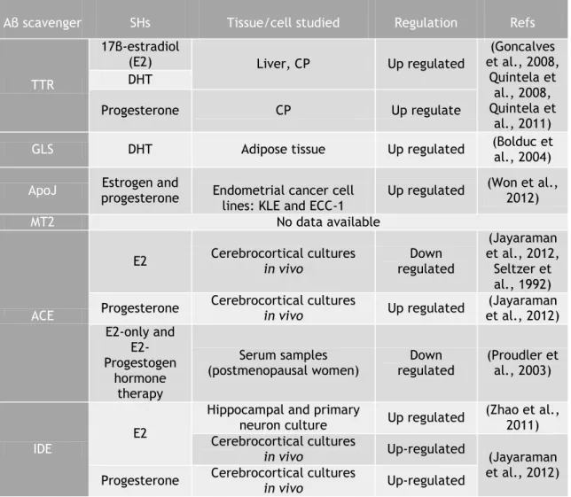
Taken into account the epidemic problematic of AD worldwide, its intrinsic association with imbalanced brain Aβ levels, and further uncountable neuroprotective effects of SHs, the regulation of Aβ scavengers by these is presented in table 2.
SHs show the ability to increase of Aβ scavengers expression is different tissues, namely in brain, such as CP and hippocampus. Therefore, SHs seem to benefit amyloid load reduction, by increasing Aβ scavenger’s levels, although some contradictory data were reported about ACE.
Table 2 - Aβ regulation by SH
Aβ scavenger SHs Tissue/cell studied Regulation Refs
TTR
17β-estradiol
(E2) Liver, CP Up regulated
(Goncalves et al., 2008, Quintela et al., 2008, Quintela et al., 2011) DHT Progesterone CP Up regulate
GLS DHT Adipose tissue Up regulated (Bolduc et al., 2004)
ApoJ progesterone Estrogen and Endometrial cancer cell
lines: KLE and ECC-1
Up regulated (Won et al., 2012)
MT2 No data available
ACE
E2 Cerebrocortical cultures in vivo regulated Down
(Jayaraman et al., 2012,
Seltzer et al., 1992) Progesterone Cerebrocortical cultures in vivo Up regulated et al., 2012) (Jayaraman
E2-only and E2-Progestogen hormone therapy Serum samples
(postmenopausal women) regulated Down (Proudler et al., 2003)
IDE
E2
Hippocampal and primary
neuron culture Up regulated (Zhao et al., 2011) Cerebrocortical cultures
in vivo Up-regulated (Jayaraman
et al., 2012) Progesterone Cerebrocortical cultures in vivo Up-regulated
1.7.2. Circadian rhythm
Organisms have developed internal timing systems to adapt to the external day and night cycles. All organisms possess endogenous daily clocks. The periods inherent of clocks, which are not exactly 24h, will then manifest themselves. This is why endogenous daily clocks are also called circadian clocks (Latin: circa diem, ‘‘approximately one day’’). Zeitgebers (e.g., light) synchronize the circadian clocks to a period of exactly 24h in the natural environment. Indeed, the circadian clock is closely related to the everyday human lives. The principal stimulus to circadian clocks is the cycle of light and darkness. Briefly, the contact of a free-running individual to light, around the time of expected nightfall and dawn, can delay and advance the clock, respectively, whereas light presented during the expected day is unsuccessful. In the case of animals, they are frequently nocturnal either diurnal in their habits (reviewed by Hastings and Maywood (2000), Kwon et al. (2011) and Helfrich-Forster (2004)).
1.7.2.1. Concept of circadian rhythm: location and components
In mammals, the brain is involved in the control and coordination of circadian rhythms. Circadian rhythms, present in almost all mammal tissues, are generated by an endogenous biological clock located in the suprachiasmatic nucleus (SCN) of the anterior hypothalamus (Figure 4). The SCN (“central clock”) is the core of the circadian system and peripheral oscillators, and contains approximately 10 000 neurons in mice and about 50 000 neurons in humans. In addition to controlling daily rhythms, the SCN is equally essential for the synchronization of an organism’s internal circadian timing to the external environmental world, integrating internal and external signals. Nuclei are strategically located for receiving visual input for light-dark entrainment through both direct and indirect retina-to-SCN pathways. The major light input pathway to the SCN is the retinohypothalamic tract (RHT), which arises from a widely distributed population of retinal ganglion cells. Circadian oscillators also exist in various areas of the brain outside the SCN such as the pineal gland, pituitary and arcuate nucleus (reviewed by Videnovic et al. (2014), Morin and Allen (2006), Reppert and Weaver (2001), Kwon et al. (2011)). More recently, the study carried out by Quintela et al. showed that CP has the capacity to express clock genes that are subjected to rhythmic expression in female and male rats (Quintela et al., 2014).
Figure 4 – Illustration of the circadian system. The timing of human biological rhythms is synchronized
to the rotation of the Earth, and influenced by numerous external and internal time cues. These stimuli are known as “zeitgebers” (germen for ‘time giver’). Light is the most important and potent zeitgeber. In addition to light, activity, feeding schedules, and the hormone melatonin also influence circadian timing. This synchronization can become disrupted, which eventually leads to misalignment or internal desynchronization. This loss of coordination of circadian rhythms can have negative consequences for sleep–wake cycles and numerous other biological functions (adapted from Videnovic et al. (2014)).
SCN neurons generate rhythmic electrical activity and produce synchronizing signals that control the phases of the oscillations called “peripheral clocks” which are present in liver, heart, lung, kidney, muscle or adipose tissue, skin and eyes. Unlike the central pacemaker, these peripheral oscillators are not directly entrained by light. Peripheral tissues produce rhythmic physiological outputs which are orchestrated by SCN and synchronized with the environment, thus providing optimal activity or response to an organism’s needs at the specific time of the day (reviewed by Green et al. (2008), Helfrich-Forster (2004)).
The mechanisms underlying circadian rhythms involve circadian oscillations in gene expression, protein modifications and secretion. These oscillations are controlled by the products of the core circadian clock genes. Despite the differences between central and peripheral clocks, both share the same molecular architecture and capacity to generate sustained circadian rhythms. Nevertheless, one key difference between central and peripheral clocks lies in the degree of their intercellular coupling (reviewed by Partch et al. (2014), Ko and Takahashi (2006)).
1.7.2.2. Molecular basis
Circadian rhythms are regulated by a set of genes, known as core “clock” genes, whose products interact to generate and maintain the rhythms (Figure 5), expressed in many peripheral tissues and in the circadian master clock. Those genes are: Period (PER1, PER2 and PER3), circadian locomotor output cycles kaput (CLOCK), aryl hydrocarbon receptor nuclear translocator-like (ARNTL, also known as BMAL1), and plant Cryptochrome gene homologues (CRY1 and CRY2). The rhythm is generated by two interconnected molecular feedback loops. In mouse main loop, PER and CRY are described as clock proteins which inhibit the transcription of their own genes after a time delay. Transcription inhibition is accomplished by blocking their transcriptional activators CLOCK and BMAL1. This negative feedback loop leads to rhythmic gene expression and rhythmic protein accumulation, as part of the pre-requisite of rhythmic behaviour. The second feedback loop reinforces the main oscillatory loop through the control of BMAL1 expression in a rhythmic manner. The entrainment of the feedback loops by light, induces transcription of mPER1 and mPER2 genes in mice. So, this
zeitgeber resets the molecular clocks, leading to its synchronization with the environment.
Many metabolic enzymes and metabolism-regulating growth factors and hormones are under the direct control of the circadian clock. When the master clock is injured, the circadian gene expression change in the peripheral tissues, by desynchronization of the peripheral oscillators (reviewed by Videnovic et al. (2014), Helfrich-Forster (2004), Kondratova and Kondratov (2012)).
Figure 5 – Molecular organization of the circadian system. PER, CLOCK, ARNTL (also known as BMAL1)
and CRY proteins interact to create a self-sustaining negative transcription–translation feedback loop. The CLOCK intracellular level remains steady throughout the 24h period. The ARNTL high level at the
beginning of the day promotes the formation of ARNTL–CLOCK heterodimers, which in turn activate PER and CRY transcription. Since PER accumulates in the cytoplasm, it becomes phosphorylated and degraded by ubiquitylation. CRY accumulates in the cytoplasm late in the subjective day, and translocates to the nucleus to inhibit ARNTL–CLOCK-mediated transcription. At night, the PER–CRY complex is degraded, and the cycle starts again. This feedback loop ensures a high level of ARNTL and low levels of PER and CRY at the beginning of a new circadian day (adapted from Videnovic et al. (2014)).
1.8. Hormonal control of circadian rhythm
Gonadal steroids modify the phase, amplitude and period of circadian rhythms. Sex differences have been identified in almost every aspect of circadian rhythms including free-running rhythms, light-induced phase shifts, sleep and activity patterns, hormonal fluctuations, and rates of re-entrainment. As mentioned, the gonadal hormones (estrogen, progesterone and androgen) control the circadian activity rhythms, and the opposite is also true (Nakamura et al., 2005, Jechura et al., 2000, Iwahana et al., 2008, Quintela et al., 2013). The presence of gonadal receptors in SCN allowing a direct influence of SHs on SCN functions. It seems that these receptors act through different mechanisms in males and females (Figure 6). The SCN has scarce ERs (ERα & ERβ), suggesting that estradiol appears to exert its effects on circadian rhythms via extra-SCN sites projecting to the SCN. Therefore, the role of steroids in females is consequently focused primarily on extra-SCN sites of action that are ER-rich and projected to the SCN (De La Iglesia et al., 1999). In contrast, ARs are prevalent in the SCN. AR expression is highly localized in the central retinorecipient “core”, which receives dense retinal input of the SCN (Karatsoreos et al., 2011). As known, the testosterone is widely aromatized to estradiol in the brain, although androgens act directly in the SCN to modulate circadian rhythms. AR activation within SCN cells is in position to impact fast cellular processes, as well as slower responses involving regulation of gene transcription (Karatsoreos et al., 2007). Hormones which act directly in the SCN may alter neural inputs, modify its structure and synaptic connectivity and modulate the responsiveness of structures that receive projections from the SCN, or some combination of these factors (Brockman et al., 2011).
Figure 6 – Differences in action mechanisms of ER and AR in SCN. ER-rich nuclei (red), including the
retina, the intergeniculate leaflet (IGL), and the dorsal raphe (DR), via the median raphe (MR), project to the SCN (top). The DR also contains androgen receptors and projects to the SCN. However, unlike the ER, ARs are densely located in the core SCN (bottom). Testosterone can be aromatized into estradiol and thus may have dual androgenic/estrogenic impacts on the system (adapted from Mong et al. (2011)).
In the absence of SHs, males and females show different temporal patterns of activity. The GDX males severely reduced activity at the start of the night, while ovariectomy (OVX) in females had no effect on period or activity (Iwahana et al., 2008). Brockman et al. documented that estradiol deficiency during development modulates the expression of circadian and daily rhythms in male and female aromatase knockout mice (Brockman et al., 2011). In this experiment, WT males, but not aromatase knockout (ArKO) males, retained the ability to respond to steroid hormones; the time of activity onset, free running period in constant darkness, and total daily activity were significantly different in GDX compared to intact males. In contrast, gonadectomy did not alter the expression of these variables in ArKO males. ArKO females had a longer free running period in constant darkness compared to WT females regardless of gonadal state. Ovariectomized ArKO females had a significantly late activity onset compared to intact ArKO females and ovariectomized WT females, despite all 3
groups being estrogen deficient. Phase shifts in response to light pulses given at different times of the day shown an interaction between genotype, sex, and circulating steroids. More recently, Butler et al. showed that the effects of androgens on circadian clock are attributable to an interaction between hormonal status and circadian parametric responses to light. The effects of androgens are a result of hormonal modulation of photic input and not due to a change in the inherent period of oscillators in the absence of light. Independent of light, androgens alter clock function and the distribution of activity during the night (Butler et al., 2012).
1.9. Circadian rhythm & Alzheimer Disease
Aging of the circadian system contributes to the decline in mental performance of aged brains, but the circadian clock may also be involved in specific age-associated neurodegenerative diseases, such as AD. The disruption of sleep and circadian rhythms is one of the most common and earliest signs of AD; when disease progresses, the abnormalities in the circadian clock and sleep worsen. Deposition of Aβ plaques and neurofibrillary tangles in patients with AD means that there is some failure in the systems responsible for the degradation of damaged proteins and other cytoplasmic components. Two major systems responsible for the degradation of cellular proteins are ubiquitin–proteasome-dependent system and autophagy. In patients with AD, the clearance of autophagic Aβ-containing vacuoles is defective. The circadian clock is one of the potential systems that have the ability to regulate autophagic activity. Although this process remains unclear, it is suggested a molecular connection between the circadian oscillator and autophagy, once the expression of several genes associated with autophagy rhythmically oscillate in a circadian manner. In the face of all these data, the circadian clock could be a novel treatment of disorders of the aging brain, namely AD (reviewed by Kondratova and Kondratov (2012)).
Aim
It has already been established that several genes of the brain clock machinery are expressed in the CP of female and male rats that may work as a peripheral clock. Since CP synthesizes several peptides and enzymes associated with Aβ metabolism/clearance and it is regulated by SHs, the objectives of this project aimed at:
1 - analyse if CP Aβ scavengers are influenced by circadian rhythmicity in both female and male Wistar Han rats;
1. Animals experiments and tissue collection
Animal experiments were carried out in Wistar Han rats. Animals were maintained with food and water ad libitum under conditions of constant temperature (20 ± 2°C) and 12 hour cycles of artificial light. All were handled in agreement with the National Institute of Health guidelines and the National and European Union rules for the care and handling of laboratory animals (Directive 2010/63/EU).
The figure 7 schematically represents the whole laboratory procedure in CP explants.
Figure 7 – Schematic illustration of experimental studies performed in order to analyse Aβ scavengers regulation by circadian rhythm and Aβ42 stimulus for 24h in rat choroid plexus. The
assessment of the influence of the circadian oscillation in Aβ scavengers expression was carried out in CPs collected from rats with two month of age through real time PCR, while Aβ42 effects in these scavengers were evaluate in CPs from newborns (PN 5-7), young (1 month) and adult male and female rats by real time PCR and Western blot techniques.
In a preliminary approach to assess the expression of Aβ scavengers, CPs were collected from 5 newborn rats (PN 5-7), frozen at -80ºC or fixed in paraformaldehyde (PFA) 4%. The next two
experiments were set to evaluate the effect of circadian rhythm and Aβ incubations on the expression of several Aβ scavengers.
1.1. Circadian rhythm
For assessing the effect of circadian rhythm, the animals (two months-old, 24 females and 24 males; n=6 of both sexes per each hour), were euthanized at 8h, 14h, 20h and 2h corresponding to ZT1, ZT7, ZT13, and ZT19, respectively. Rats euthanized at 20h and 2h were completely deprived of light. CP of lateral ventricles were collected and frozen in liquid nitrogen for future procedures.
1.2. Aβ stimulus 24h
Another experiment aimed at evaluating the effect of Aβ on the expression of Aβ scavengers. For these studies CPs were collected from newborns (PN 5-7), young (1 month), and adult female and male rats (three month of age) and incubated in Dulbecco’s Modified Eagle Medium (DMEM) completed with 10% fetal bovine serum (FBS) and 1% penicillin-streptomycin for 24h at 37ºC and 5% CO2 atmosphere, with or without Aβ42 (AnaSpec) at 1 or 3 µg/mL (n=6
per each condition). At the end of the stimuli, culture medium and tissues were collected. Culture media was frozen at -20ºC, while CP tissue was frozen in liquid nitrogen.
2. Total RNA extraction
Total RNA (tRNA) was extracted from all CPs collected. All procedures were carried out on ice since RNA is temperature sensitive, and due to the easy enzymatic degradation of RNA it is necessary to use agents which inactivate RNases, like water treated with diethylpyrocarbonate (DEPC).
The extraction of tRNA was carried out by homogenizing each CP in 300 µL TripleXTractor reagent from (TRIzol, Grisp) using a pestle, to allow the separation of the cellular components, followed by an incubation of 5 minutes at room temperature (RT) for complete dissociation of nucleoprotein complexes. Later, 60 µL of chloroform (200 µL chloroform/1 mL TRIzol) was added and tubes were inverted for homogenization. Afterwards, samples were incubated again at RT for 10 minutes and then centrifuged at 4ºC for 15 minutes at 12 000 g.