R E V I E W
Open Access
Heterologous fibrin sealant derived from
snake venom: from bench to bedside
–
an
overview
Rui Seabra Ferreira Jr
1,2,4*, Luciana Curtolo de Barros
2, Luciana Patrícia Fernandes Abbade
3,
Silvia Regina Catharino Sartori Barraviera
3, Maria Regina Cavariani Silvares
3, Leticia Gomes de Pontes
1,2,
Lucilene Delazari dos Santos
1,2and Benedito Barraviera
1,2Abstract
Hemostatic and adhesive agents date back to World War II, when homologous fibrin sealant came onto scene.
Considering that infectious diseases can be transmitted via human blood, a new heterologous fibrin sealant was
standardized in the 1990s. Its components were a serine protease (a thrombin-like enzyme) extracted from the venom
of
Crotalus durissus terrificus
snakes and a fibrinogen-rich cryoprecipitate extracted from the blood of
Bubalus bubalis
buffaloes. This new bioproduct has been used as a coagulant, sealant, adhesive and recently as a candidate scaffold for
mesenchymal stem cells and bone and cartilage repair. This review discusses the composition of a new heterologous
fibrin sealant, and cites published articles related to its preclinical applications aiming at repairing nervous system
traumas and regenerating bone marrow. Finally, we present an innovative safety trial I/II that found the product to be a
safe and clinically promising candidate for treating chronic venous ulcers. A multicenter clinical trial, phase II/III, with a
larger number of participants will be performed to prove the efficacy of an innovative biopharmaceutical product
derived from animal venom.
Keywords:
Fibrin sealant, Snake venom, Cryoprecipitate coagulum, Thrombin-like enzyme, Buffaloes
Background
The first research studies on hemostatic agents and
adhe-sives date back to World War II, when fibrin glue was
pro-posed. In that time, a mixture of human fibrinogen and
thrombin was applied to the affected aera. In 1970, given
that the basic principles for extracting fibrinogen-rich
cryo-precipitate and coagulation factors were already known, the
concept of fibrin glue was reevaluated. Since that point, a
new adhesive has been standardized with the following
composition: fibrinogen-rich human cryoprecipitate, bovine
thrombin and calcium chloride as the diluent. This sealant
was successfully commercialized for years [1, 2].
In 1978, the U.S. Food and Drug Administration
(FDA) suspended its commercialization due to the
possibility of the transmission of infectious diseases,
car-ried via products derived from human blood [3, 4].
In order to overcome these difficulties, in the 1990s
the Center for the Study of Venoms and Venomous
Ani-mals (CEVAP) at São Paulo State University (UNESP)
initiated studies to achieve the standardization of a new
heterologous fibrin sealant (HFS). After several
experi-ments, a new sealant was proposed, which was
com-posed of a fibrinogen-rich cryoprecipitate extracted from
the blood of the buffalo
Bubalus bubalis
in association
with a serine protease (a thrombin-like enzyme)
ex-tracted from
Crotalus durissus terrificus
venom [5
–
8].
The active ingredient of this new heterologous fibrin
sealant mimics the final step of the coagulation cascade.
So, a thrombin-like enzyme acts upon the fibrinogen
molecule, transforming it into fibrin monomers that
polymerize in the presence of calcium to form a stable
clot with adhesive, hemostatic and sealant effects [8, 9].
Figure 1 shows the blood-clotting cascade in three
dif-ferent pathways, with human thrombin, bovine thrombin
* Correspondence:rseabra@cevap.unesp.br
1Graduate Program in Tropical Diseases, Botucatu Medical School, São Paulo
State University (UNESP–Univ Estadual Paulista), Botucatu, SP, Brazil
2Center for the Study of Venoms and Venomous Animals (CEVAP), São Paulo
State University (UNESP–Univ Estadual Paulista), Botucatu, SP, Brazil Full list of author information is available at the end of the article
and serine protease extracted from
Crotalus durissus
terrificus
venom [9]. Figure 2 shows a stable network of
fibrin formed from a mixture of animal cryoprecipitate
with serine protease extracted from snake venom,
ob-served by electron microscopy [10].
Composition of the heterologous fibrin sealant
Fraction I: serine protease (gyroxin)
Molecular structure
The composition of the venom from
Crotalus
duris-sus terrificus
snakes is complex and constituted of
enzymes, toxins and peptides. Since the 1980s,
several authors have studied, isolated and purified
serine proteases including gyroxin, a thrombin-like
enzyme extracted from the venom of
Crotalus
duris-sus terrificus
[11
–
15]. Electrophoretic analysis
veri-fied that this enzyme is a single-chain type, with an
estimated molecular mass of 34 kDa and maximum
stability at pH 8.0, and does not present alteration
by freezing or thawing. Its maximum enzymatic
ac-tivity occurs at pH 4.0, being resistant to treatment
at 40 ° C for 15 min.
Theoretical molecular modeling of this serine protease
extracted from
Crotalus durissus terrificus
venom was
ac-complished via the program Modeller and visualization of
the model by the program PyMOL. In this manner, Fig. 3
shows the structural model, which was revealed as a
monomeric globular structure, presenting two
α
-helix
structures (red) containing the residues 146
–
152 and
215
–
227, two
β
-barris structures formed by six antiparallel
sheets and loops (green), five disulfide bridges (blue) and a
catalytic triad (orange) [16, 17].
Due to its enzymatic activity, similar to thrombin, the
serine protease acts on human and animal fibrinogen,
cleaving the
α
-chain proximal to the N-terminal. The
re-sultant fibrin monomers were polymerized in an intense
stable network (Fig. 2) in contrast to that traditionally
produced by thrombin.
Isolation and structural elucidation
Venom from
Crotalus durissus terrificus
snakes (Fig. 4)
was milked at CEVAP and pooled according to good
manufacturing practices (GMP). All the snakes are
micro-chipped to ensure the traceability of the venom lots used
in the composition of heterologous fibrin sealant. After
fil-tration and lyophilization, the venoms are stored in the
Venoms Bank of CEVAP.
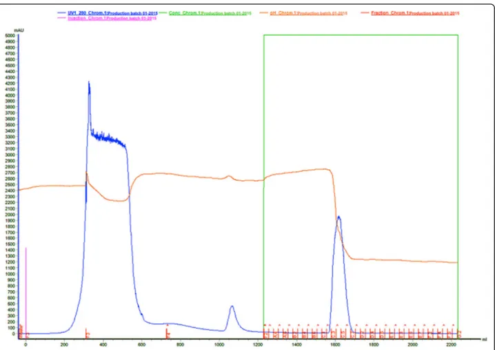
To isolate the serine protease (gyroxin), a low-pressure
liquid chromatography system was employed, specifically
the model Äkta Pilot®
(GE HealthCare Life Science,
Sweden) and the software Unicorn®
6.3 controlled the
data acquisition. All reagents and salts utilized were of
HPLC grade, and the Milli-Q water used was obtained
in a Milipore® ultra-purifier (Fig. 5).
Fifteen grams of lyophilized venom from
Crotalus
duris-sus terrificus
was suspended in 200 mL of the buffer 0.05
M Tris-HCl pH 7.4. This was applied in an AxiChrom
100/300® chromatographic column (100 mm x 300 mm x
350 mL) (GE HealthCare Life Science, Sweden) encased
with affinity resin Benzamidine Sepharose 6B® (GE
HealthCare Life Science, Sweden) previously equilibrated
with 525 mL of 0.05 M Tris-HCl pH 7.4 buffer (buffer 1).
The sample was eluted with 525 mL of 0.05 M Tris-HCl
pH 7.4 (buffer 1), followed by 525 mL of 0.05 M
Tris-HCl/0.5 M NaCl pH 7.4 (Buffer 2) and 1,050 mL of
gly-cine 0.02 M pH 3.2 (buffer 3). The flow utilized was 10
mL/min and collected 25 mL/tube. The elution was
moni-tored at an absorbance of 280 nm.
This purification process generates a single fraction
whose purity is evaluated by N-terminal sequences
(EDMAN) and mass spectrometry.
Fig. 3Theoretical molecular modeling of gyroxin accomplished using Modeller and PyMOL programs. This serine protease has two
α-helix structures (red) containing the residues 146–152 and 215–
227, twoβ-barris structures formed by six antiparallel sheets and loops (green), five disulfide bridges (blue) and a catalytic
triad (orange) Fig. 4Crotalus durissus terrificussnake
Fig. 2Stable fibrin network visualized in an electron microscope (4,000×). Reprinted from“A new fibrin sealant as a three-dimensional scaffold candidate for mesenchymal stem cells”by VPO Gasparotto et al.,
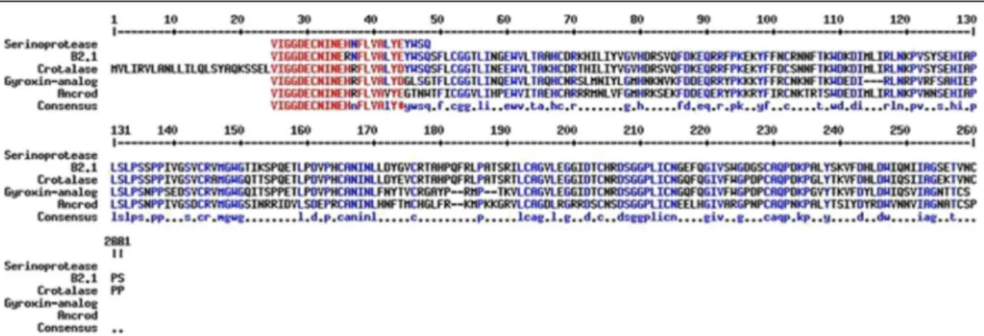
Figures 6 and 7 show, respectively, the comparison of
N-terminal sequence of gyroxin with other
thrombin-like snake toxins and their molecular mass by ESI mass
spectrometry.
Biological activity
In 2011, Barros et al
.
[9, 18] evaluated the
coagu-lant activity of a serine protease isolated from
Cro-talus durissus terrificus
venom, which was able to
induce the formation of a fibrin network and
con-sequently the formation of a stable clot at different
concentrations.
Coagulant activity was studied at three different pH,
namely: 4.0, 6.0 and 7.4. At each of them, the minimum
coagulant dose (MCD) was verified and defined as the
quantity at which a certain enzyme is capable of
coagu-lating 200
μ
L of plasma in 60 s [15]. At pH 4.0, the
MCD was 0.037
μ
g/
μ
L of plasma, versus 0.015
μ
g/
μ
L at
pH 6.0 and 0.021
μ
g/
μ
L at pH 7.4. Table 1 and Fig. 8
display the MCD at pH 7.4.
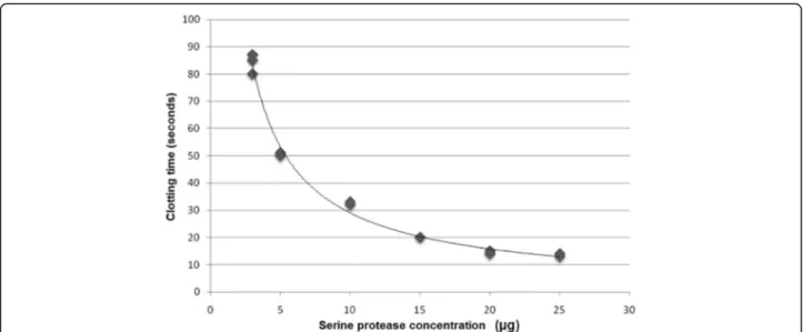
The serine protease coagulant activity at pH 7.4 was
also confirmed through dose-dependent activity
evalu-ated to obtain the MCD. For potential regression
ana-lysis, the MCD was determined at 0.021
μ
g/
μ
L of
human plasma, as shown in Fig. 9.
It must be emphasized that the statistical analysis did
not present a difference in the comparison of serine
pro-tease activity at pH 6.0 versus pH 7.4. These results lead
to the conclusion that the best activity of the enzyme is
found at between pH 6 and 7.4, values close to the
optimum pH for blood-thrombin activity, which is 7.3
and varies in blood between 7.35 and 7.45 [18].
Fraction II: cryoprecipitate
unit has a volume from 10 to 20 mL, which must be
stored at
–
20 ° C and has a shelf life of one year.
In 1995, Iuan et al. [5] proposed for the first time a new
fibrin sealant constituted of a serine protease extracted
from the venom of
Crotalus durissus terrificus
and human
cryoprecipitate. The new product was compared with the
commercial sealant in relation to the repair of sciatic
nerves in Wistar rats. The anatomopathological analyses
found similar results between the two products. Aiming at
preventing infectious diseases transmitted by human
Fig. 7LC-MS by ESI-ToF mass spectra (MicroQ-ToF III, Bruker Daltonics®).aDifferent protonated forms of gyroxin fromCrotalus durissus terrificus.b
Deconvoluted mass spectrum showing [M + H]+1= 29.472 m/z. Molecular mass of this serine protease is 29.473 Da
blood, Thomazini-Santos et al
.
[21] in 1998 proposed for
the first time to replace fibrinogen extracted from human
blood with that from buffaloes. These same authors [21]
evaluated the cryoprecipitate level of diverse animals and
compared them with that extracted from human blood.
They observed that buffaloes presented the highest
fi-brinogen levels, as shown in Table 2. Due to the good
per-formance of the cryoprecipitate extracted from buffalos,
these animals have been selected as the ideal donors.
In Brazil, the following four buffalo breeds are
recog-nized by the Brazilian Association of Buffalo Breeders:
mediterrâneo
,
murrah
,
jafarabadi
(river buffalo) and
carabao
(swamp buffalo). The
murrah
breed,
Bubalus
bubalis
, of Indian origin has been raised at the Lageado
Experimental Farm, UNESP campus in Botucatu, for
more than 30 years [22].
In order to ensure that this bioproduct contains no
sub-stance foreign to the human body, it is necessary to select
and certify the donors. Therefore, sanitary management is
mandatory for good economic results, which includes the
following actions: annual vaccination against
foot-and-mouth disease, brucellosis and rabies; systematic
deworm-ing; measures for hygiene and asepsis; practices of isolation
and quarantine; protection of animals against vectors of
transmissible diseases; diagnostic serological tests against
brucellosis and leptospirosis; an annual hypersensitivity test
against tuberculosis (tests of tuberculinization and of
Man-toux or PPD); as well as frequent clinical exams performed
by an experienced veterinary physician. These actions are
recommended by the Department of Animal Health of the
Secretariat for the Defense of Agribusiness and Livestock
Raising in the Ministry of Agriculture, Livestock and Food
Supply (MAPA) and by the World Health Organization,
and are in continuous execution by the abovementioned
team [23
–
29].
Despite all these precautions, these animals can still
pose a risk to human health due to transmission of
spongiform encephalopathies (TSEs), also known as prion
diseases, or as
“
mad cow
”
[29]. They are fatal
neurodegen-erative diseases that include
scrapie
in sheep, a bovine
spongiform encephalopathy (BSE) and Creutzfeldt-Jakob
disease (CJD) in humans. In buffalos, the transmission can
occur through the consumption of previously infected
tis-sue that is used in the feed manufacturing, particularly
nerve tissue. In suspicious cases, the necropsy becomes
the highest priority, followed by anatomical-pathological
analysis. Researchers at CEVAP in partnership with the
Center for Stable Environmental Isotopes, in the Botucatu
Biosciences Institute of UNESP, developed a globally
pio-neering technique of isotopic tracers based on the dosing
of carbon isotopes (
13C) and nitrogen (
15N) in sheep and
buffaloes [30, 31]. After its standardization, this technique
was tested in animals of the abovementioned herd,
show-ing an absence of animal protein show-ingestion, which
indir-ectly indicates that the buffalo donors of cryoprecipitate
were potentially free of mad cow disease.
The growing concern with the rapid identification and
resolution of sanitary problems in livestock has increased
the interest in the study of biomarkers. Recent research
has shown that the quantification of acute-phase proteins
in blood can offer useful information for early diagnosis,
prognosis and monitoring of diseases [32]. These proteins
are considered not only potential indicators of
inflamma-tory disease or contagious infections, but also an
import-ant tool in slaughterhouses to ensure food safety [33, 34].
Fig. 8Coagulant activity:afibrin clot formed after incubation of human plasma with serine protease;bdetail of fibrin network
Table 1
Serine protease concentrations employed to evaluate
the clotting time, the mean of three measures, the standard
deviation and standard error of the mean for a 95% confidence
interval at pH 7.4
Concentration (μg) Mean (seconds) Standard deviation Standard error of the mean
3 84 ±3.60 2.08
5 50.7 ±0.57 0.33
10 32.7 ±0.57 0.33
15 20 ±0.0 0.0
20 14.7 ±0.57 0.33
The concentrations of these proteins, which remain
circulating for long periods, depend on the severity of
the dysfunction. Therefore, their quantification is an
es-sential tool to evaluate the presence and severity of the
inflammatory process, in contrast to the cytokines that
remain circulating for short periods and whose
measure-ment is onerous [35].
The first response of the organism to immunological
stress is a non-specific release of cytokines that are
me-diators in the variation of acute-phase proteins [34, 35].
Through the influence of interleukins 1 and 6 (1,
IL-6) and of tumor necrosis factor (TNF-
α
), the hepatic
cells augment or diminish the synthesis and the
secre-tion of certain proteins. The response occurs
immedi-ately after a lesion or disease, declining within one or
two days. The acute-phase proteins can be divided into
two groups: negative and positive. The negative proteins
are those that diminish the concentration when an
acute-phase response occurs
–
and include albumin and
transferrin, while the positive ones have their level
ele-vated when there is an acute-phase response. In the
lat-ter case we have an increase in circulating C-reactive
protein
(CRP),
glycoprotein-1
acid,
antitrypsin-1,
antichemotrypsin-1, serum amyloid A, ceruloplasmin,
haptoglobin, macroglobin-2, fibrinogen and component
C
3of the complement system [34, 36, 37].
For ruminants, haptoglobin has been described as the
most important and reliable marker [32, 33]. Thus, the
standardization of acute-phase biomarkers (fibrinogen
and haptoglobin) and the clinical evaluation prior to
blood donation permit the presumptive diagnosis of
pos-sible diseases and the removal of the donor animal to
ensure the extraction of a safe bioproduct.
A rigorous protocol was applied in order to maintain
biosecurity and the traceability of cryoprecipitate, as
follows:
In the herd of buffalos:
➢
microchipping permitting traceability
a
posteriori
;
➢
annual vaccination control against rabies,
brucellosis and foot-and-month disease;
➢
application and annual evaluation of the
tuberculin;
➢
control of spongiform encephalopathy (mad
cow disease) by means of isotopic analysis;
➢
nonspecific presumptive diagnosis of diseases
for selection of ideal donors by means of
haptoglobin and fibrinogen biomarkers.
In blood collection:
➢
utilizing quadruple bag with filters in a line
similar to those employed for humans;
➢
transporting the bags with blood in refrigerated
boxes to the processing laboratory;
➢
applying techniques to evaluate fibrinogen
levels and the factors V, VIII and von Willebrand;
Table 2
Comparison of fibrinogen concentration in mg% in human, bovine, equine, ovine and buffalo blood
Mean fibrinogen concentration in cryoprecipitate (mg%)Human (N= 12) G1 Bovine (N= 9) G2 Equine (N= 10) G3 Ovine (N= 10) G4 Buffalo (N= 7) G5
375.50 ± 70.95 218.33 ± 9.76 240.80 ± 72.03 267.70 ± 25.42 664.00 ± 11.96
Statistics: G1 × G2 × G3 × G4 × G5, F = 120.26,p< 0.001, G5 > G1 > (G2 = G3 = G4)
➢
preventing possible contaminations of the bags
utilizing for quality control animal blood culture
and of the bags in Bactec
®
for aerobic and
anaerobic bacteria and Bactec Myco-F Lytic
®
for
fungi.
Finally, analytical methods with singular characteristics,
such as higher sensitivity, resolution and reproducibility
were employed with a clinical proteomic approach [38].
Two-dimensional electrophoresis (2D) was used for
isolat-ing and identifyisolat-ing proteins by means of their molecular
masses and isoelectric points in polyacrylamide gel, and
electrospray-type mass spectrometry was used for
sequen-cing peptides and proteins and identifying their biological
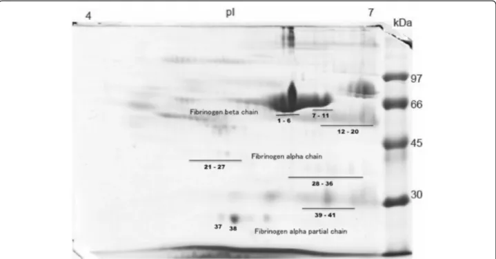
function. Figure 10 shows the total protein profile of
cryo-precipitate extracted from buffalos presenting the different
forms of fibrinogen, evidenced for better visualization,
since this protein is the main molecule responsible for
stable fibrin clot formation. There are three observable
classes of fibrinogen denominated:
β
-chain fibrinogen,
from
α
and partial forms of
α
-chain fibrinogen, totaling 40
different forms of the molecule.
Fig. 10Bubalus bubaliscryoprecipitate protein profile (2D-SDS-PAGE) showing 40 different forms of fibrinogen molecules
In sum, the cryoprecipitate extracted to be applied as
a new heterologous fibrin sealant standardized by
CEVAP is a product that is safe and free from
undesir-able substances. The formulation, as well as its storage,
handling and dosage are described in detail in the
inter-nationally required patents (PCT/BR2015/000065 and
PCT/BR2015/000064) [39].
The human use of heterologous fibrin sealant
The heterologous fibrin sealant, widely studied
experimen-tally, is now in a phase I/II clinical trial for the treatment of
chronic venous ulcers. Herein, we briefly describe the
methodology utilized for manufacturing this new
biomedi-cine that possesses vast potential to replace the human
con-stituents utilized in commercial sealants currently available
on the market. This product has undergone more than 20
years of development and due to its novelty and originality,
it represents a success story in the context of World
Toxinology, mostly in the southern hemisphere.
Until the present, two phase I/II clinical trials (called
Sealant I and Sealant II) have been proposed for
evalu-ating the treatment of chronic venous ulcers. For this
purpose, four batches of sealant were produced for
ap-plication in ten participants in the first project already
concluded (Sealant I) and, in 30 participants in the
sec-ond, which is now underway (Sealant II).
For the determination of the protein concentration of
serine protease (gyroxin) utilized in the finished product,
protein dosing was performed via direct reading at 280
nm utilizing a NanoView® spectrophotometer (GE
Health-care, USA). This apparatus quantifies the concentration of
proteins according to the law of Lambert-Beer [40]. In
this manner, the gyroxin quantity sufficient for
poly-merizing the fibrin contained in 1 mL of cryoprecipitate
was defined for each 2 mL-dose of fibrin sealant. This
quantity of polymer should cover an ulcer with a
max-imum size of 60 cm
2. A 1 mL vial of cryoprecipitate
contains, in addition to fibrinogen, the following
coagu-lation factors: factor V, VIII and von Willebrand. The
diluent vial contains 0.6 mL of a stable solution of
cal-cium chloride. The details of this composition are
de-scribed in the submitted patents [39].
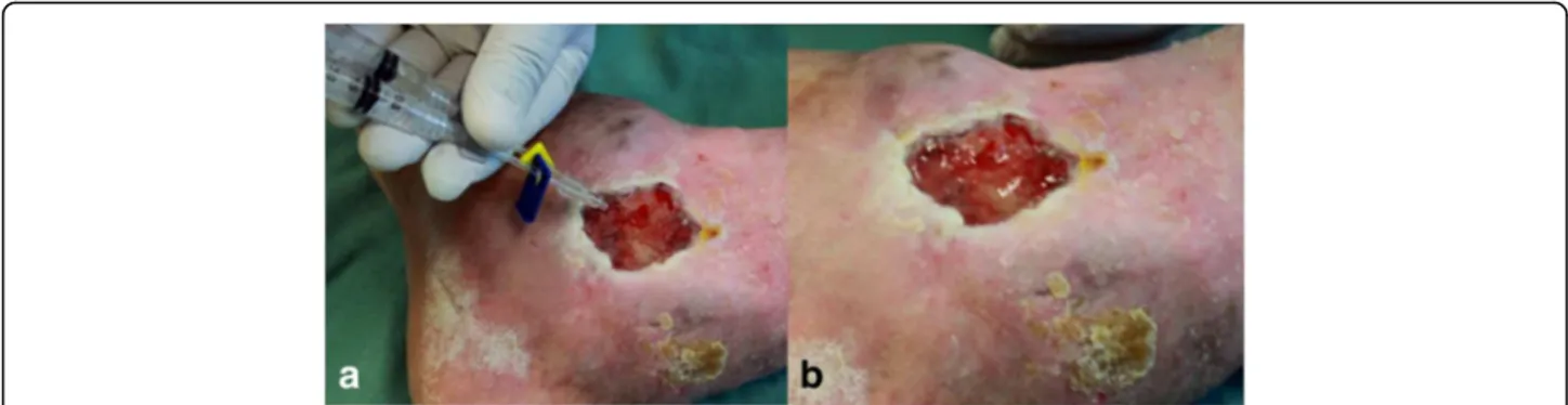
Figures 11 and 12 demonstrate the product packaged
for clinical research, with attention to the protocols of
the National Health Surveillance Agency (ANVISA) in
Brazil, as well as its preparation for application in ulcers
of the participants (ethics approval CONEP-CAAE:
19006813.4.1001.5411).
Fig. 12aApplication of the product utilizing a double-outlet syringe with mixer at its end.bPolymerized product covering an ulcer
The objectives of the Sealant I project were already
achieved, namely: to study safety and most appropriate
dose of the new heterologous fibrin sealant for treating
chronic venous ulcers.
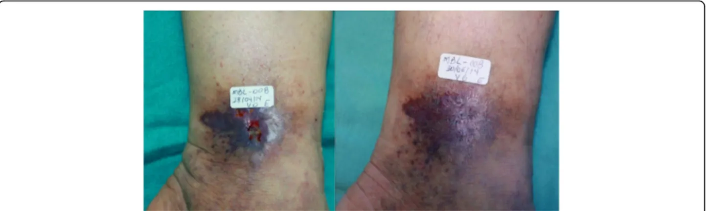
Figures 13 and 14 show the evolution and ulcers healing in
two participants, before (V0) and at the end of treatment.
The new heterologous fibrin sealant is a safe and
clin-ically promising candidate for treating chronic venous
ulcers. A multicenter clinical trial, phase II/III, with a
larger number of participants will be performed to prove
the efficacy of the product [41]. A six-minute video is
provided showing an overview of the production and
ap-plication of the fibrin sealant (Additional file 1)
(avail-able at: https://youtu.be/y6ho6M0amA8).
Conclusions
The homologous commercial fibrin sealant has been
used with success since the 1970s. Nowadays, its
appli-cation has been consolidated in surgical procedures as
an efficient method to avoid suturing, decrease
recov-ery time and increase the success rate. Its indications
are well defined and proven through a systematic
re-view of studies and meta-analysis [42
–
45]. Among the
unsolved problems, especially in biopharmaceutical
production, are the high costs and transmission of
in-fectious diseases by human blood [3, 4]. The new
heter-ologous fibrin sealant, composed primarily of extracted
animal products, has low production costs and does
not transmit infectious diseases. Standardized for over
20 years by a consortium of Brazilian researchers,
sev-eral preclinical studies and clinical trials have already
been completed. Thus, preclinical trials applying the
product in the peripheral nervous and musculoskeletal
systems [46
–
56] and as a scaffold for stem cells have
been studied extensively [57
–
60]. Trials in plastic
surgery skin repair [61], periodontal surgery [62
–
64]
and in chronic venous ulcers have also been performed
[41, 65]. In addition to treating chronic venous ulcers,
further clinical trials, especially ones linked to the
ner-vous system and to skeletal muscle, will allow for a
more precise use.
Additional file
Additional file 1:The video shows a six-min overview of the production and application of the fibrin sealant derived from snake venom and buffalo blood (available at https://youtu.be/y6ho6M0amA8). (DOCX 11 kb)
Abbreviations
2D:Two-dimensional electrophoresis; ANVISA: National Health Surveillance Agency; BSE: Bovine spongiform encephalopathy; CEVAP: Center for the Study of Venoms and Venomous Animals; CJD: Creutzfeldt-Jakob disease (); CRP: C-reactive protein; FDA: U.S. Food and Drug Administration; FFP: Frozen fresh plasma; GMP: Good manufacturing practices; HFS: Heterologous fibrin sealant; IL-1: Interleukin 1; IL-6: Interleukin 6; MCD: Minimum coagulant dose; TNF: Tumor necrosis factor; TSEs: Transmission of spongiform
encephalopathies
Acknowledgments
The authors would like to thank Guilherme Shin Iwamoto Haga, Aristides Pavan, Carlos Antonio Caramori, Márcia Tonin Rigotto Carneiro and Natália Bronzato Medolago for their assistance in the present study. Thanks are also due to the Center for the Study of Venoms and Venomous Animals (CEVAP) of UNESP for enabling the publication of this paper (Edital Toxinologia CAPES no. 063/2010, process no. 230.38.006285/2011-21, AUXPE Toxinologia 1219/2011).
Funding
This study was supported by the São Paulo Research Foundation (FAPESP) (process no. 2009/53846-9 granted to BB and RSF Jr; process no. 2012/08101-8 granted to RSF Jr and process no. 2014/13299-7 granted to LDS), and to the National Council for Scientific and Technological Development (CNPq) (process no. 563582/2010-3 granted to BB). This work was also supported by the Coordination for the Improvement of Higher Education Personnel (CAPES) through Edital Toxinologia CAPES no. 063/2010, process no. 230.38.006285/2011-21, AUXPE Toxinologia 1219/2011 and by the Department of Science and Technology (DECIT) and the Secretariat of Science, Technology and Strategic Inputs (SCTIE), CNPq process n. 401170/ 2013-6, of the Brazilian Ministry of Health. RSF Jr is a CNPq DTI research fellow (310395/2014-3).
Authors’contributions
RSF Jr., LCB, SRCSB, MRCS, LDS and LGP searched the databases (PubMed, Scopus, Scielo and Web of Science) and wrote the review. RSF Jr., LPFA and BB corrected the manuscript and prepared the images. All authors read and approved the final manuscript.
Fig. 14The 50 year-old patient had an ulcer for 4 months.aVisit 0–area of the ulcer was 0.3 cm2.bVisit 6
Competing interests
One of the authors of this article, Dr. Benedito Barraviera, is the Editor-in-Chief ofJournal of Venomous Animals and Toxins including Tropical Diseases. He did not get involved in the peer review process of this manuscript.
Consent for publication
Written informed consent was obtained from the patients for publication of this study.
Ethics approval and consent to participate
Part of the research herein presented was approved by the Research Ethics Committee of the Botucatu Medical School (CEP protocol n. 501.232). Moreover, the clinical trials described in the section“The human use of heterologous fibrin sealant”were approved by the National Commission for Ethics on Research (CONEP) and received the Certificate of Presentation for Ethical Appreciation (CAAE)–19006813.4.1001.5411.
Publisher
’
s Note
Springer Nature remains neutral with regard to jurisdictional claims in published maps and institutional affiliations.
Author details
1Graduate Program in Tropical Diseases, Botucatu Medical School, São Paulo
State University (UNESP–Univ Estadual Paulista), Botucatu, SP, Brazil.2Center for the Study of Venoms and Venomous Animals (CEVAP), São Paulo State University (UNESP–Univ Estadual Paulista), Botucatu, SP, Brazil.3Department of Dermatology and Radiology, Botucatu Medical School, São Paulo State University (UNESP–Univ Estadual Paulista), Botucatu, SP, Brazil.4CEVAP/ UNESP, Avenida José Barbosa de Barros, 1780, Botucatu, SP CEP 18610-307, Brazil.
Received: 14 January 2017 Accepted: 16 March 2017
References
1. Spotnitz WD, Burks S. Hemostats, sealants, and adhesives III: a new update as well as cost and regulatory considerations for components of the surgical toolbox. Transfusion. 2012;52(10):2243–55. doi:10.1111/j.1537-2995. 2012.03707.x.
2. Spotnitz WD. Fibrin sealant: The only approved hemostat, sealant, and adhesive—a laboratory and clinical perspective. ISRN Surg. 2014;2014:1–28. doi:10.1155/2014/203943.
3. Hino M, Ishiko O, Honda KI, Yamane T, Ohta K, Takubo T, et al. Transmission of symptomatic parvovirus B19 infection by fibrin sealant used during surgery. Br J Haematol. 2000;108(1):194–5.
4. Kawamura M, Sawafuji M, Watanabe M, Horinouchi H, Kobayashi K. Frequency of transmission of human parvovirus B19 infection by fibrin sealant used during thoracic surgery. Ann Thorac Surg. 2002;73(4):1098–100. 5. Iuan FC, Thomazini IA, Giannini MJM, Viterbo F, Toscano E, Moraes RA, et al.
Reparation of peripheral nerves with fibrin glue prepared from snake venom: preliminary results. Sao Paulo Med J. 1995;113(5):1000–2. 6. Biscola NP, Cartarozzi LP, Ulian-Benitez S, Barbizan R, Castro MV, Spejo AB, et
al. Multiple uses of fibrin sealant for nervous system treatment following injury and disease. J Venom Anim Toxins incl Trop Dis. 2017;23:13. doi:10. 1186/s40409-017-0103-1.
7. Thomazini-Santos IA. Fibrin adhesive from snake venom: the effect of adding eaminocaproic acid, tranexamic acid and aprotinin for coaptation of wound in rat skin incisions. J Venom Anim Toxins. 2001;7(1):148–9. 8. Thomazini-Santos IA, Barraviera SRCS, Mendes-Giannini MJS, Barraviera B.
Surgical adhesives. J Venom Anim Toxins. 2001;7(2):159–71.
9. Barros LC, Ferreira Jr RS, Barraviera SRCS, Stolf HO, Thomazini-Santos IA, Mendes-Giannini MJS, et al. A new fibrin sealant fromCrotalus durissus terrificusvenom: applications in medicine. J Toxicol Environ Health B Crit Rev. 2009;12(8):553–71.
10. Gasparotto VPO, Landim-Alvarenga FC, Oliveira ALR, Simões GF, Lima-Neto JF, Barraviera B, et al. A new fibrin sealant as a three-dimensional scaffold candidate for mesenchymal stem cells. Stem Cell Res Ther. 2014;5(3):78. 11. Seki C, Vidal JC, Barrio A. Purification of gyroxin from a South American rattlesnake (Crotalus durissus terrificus)venom. Toxicon. 1980;18(3):235–47.
12. Raw I, Rocha MC, Esteves MI, Kamiguti AS. Isolation and characterization of a thrombin-like enzyme from the venom ofCrotalus durissus terrificus. Braz J Med Biol Res. 1986;19(3):333–8.
13. Bercovici D, Chudziniski AM, Dias WO, Esteves MI, Hiraichi E, Oishi NY, et al. A systemic fractionation ofCrotalus durissus terrificusvenom. Mem Inst Butantan. 1987;49(3):69–78.
14. Rawling ND, Tolle DP, Barret AJ. Evolutionary families of peptidase inhititors. Biochem J. 2004;378(Pt 3):705–16.
15. Andrews RK, Gardiner EE, Berndt MC. Snake venom toxins affecting platelet function. Methods Mol Biol. 2004;273:335–48.
16. de Oliveira DGL, Murakami MT, Cintra ACO, Franco JJ, Sampaio SV, Arni RK. Functional and structural analysis of two fibrinogen-activating enzymes isolated from the venoms ofCrotalus durissus terrificusandCrotalus durissus collilineatus. Acta Biochim Biophys Sin Shanghai. 2009;41(1):21–9. doi:10. 1093/abbs/gmn003.
17. Buchi AT. Purification, characterization, crystallization and theoretical molecular modeling of gyroxin fraction fromCrotalus durissus terrificusvenom (Laurenti, 1768). J Venom Anim Toxins incl Trop Dis. 2010;16(2):389–90.
18. Barros LC, Soares AM, Costa FL, Rodrigues VM, Fuly AL, Giglio JR, et al. Biochemical and biological evaluation of gyroxin isolated fromCrotalus durissus terrificusvenom. J Venom Anim Toxins incl Trop Dis. 2011;17(1):23–33. 19. O’Shaughnessy DF, Atterbury C, Bolton Maggs P, Murphy M, Thomas D,
Yates S, et al. Guidelines for the use of fresh-frozen plasma, cryoprecipitate and cryosupernatant. Br J Haematol. 2004;126(1):11–28. doi:10.1111/j.1365-2141.2004.04972.x.
20. Nascimento B, Goodnough LT, Levy JH. Cryoprecipitate therapy. Br J Anaesth. 2014;113(6):922–34. doi:10.1093/bja/aeu158.
21. Thomazini-Santos IA, Giannini MJSM, Toscano E, Machado PEA, Lima CRG, Barraviera B. The evaluation of clotting time in bovine thrombin, Reptilase®,
and thrombin-like fraction ofCrotalus durissus terrificusvenom using bovine, equine, ovine bubaline and human cryoprecipitates. J Venom Anim Toxins. 1998;4(2):120–36.
22. Associação Brasileira de Criadores de Búfalos. Available in 10 Aug 2016 at http://www.bufalo.com.br/home/
23. Ministério da Agricultura, Pecuária e Abastecimento. Plano de ação para febre aftosa. Ministério da Agricultura, Pecuária e Abastecimento. Secretaria de Defesa Agropecuária - Brasília: MAPA/SDA/DAS; 2009. p. 96.
24. Ministério da Agricultura, Pecuária e Abastecimento. Programa Nacional de Controle e Erradicação da Brucelose e da Tuberculose Animal (PNCEBT), Brasília: MAPA/SDA/DAS; 2006. p. 188.
25. Ministério da Agricultura, Pecuária e Abastecimento Controle da Raiva dos herbívoros–Brasília; 2005. p. 104.
26. Barros CSL, Marques GHF. Procedimentos para o diagnóstico das doenças do sistema nervoso central de bovinos. Brasília; 2003. p. 50.
27. Ministério da Agricultura, Pecuária e Abastecimento. Manual de Legislação: programas nacionais de saúde animal do Brasil. Ministério da Agricultura, Pecuária e Abastecimento. Secretaria de Defesa Agropecuária. Brasília: Departamento de Saúde Animal; 2009.
28. Manual de procedimentos para a atenção às ocorrências de febre aftosa e outras enfermidades vesiculares. Projeto BID/PANAFTOSA - OPAS/OMS para os países do MERCOSUL Ampliado. Rio de Janeiro: PANAFTOSA - OPAS/ OMS; 2007. p. 144.
29. Zoonoses and veterinary public health. World Health Organization. Available in 10 Aug 2016 at http://www.who.int/zoonoses/diseases/prion_diseases/en/ 30. da Silva DA F, Biscola NP, Souza RMF, Caetano DA, Denadai JC, Sartori MMP,
et al. Carbon-13 and Nitrogen-15 turnover in serum of bubaline donors of biological material for medical use. Toxicon. 2012;60(2):117.
31. Fossato da Silva D, Biscola NP, dos Santos LD, Sartori MMP, Denadai JC, Silva ET, et al. Detecting animal by-product intake using stable isotope ratio mass spectrometry (IRMS). Vet J. 2016;217:119–25. doi:10.1016/j.tvjl. 2016.10.002.
32. Petersen HH, Nielsen JP, Heegaard PMH. Application of acute phase protein measurements in veterinary clinical chemistry. Vet Res. 2004;35(2):163–87. 33. Petersen HH, Ersbøll AK, Jensen CS, Nielsen JP. Serum-haptoglobin
concentration in Danish slaughter pigs of different health status. Prev Vet Med. 2002;54(4):325–35.
34. Gruys E, Toussaint MJM, Niewold TA, Koopmans SJ. Acute phase reaction and acute phase proteins. J Zhejiang Univ Sci B. 2005;6(11):1045–56. 35. Gutierrez AM, Martinez-Subiela S, Eckersall PD, Cerón JJ. C-reactive protein
36. Ferreira Jr RS, Almeida RAMB, Barraviera SRCS, Barraviera B. Historical perspective and human consequences of africanized bee stings in the Americas. J Toxicol Environ Health B Crit Rev. 2012;15(2):97–108. 37. Barraviera B, Lomonte B, Tarkowski A, Hanson LA, Meira DA. Acute-phase
reactions, including cytokines, in patients bitten byBothropsandCrotalus
snakes in Brazil. J Venom Anim Toxins. 1995;1(1):11–22.
38. Tabb DL. Quality assessment for clinical proteomics. Clin Biochem. 2013; 46(6):411–20.
39. Ferreira Jr RS. Autologous or heterologous fibrin sealant scaffold: which is the better choice? J Venom Anim Toxins incl Trop Dis. 2014;20:31. doi:10. 1186/1678-9199-20-31.
40. Mäntele W, Deniz E. UV–VIS absorption spectroscopy: Lambert-Beer reloaded. Spectrochim Acta A Mol Biomol Spectrosc. 2017;173:965–8. 41. Abbade LPF, Barraviera SRCS, Silvares MRC, Carneiro MTR, Medolago NB,
Ferreira Jr RS, et al. A new fibrin sealant derived from snake venom candidate to treat chronic venous ulcers. J Am Acad Dermatol. 2015;72(5): AB271. Supplement 1.
42. Li J, Li HB, Zhai XC, Qin-Lei, Jiang XQ, Zhang ZH. Topical use of topical fibrin sealant can reduce the need for transfusion, total blood loss and the volume of drainage in total knee and hip arthroplasty: A systematic review and meta-analysis of 1489 patients. Int J Surg. 2016;36(Pt A):127–37. doi:10. 1016/j.ijsu.2016.10.022.
43. Rogers AC, Turley LP, Cross KS, McMonagle MP. Meta-analysis of the use of surgical sealants for suture-hole bleeding in arterial anastomoses. Br J Surg. 2016;103(13):1758–67. doi:10.1002/bjs.10308.
44. Brustia R, Granger B, Scatton O. An update on topical haemostatic agents in liver surgery: systematic review and meta analysis. J Hepatobiliary Pancreat Sci. 2016;23(10):609–21. doi:10.1002/jhbp.389.
45. Weldrick C, Bashar K, O’Sullivan TA, Gillis E, Clarke Moloney M, Tang TY, et al. A comparison of fibrin sealant versus standard closure in the reduction of postoperative morbidity after groin dissection: A systematic review and meta-analysis. Eur J Surg Oncol. 2014;40(11):1391–8. doi:10.1016/j.ejso.2014.07.034. 46. Barbizan R, Castro MV, Rodrigues AC, Barraviera B, Ferreira Jr RS, Oliveira
ALR. Motor recovery and synaptic preservation after ventral root avulsion and repair with a fibrin sealant derived from snake venom. PLoS One. 2013; 8(5), e63260.
47. Spejo AB, Carvalho JL, Goes AM, Oliveira AL. Neuroprotective effects of mesenchymal stem cells on spinal motoneurons following ventral root axotomy: synapse stability and axonal regeneration. Neuroscience. 2013; 250:715–32.
48. Benitez SU, Barbizan R, Spejo AB, Ferreira Jr RS, Barraviera B, Goes AM, et al. Synaptic plasticity and sensory-motor improvement following fibrin sealant dorsal root reimplantation and mononuclear cell therapy. Front Neuroanat. 2014;8:96.
49. Barbizan R, Castro MV, Barraviera B, Ferreira Jr RS, Oliveira ALR. Influence of delivery method on neuroprotection by bone marrow mononuclear cell therapy following ventral root reimplantation with fibrin sealant. PLoS One. 2014;9(8), e105712.
50. Barbizan R, Castro MV, Ferreira Junior RS, Barraviera B, Oliveira ALR. Long-term spinal ventral root reimplantation, but not bone marrow mononuclear cell treatment, positively influences ultrastructural synapse recovery and motor axonal regrowth. Int J Mol Sci. 2014;15(11):19535–51.
51. Buchaim RL, Andreo JC, Barraviera B, Ferreira Jr RS, Buchaim DV, Rosa Jr GM, et al. Effect of low-level laser therapy (LLLT) on peripheral nerve regeneration using fibrin glue derived from snake venom. Injury. 2015;46(4):655–60. 52. Castro MV, Barbizan R, Ferreira Jr RS, Barraviera B, Oliveira ALR. Direct spinal
ventral root repair following avulsion: effectiveness of a new heterologous fibrin sealant on motoneuron survival and regeneration. Neural Plast. 2016; 2016:2932784.
53. Biscola NP, Cartarozzi LP, Ferreira Jr RS, Barraviera B, Oliveira ALR. Long-standing motor and sensory recovery following acute fibrin sealant based neonatal sciatic nerve repair. Neural Plast. 2016;2016:9028126.
54. Buchaim DV, Rodrigues A de C, Buchaim RL, Barraviera B, Ferreira Jr RS, Jr GM, et al. The new heterologous fibrin sealant in combination with low-level laser therapy (LLLT) in the repair of the buccal branch of the facial nerve. Lasers Med Sci. 2016;31(5):965–72.
55. de Oliveira Gonçalves JB, Buchaim DV, de Souza Bueno CR, Pomini KT, Barraviera B, Ferreira Jr RS, et al. Effects of low-level laser therapy on autogenous bone graft stabilized with a new heterologous fibrin sealant. J Photochem Photobiol B. 2016;162:663–8.
56. Spejo AB, Chiarotto GB, Ferreira Jr RS, Barraviera B, Oliveira ALR. Effects of mesenchymal stem cell and fibrin sealant treatment following spinal cord injury. 12th International Congress of Cell Biology (ICCB 2016), Vol. 1, Praga, República Tcheca; 2016. p.1-3
57. de Barros CN, Miluzzi Yamada AL, Ferreira Jr RS, Barraviera B, Hussni CA, de Souza JB, et al. A new heterologous fibrin sealant as a scaffold to cartilage repair - Experimental study and preliminary results. Exp Biol Med (Maywood). 2016;241(13):1410–5. doi:10.1177/1535370215597192. 58. Machado EG, Issa JPM, Figueiredo FAT, Santos GR, Galdeano EA, Alves MC,
et al. A new heterologous fibrin sealant as scaffold to recombinant human bone morphogenetic protein-2 (rhBMP-2) and natural latex proteins for the repair of tibial bone defects. Acta Histochem. 2015;117(3):288–96. 59. Cunha MR, Menezes FA, Santos GR, Pinto CAL, Barraviera B, Martins VCA, et
al. Hydroxyapatite and a new fibrin sealant derived from snake venom as scaffold to treatment of cranial defects in rats. Mat Res. 2015;18(1):196–203. 60. Cartarozzi LP, Spejo AB, Ferreira Jr RS, Barraviera B, Duek D, Carvalho JL, et
al. Mesenchymal stem cells engrafted in a fibrin scaffold stimulate Schwann cell reactivity and axonal regeneration following sciatic nerve tubulization. Brain Res Bull. 2015;112:14–24.
61. Stolf HO. The use of fibrin adhesive derived from snake venom and the evaluation of skin grafting using skin from the patient’s nasolabial fold. J Venom Anim Toxins. 1999;5(2):227.
62. Barbosa MDS, Greghi SLA, Passanezi E. Fibrin adhesive derived from snake venom in periodontal surgery. J Periodontol. 2007;78(10):2026–31. 63. Barbosa MD, Stipp AC, Passanezi E, Greghi SL. Fibrin adhesive derived from
snake venom in periodontal surgery: histological analysis. J Appl Oral Sci. 2008;16(5):310–5.
64. Chiquito GCM. Comparison between suture and fibrin adhesive derived from snake venom for fixation of connective tissue graft in correction of marginal tissue recession. J Venom Anim Toxins incl Trop Dis. 2007;13(2):559. 65. Gatti MAN, Vieira LM, Barraviera B, Barraviera SRCS. Treatment of venous
ulcers with fibrin sealant derived from snake venom. J Venom Anim Toxins incl Trop Dis. 2011;17(2):226–9.
• We accept pre-submission inquiries
• Our selector tool helps you to find the most relevant journal
• We provide round the clock customer support
• Convenient online submission
• Thorough peer review
• Inclusion in PubMed and all major indexing services
• Maximum visibility for your research Submit your manuscript at
www.biomedcentral.com/submit

![Fig. 2 Stable fibrin network visualized in an electron microscope (4,000×). Reprinted from “ A new fibrin sealant as a three-dimensional scaffold candidate for mesenchymal stem cells ” by VPO Gasparotto et al., Stem Cell Res Ther, 2014, 5(3), 78 [10]](https://thumb-eu.123doks.com/thumbv2/123dok_br/15915083.674207/3.892.86.434.697.994/visualized-electron-microscope-reprinted-dimensional-candidate-mesenchymal-gasparotto.webp)