Objective: To describe a case of cholesteryl ester storage disease (CESD) and discuss the importance of liver biopsy for diagnosis.
Case description: A female patient, aged two years and ten months, presented with an increased abdominal volume following hepatomegaly for four months. Abdominal ultrasound demonstrated hepatomegaly and hepatic steatosis. Laboratory tests showed elevated liver serum enzymes and dyslipidemia. Liver biopsy was consistent with CESD.
Comments: Although measuring enzyme activity is the gold standard for CESD diagnosis, liver biopsy is very helpful when investigating suspected cases of CESD, particularly upon other diferential diagnoses to be considered.
Keywords: Biopsy; Cholesteryl ester storage disease; Fatty liver; Dyslipidemias; Sterol esterase.
Objetivo: Descrever a doença de depósito de ésteres de colesterol (DDEC) e a importância da biópsia hepática na realização do diagnóstico.
Descrição do caso: Paciente feminina, dois anos e dez meses de idade, com queixa de aumento do volume abdominal secundário à hepatomegalia há quatro meses. Ultrassonograia abdominal demonstrou hepatomegalia e esteatose hepática. Exames laboratoriais mostraram aumento de enzimas hepáticas e dislipidemia. A biópsia hepática foi compatível com DDEC.
Comentários: Embora a medida da atividade enzimática seja o padrão-ouro para o diagnóstico de DDEC, a biópsia hepática é muito útil na investigação de casos suspeitos, particularmente quando há outros diagnósticos diferenciais a serem considerados. Palavras-chave: Biópsia; Doença do armazenamento de éster de colesterol; Fígado gorduroso; Dislipidemias; Esterol esterase.
ABSTRACT
RESUMO
*Corresponding author. E-mail: [email protected] (A.M.A. Tommaso).
aUniversidade Estadual de Campinas, Campinas, SP, Brazil.
bUniversidade Federal do Rio Grande do Sul, Porto Alegre, RS, Brazil.
Received on January 22, 2017; approved on March 27, 2017; available online on October 17, 2017.
IMPORTANCE OF LIVER BIOPSY IN
THE DIAGNOSIS OF LYSOSOMAL
ACID LIPASE DEFICIENCY: A CASE REPORT
Importância da biópsia hepática no diagnóstico
da deficiência de lipase ácida lisossomal: relato de caso
Adriana Maria Alves De Tommaso
a,*, Flávia Fonseca de Carvalho Barra
a,
Gabriel Hessel
a, Carolina Araújo Moreno
a, Roberto Giugliani
b,
Cecília Amélia Fazzio Escanhoela
aLiver biopsy in deficiency of LAL-D
114
Rev Paul Pediatr. 2018;36(1):113-116
INTRODUCTION
Wolman Disease and cholesteryl ester storage disease (CESD) are rare entities caused by complete or partial lysosomal acid lipase (LAL) deiciency, which is encoded by the LIPA gene located at 10q23.31. his enzyme mediates the intracellular hydrolysis of cholesteryl esters and triglycerides.1,2
In Wolman Disease, there is total enzyme deiciency, char-acterized by abdominal distension, hepatosplenomegaly, ascites, intestinal malabsorption with diarrhea, adrenal calciication, and failure to thrive. he disease progresses rapidly, causing death before the irst year of life. In CESD, enzyme deiciency is partial and signs and symptoms vary widely. Hepatosplenomegaly, elevated serum transaminase levels, hyperlipidemia, hypercholesterolemia, low serum levels of high-density lipoprotein cholesterol (HDL), vomiting, diarrhea, growth failure, and liver steatosis can occur.1,3
Fatty liver in childhood develops for several reasons,4 and
CESD has a broad clinical and laboratory proile.1,3,5 he aim
of this paper is to describe the irst case of CESD identiied at UNICAMP Clinics Hospital and the importance of liver biopsy for diagnosis.
CASE REPORT
Female patient, aged 2 years and 10 months, presented with complaints of enlarged abdominal volume for four months. At physical examination, she weighed 12.3 kg (P3-P10), and was 86-cm height (P25), with body mass index (BMI) of 16.8. he liver was palpable approximately 6 cm below the right costal margin, without splenomegaly. he abdominal ultrasonography demonstrated hepatomegaly and liver steatosis. Laboratory tests showed: ALT=57 U/L (normal value: up to 39); AST=65 U/L (normal value: up to 56); cholesterol=373 mg/dL (normal value: up to 170); HDL=19 mg/dL (normal value >40); and triglycerides=217 mg/dL (normal value up to 100). Glucose, albumin, INR values, and complete CBC were normal.
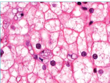
he clinical hypothesis was glycogen storage disease (GSD) with good fasting tolerance. Liver biopsy was indicated and showed difuse enlargement of hepatocyte volume with intense microvesicular steatosis and ballooning, as well as triglyceride deposits in Kupfer cells and portal macrophages, associated with occasional deposition of cholesterol crystals (Figure 1). Histologic indings were suggestive of Wolman Disease/CESD. Adrenal calciication was not present. Liver function was pre-served and the patient’s general health was good. hus, if LAL deiciency existed, clinical presentation would be consistent with CESD.
A sample sent to laboratory analysis of acid lipase activ-ity in leukocytes tested 2.8 nmol/h/mg (reference value: 112-378), which is consistent with biochemical diagnosis of LAL
deiciency. LAL activity was measured according to criteria sug-gested by Civallero et al.6 at the Laboratory of Inborn Errors of
Metabolism, Medical Genetics Service, Hospital de Clínicas de Porto Alegre—the main reference center for diagnosis of lyso-somal diseases in Brazil.
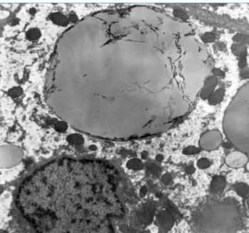
Upon electron microscopy, we noted massively distended hepatocytes due to large amount of round triglyceride depos-its, varying in size and lysosomes or lying freely in cytoplasm. hese cells were sometimes fragmented with irregular stria-tions, likely corresponding to the removal of cholesteryl esters. Needle-shaped spaces were overlapping triglyceride vacuoles (“bitten apple” aspect). here were also triglyceride depos-its in cytoplasm of macrophages (portal and Kupfer cells). he ultrastructural indings were consistent with the previous morphological hypothesis of Wolman Disease/CESD (Figure 2). he patient’s parents signed an informed consent form allowing the publication of this case.
DISCUSSION
Diagnosis of CESD is challenging, for it shares clinical and laboratory characteristics with conditions such as nonalcoholic fatty liver disease, familial combined hyperlipidemia, and several metabolic disorders.4,5,7 In our case, the initial hypothesis was
hepatic GSD (incidence of approximately 1/30,000), condi-tion that is more common than CESD (~1/130,000).8,9 In both
conditions, hepatomegaly and hyperlipidemia can progress with no signiicant changes in neuromotor development or without being notable at physical examination. However, a routine his-tological examination was important to distinguish it. In GSD, hepatocytes become turgescent due to glycogen accumulation,
Figure 1 Intense microvesicular steatosis with focal deposition of cholesterol crystals in Kupffer cell at
Tommaso AMA et al.
115
Rev Paul Pediatr. 2018;36(1):113-116
as shown by periodic acid Schif (PAS) staining and the large lipid vacuoles eventually found in cytoplasm. Glycogen dis-places mitochondria and other cytoplasmic organelles toward the cell membrane, resulting in the appearance of a pseudo-thickening, plant-like hepatocyte membrane.10
he following histological signs have been described in CESD: predominantly microvesicular steatosis involving hepato-cytes, Kupfer cells, and portal macrophages, with progression to ibrosis and micronodular cirrhosis.11 Hůlková and Elleder12
evaluated liver biopsy specimens from 19 patients with CESD to determine the histopathological criteria that distinguishes this condition from other forms of microvesicular steatosis, such as nonalcoholic fatty liver disease and other nonlyso-somal metabolic conditions. hey reported a histochemical
test with cathepsin D readily able to identify accumulation of lipids in lysosomes, facilitating the diagnosis of CESD. Furthermore, pathognomonic birefringent cholesteryl crystals were seen in fresh tissue under polarized light.
In this case report, clear needle-shaped spaces observed in Kupfer cells and portal macrophages by optic microscopy, corresponding to cholesterol crystals that were eliminated during histological processing, guided the diagnosis. By elec-tron microscopy, lipid deposits in the form of triglycerides in Kupfer cells and portal macrophages, associated with storage of cholesterol crystals in hepatocytes, was considered suicient for the diagnosis of Wolman Disease/CESD.
Enzyme replacement therapy has been used efectively in clinical trials,13 and has already been approved in European
Union and USA since 2015, Although it still pends approval in Brazil. hus, early diagnosis of this morbid condition is critical. Histopathological alterations can be suggestive of this disease and lead to conclusive testing. Although measuring enzyme activity is the preferred method for diagnosing CESD, liver biopsy is fundamental when suspecting it, particularly when other diferential diagnoses are considered.
Acknowledgements
he authors wish to express their gratitude to the Laboratory of Inborn Errors of Metabolism, Medical Genetics Service, Hospital de Clínicas de Porto Alegre, particularly to biochem-ists Jurema de Mari, Gabriel Civallero, Kristiane Michelin, and Maira Burin, for the essay about the activity of lysosomal acid lipase in leukocytes.
Funding
his study did not receive funding.
Conflict of interests
he authors declare no conlict of interests.
REFERENCES
1. Bernstein DL, Hülkova H, Bialer MG, Desnick RJ. Cholesteryl ester storage disease: review of the indings in 135 reported patients with an underdiagnosed disease. J Hepatol. 2013;58:1230-43.
2. Anderson RA, Rao N, Byrum RS, Rothschild CB, Bowden DW, Hayworth R, et al. In situ localization of the genetic locus encoding the lysosomal acid lipase/cholesterol esterase (LIPA) deicient in Wolman disease to chromosome 10q23. 2-q23.3. Genomics. 1993;15:245-7.
3. Zhang B, Porto AF. Cholesterol ester storage disease: protean presentations of lysosomal acid lipase deiciency. J Pediatr Gastroenterol Nutr. 2013;56:682-5.
4. Vajro P, Lenta S, Socha P, Dhawan A, McKiernan P, Baumann U, et al. Diagnosis of nonalcoholic fatty liver disease in children and adolescents: position paper of the ESPGHAN Hepatology Committee. J Pediatr Gastroenterol Nutr. 2012;54:700-13.
5. Freudenberg F, Buler P, Ensenauer R, Lohse P, Koletzko S. Cholesterol ester storage disease: an easily missed diagnosis in oligosymptomatic children. Z Gastroenterol. 2013;51:1184-7.
6. Civallero G, De Mari J, Bittar C, Burin M, Giugliani R. Extended use of a selective inhibitor of acid lipase for the diagnosis of Wolman disease and cholesteryl ester storage disease. Gene. 2014:539:154-6.
Figure 2 Triglyceride deposits were sometimes fragmented
Liver biopsy in deficiency of LAL-D
116
Rev Paul Pediatr. 2018;36(1):113-116
7. Reiner Z, Guardamagna O, Nair D, Soran H, Hovingh K, Bertolini S, et al. Lysosomal acid lipase deiciency--an under-recognized cause of dyslipidaemia and liver dysfunction. Atherosclerosis. 2014;235:21-30.
8. Alagille D. Inborn errors of metabolism. In: Roy CC, Silverman A, Alagille D, editors. Pediatric Clinical Gastroenterology. USA: Mosby; 1995. p. 812-76.
9. Scott SA, Liu B, Nazarenko I, Martis S, Kozlitina J, Yang Y, et al. Frequency of the cholesterol ester storage disease common LIPA E8SJM mutation (c.894G>A) in various racial and ethnic groups. Hepatology. 2013;58:958-65.
10. Ovchinsky N, Moreira RK, Lefkowitch JH, Lavine JE. Liver biopsy in modern clinical practice: a pediatric point-of-view. Adv Anat Pathol. 2012;19:250-62.
11. Boldrini R, Devito R, Biselli R, Filocamo M, Bosman C. Wolman disease and cholesterol ester storage disease diagnosed by histological and ultrastructural examination of intestinal and liver biopsy. Pathol Res Pract. 2004;200:231-40.
12. Hůlková H, Elleder M. Distinctive histopathological features that support a diagnosis of cholesterol ester storage disease in liver biopsy specimens. Histopathology. 2012;60:1107-13.
13. Valayannopoulos V, Malinova V, Honzík T, Balwani M, Breen C, Deegan PB, et al. Sebelipase alfa over 52 weeks reduces serum transaminases, liver volume and improves serum lipids in patients with lysosomal acid lipase deficiency. J Hepatol. 2014;61:1135-42.