A functional study of caustic strictures
of the esophagus in children
Departamentos de 1Pediatria, 2Anatomia Patológica, and Disciplinas de 3Gastrocirurgia and 4Cirurgia Pediátrica, Departamento de Cirurgia,
Faculdade de Ciências Médicas, Universidade Estadual de Campinas, Campinas, SP, Brasil
E.A.L. Da-Costa-Pinto1,
T.K. Dorsa1, A. Altimani2,
N.A. Andreollo3,
S.R. Cardoso1,
D.J. Morais3
and J.M. Bustorff-Silva4
Abstract
The objective of the present study was to assess esophageal motor function in 21 children (7.5 ± 2.9 years) with caustic strictures. Esophageal manometry was performed using a water-infusion system interfaced with a polygraph and displayed on a computer screen. The data were compared with those obtained from 9 healthy children. Radionuclide transit was determined by studying deglutition of a single bolus of 99mTc pertechnetate in 10 ml of water. Non-peristaltic
low-amplitude and long-duration waves were the most common find-ings detected in patients with strictures longer than 20% of esophageal length (N = 11). Compared with the control group, these patients presented lower mean amplitude and longer mean duration of waves (24.4 ± 11.2 vs 97.9 ± 23.7 mmHg, P < 0.05, and 6.7 ± 2.4 vs 1.6 ± 0.1
s, P < 0.05, respectively). Six patients presented low-amplitude waves just below the constricted site. Ten children presented delayed esoph-ageal transit. There was an association between dysphagia and abnor-malities on manometry (P = 0.02) and between symptoms and scintig-raphy data (P = 0.01). Dysphagia in caustic strictures is due to esophageal motility abnormalities, which are closely related to the scarred segment.
Correspondence
E.A.L. Da-Costa-Pinto Condomínio Lagoa Serena Estrada da Rhodia, km 15, Casa 28 13085-902 Campinas, SP Brasil
Fax: +55-19-3287-0432 E-mail: [email protected]
Research supported by FAEP(No. 1282/02). T.K. Dorsa is the recipient of a PIBIC/CNPq fellowship.
Publication supported by FAPESP.
Received September 16, 2003 Accepted July 12, 2004
Key words
•Caustic esophagitis •Esophageal dysfunction •Manometry
•Esophageal transit
Introduction
Infants and young children often dentally ingest caustic agents. Most acci-dents occur because children have access to strong household cleaners that were stored inappropriately in food and drink containers. Latin American children are more vulner-able because caustics are commercially avail-able and there is no legislation requiring the manufacture of less concentrated products. A recent survey has reported up to 100
acci-dents per year at a single Health Unit in Montevideo, Uruguay, with most accidents involving children who had ingested sodium hydroxide or lye solutions (1).
give origin to a long-term health problem. In Brazil, most endoscopic therapeutic proce-dures at Pediatric Centers consist of esoph-ageal dilatations of chronic caustic strictures (4).
Dysphagia is the most common symptom and in many cases it is a consequence of the esophageal narrowing. However, it has been observed that swallowing difficulties are not always related to the esophageal stricture and that motility abnormalities also play an important role as a cause of the symptom (5,6).
We report here the results of esophageal motility studies carried out on children with esophageal strictures caused by severe caus-tic accidents.
Patients and Methods
Fifty-eight children with esophageal caus-tic burns were admitted to the University Hospital of the School of Medical Sciences, State University of Campinas, Campinas, SP, Brazil, over a 10-year period (1991-2000). Of these, 52% (N = 30) have been treated with a dilatation protocol involving upper digestive endoscopy for stricture for-mation and obstruction of the esophageal lumen. Esophageal motility was studied in 21 of these children, 6 patients were not studied for having a guide thread for bougi-nage treatment and three children were lost follow-up because of moving to another city during the study period.
All patients had ingested sodium hydrox-ide. Age at the time of the accident ranged from 1.2 to 13.4 years (2.8 ± 2.6 years, mean ± SD). At the time of the functional study, patients were aged 4.3 to 15.5 years (7.5 ± 2.9 years, mean ± SD). All occurrences were accidental, except for one girl who had at-tempted suicide.
Immediately after the accident, patients were hospitalized and intravenous fluid and a prophylactic antibiotic were administered. Those who were unable to swallow received
total parenteral nutrition, had a nasogastric tube placed under endoscopy guidance, or underwent emergency gastrostomy. An esophagogastroduodenoscopy was per-formed within 48 h of caustic ingestion to determine the severity of injury. Damage was considered slight when it consisted of mucosal erythema, moderate when there were non-circumferential superficial ulcerations, and severe when erosions or ulcerations were confluent, deeper than in moderate cases, and included the entire esophageal circum-ference.
During the study period, 9 patients un-derwent regular esophageal dilatations. The remaining children had undergone esoph-ageal dilatations in the past. Sixteen patients had undergone a fundoplication before the functional studies because of a reflux index above 4% in prolonged pH monitoring and/ or erosive peptic esophagitis in an upper digestive endoscopy. Two patients (number 1 and 6 in Table 1) needed esophageal re-placement surgery performed after the func-tional study. Esophageal fragments from these two patients were fixed in formol, embedded in paraffin, sectioned transversely, and stained with hematoxylin-eosin.
Functional studies were performed 2 days apart. In patients undergoing dilatation, the studies were scheduled 2 weeks after the procedure. An upper digestive endoscopy was performed during the two-week period before the functional studies for an eventual diagnosis of peptic esophagitis. Anatomical or histological signs of peptic esophagitis were not diagnosed in these upper digestive endoscopy procedures.
Functional studies
with an outer diameter of 3.0 mm (orifices placed 3 cm apart) used for children under 5 years old, or of 3.5 mm (orifices placed 5 cm apart) for older children. Each capillary was connected to a polygraph and data were displayed on a PC (IBM™), using a specific software (Gastrosoft™, Polygram Upper GI™, version 6.40, Synectics Medical™, Stockholm, Sweden).
Esophageal manometry was performed after a 6-h fast. Patients were studied while lying on their left side and received no seda-tives. The catheter was introduced through the nostril until the four side holes were inside the stomach. After 15 min for patient adaptation, the catheter was pulled out in 0.5-cm steps and the lower esophageal sphincter pressure was measured (through the four orifices) using the station pull-through technique. Sphincter pressure was measured from gastric baseline to end-expi-ration pressure at the highest resting sure (usually immediately before the sure inversion point). The mean of the pres-sures measured at the four orifices was taken to be the lower esophageal sphincter pres-sure.
To evaluate the esophageal body, con-traction waves were induced by swallowing 3 to 5 ml of water. The amplitude and dura-tion of contracdura-tion waves for each individual were calculated as the mean of 10 swallows. A swallowing complex was considered to be measured only when it started at least 20 s after the previous one. Contraction wave amplitude was measured 3 cm above the proximal end of the lower esophageal sphinc-ter. Non-peristaltic contractions were de-fined as the simultaneous onset of contrac-tion waves recorded 3 or 5 cm apart. Refer-ence values were obtained from nine healthy children (mean age ± SD: 8.9 ± 1.7 years) undergoing esophageal manometry due to unconfirmed gastroesophageal reflux disease (control group).
Esophageal transit scintigraphy for de-tecting esophageal transit time was carried
out using 10 ml of water containing 99mTc
pertechnetate. All children were studied in the sitting position after a fast of at least 6 h. The child was asked to swallow a single bolus and the progression of the bolus through the esophagus was monitored with a compu-terized gamma camera. Esophageal transit was considered to be normal when the entire radioactive bolus passed through the esopha-gus and entered the stomach in less than 8 s, leaving no residual radioactivity in the esoph-ageal area. Three scores were used in the presence of residual radioactivity (7): 1) slight esophageal transit delay = residual activity less than 5% for more than 8 s and less than 15 s; 2) moderate esophageal transit delay = less than 5% of residual activity for more than 15 s or more than 5% residual activity for less than 20 s; 3) severe esophageal transit delay = more than 5% residual activ-ity for more than 20 s.
Results are reported as means ± SD. Data were analyzed statistically by the unpaired Student t-test or Fisher exact test, with the level of significance set at 5%.
The Hospital Ethics Committee approved the study, and informed consent was ob-tained from the parents of all patients.
Results
The clinical and laboratory data of the patients are presented in Table 1. The mean of the interval from the accident to the func-tional studies was 4.6 ± 2.6 years. There was no association between severity of acute esophageal injury and late dysphagia (P = 0.51, two tailed Fisher’s exact test).
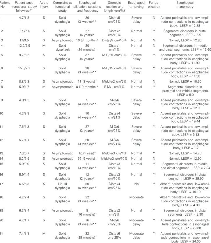
Table 1. Clinical data for 21 patients presenting esophageal strictures caused by sodium hydroxide ingestion.
Patient Patient ages Acute Complaint at Esophageal Stenosis Esophageal Fundo- Esophageal
No. (functional study/ injury functional dilatation sessions location and emptying plication manometry
follow-up*) study and frequency length (cm/%)
1 4.7/1.8 S Solid 26 Distal/5 Severe N Absent peristalsis and
low-ampli-dysphagia (3 weeks)** cm/25% delay tude contractions in esophageal
body, LESP = 12.88
2 9.7 /7.4 S Solid 27 Distal/2 Normal Y Segmental disorders in distal
dysphagia (4 years)+ cm/10% segment, LESP = 5.9
3 11/9.5 S Asymptomatic 16 (8 months)+ Distal/5 cm/15% Normal N Normal, LESP = 12.80
4 12.2/9.5 M Solid 20 Distal/1 Normal N Segmental disorders in middle
dysphagia (24 months)+ cm/4% and distal segments, LESP = 13.60
5 9.7/8.2 S Solid 37 M-D/25 cm/80% Severe Y Absent peristalsis and
low-ampli-dysphagia (4 years)+ delay tude contractions in esophageal
body, LESP = 11.90
6 15.5/2.1 S Solid 28 M-D/15 cm/40% Severe Y Absent peristalsis and
low-ampli-dysphagia (3 weeks)** delay tude contractions in esophageal
body, LESP = 11.90
7 8.8/5.3 S Asymptomatic 11 (3 years)+ Middle/2 cm/6% Normal N Normal, LESP = 10.50
8 5.9/4.7 M Asymptomatic 8 (10 months)+ P-M/1 cm/4% Normal Y Segmental disorders in
proximal and middle segments, LESP = 5.0
9 4.8/1.5 S Solid 5 M-D/6 Severe Y Absent peristalsis and
low-ampli-dysphagia (4 weeks)** cm/25% delay tude contractions in esophageal
body, LESP = 12.0
10 4.3/2.9 S Solid 10 Distal/4 Severe Y Absent peristalsis and
low-ampli-dysphagia (4 weeks)** cm/21% delay tude contractions in esophageal
body, LESP = 19.44
11 7.5/5.3 S Solid 27 M-D/6 Severe Y Absent peristalsis and
low-ampli-dysphagia (3 years)+ cm/23% delay tude contractions in esophageal
body, LESP = 9.13
12 5.7/4.1 S Solid 50 M-D/5 Severe Y Absent peristalsis and
low-ampli-dysphagia (3 weeks)** cm/21% delay tude contractions in esophageal
body, LESP = 1.7
13 7.3/5.7 S Asymptomatic 10 (1 year)+ Middle/3 cm/4% Normal Y Normal, LESP = 14.70
14 8.2/6.9 S Asymptomatic 56 (5 years)+ Middle/3 cm/10% Normal N Normal, LESP = 12.90
15 5.9/0.9 S Solid 11 Distal/3 Normal Y Segmental disorders in middle
dysphagia (3 weeks)** cm/10% and distal segment, LESP = 7.94
16 5.9/4.4 S Solid 12 Distal/3 Normal Y Segmental disorders in distal
dysphagia (2 years)+ cm/10% segment, LESP = 29.90
17 6.6/5.3 S Liquid 50 Distal/4 Np Y Absent peristalsis and
low-ampli-dysphagia (6 weeks)** cm/25% tude contractions in esophageal
body, LESP = 19.10
18 4.7/2.4 S Solid 22 Moderate Absent peristalsis and
low-ampli-dysphagia (3 weeks)** tude contractions in esophageal
body, LESP = 4.90
19 6.3/3.4 M Asymptomatic 8 Distal/2 Normal Y Segmental disorders in distal
(16 months)+ cm/8% segments, LESP = 8.90
20 4.7/1.7 S Solid 16 M-D/6 Moderate Y Absent peristalsis and
low-ampli-dysphagia (3 weeks)** cm/25% delay tude contractions in esophageal
body, LESP = 29.00
21 7.4/3.8 M Solid 22 Distal/6 Moderate Y Absent peristalsis and
low-ampli-dysphagia (29 months)+ cm/ 25% delay tude contractions in esophageal
body, LESP = 24.00
*Follow-up refers to the period from accident to functional study; **total number of dilatations performed in the past and current frequency;
+total number of dilatation sessions performed in the past and total period of dilatation therapy. LESP = lower esophageal sphincter pressure in
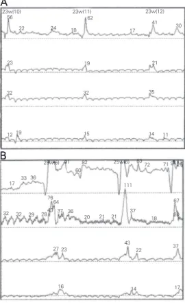
In 11 patients displaying long strictures (>20% of esophageal length), deglutition complexes also presented aperistalsis. The results of manometry recordings are pre-sented in Table 1. Most of these recordings presented elevated basal pressure, low-am-plitude waves and exaggerated transmission of aortic pulse (Figure 1A). Manometry data from this group were compared with the con-trol group data and are presented in Table 2.
Data on esophageal manometry record-ings from children who underwent fundopli-cation were compared to those from non-operated children. The results related to esophageal body or lower esophageal sphinc-ter pressure and upper esophageal sphincsphinc-ter pressure did not differ significantly between groups (Table 2).
Six patients presented esophageal motor abnormality characterized by low-amplitude, broad-based and double-peak contractions localized only in the constricted esophageal segment (Figure 1B). Four patients presented peristaltic contractions with mean amplitude and duration similar to the control group (101 ± 36.1 and 97.9 ± 23.7 mmHg, respec-tively; P > 0.05). There was no significant difference in wave duration between these patients and controls (1.7 ± 0.9 vs 1.6 ± 0.1 s, respectively; P > 0.05).
Upper esophageal function was consid-ered normal when compared to healthy con-trols. Upper esophageal sphincter pressure could be measured in 15 patients and the mean value was not different from the con-trol group (88.9 ± 21.7 vs 80.2 ± 16.7 mmHg, P > 0.05). Normal coordination with the pharynx and complete relaxation occurred following swallowing. Six patients did not tolerate the water perfusion of the upper esophageal sphincter during the catheter pull-through technique.
There was a significant association be-tween the presence of dysphagia and abnor-malities on manometry (P = 0.02, Fisher) and between dysphagia and scintigraphy re-sults (P = 0.01, Fisher). There was a strong
Figure 1. Manometry recording obtained at the level of the esophageal body after a 5-ml water swallow (W). Each record shows three deglutition com-plexes. Time elapsed between two dotted vertical lines = 4 s. Each horizontal line indicates a pressure sensor placed 3 cm apart for patient 1 and 5 cm apart for patient 15. A, Patient number 1: low amplitude con-tractions and esophageal aperi-stalsis, elevated basal pressure, low-amplitude waves, and exag-gerated transmission of aortic pulse. B, Patient number 15: segmental disorders in middle and distal segment.
23w(10) 23w(11) 23w(12)
25w(5) 25w(6)
56
22 24 18
62
17
41 30
23 19 21
32 32 35
12 19 15 14 11
94 25 67 30 18 37 111 36 33 17 91 60
82 8372 71
21 21 20 36 37 64 76 43 28 29 32 32 27 23 43 22 37
16 14 17
A A A A A B BB BB
Table 2. Results of manometry, with comparison of the caustic group with absent peristalsis and the control group and between operated and non-operated children from the caustic group.
Caustic group Control Caustic Caustic
with absent group operated non-operated
peristalsis (N = 9) group group
(N = 11) (N = 16) (N = 5)
Amplitude of waves (mmHg) 24.4±11.2* 97.9±23.7 38.6±38.4 54.6±19.7
Duration of waves (s) 6.7±2.4* 1.6±0.1 5.6±3.4 4.9±4.2
LESP (mmHg) 15.0±8.0 15.1±1.7 13.2±7.4 12.5±1.2
UESP (mmHg) 78.4±26.8 88.9±21.7 89.9±34.1 83.3±24.4
Data are reported as means ± SD. LESP = lower esophageal sphincter pressure; UESP = upper esophageal sphincter pressure. *P < 0.05 compared to control group (Student t-test).
association between esophageal transit de-lay and aperistalsis on manometry. Record-ings on manometry which showed abnor-malities localized in a portion of the esoph-ageal body did not present a correlation with the scintigraphy findings (P = 0.09).
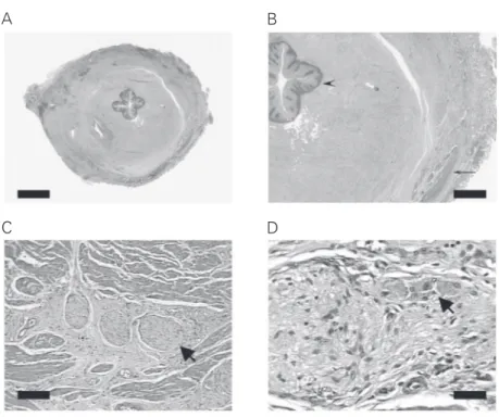
replacement procedure showed extensive fi-brosis totally replacing the mucosal and sub-mucosal layers and partially replacing the muscular layer. A histologically normal my-enteric plexus was identified in all segments examined (Figure 2).
Discussion
Alkali ingestion in childhood is usually accidental and involves smaller amounts than those ingested by adults with a suicidal in-tent. However, esophageal injuries are equally deep and result in strictures in about 30% of all accidents (8-10). Recent studies have reported motor dysfunction after sec-ond- or third-degree burns and complete ab-sence of peristalsis, and low-amplitude waves after swallows were found in 70% of record-ings on manometry (5,11). Low-amplitude aperistaltic contractions and narrowing and
shortening of the esophagus have been con-sidered to account for impaired esophageal motor function (5,6,11-13). These studies indicated that development of esophageal stricture, impaired motility, and gastroesoph-ageal reflux are very frequent outcomes of caustic esophageal burns and should be con-sidered when evaluating symptoms and de-fining therapeutic strategies. The children in the present series continued to display dys-phagia even after being discharged from di-latation therapy, as also reported by Bautista et al. (5), who concluded that the patients’ complaints do not always correspond to the degree of severity of the stenosis but indicate disturbances of esophageal motility.
Esophageal dysmotilities were observed frequently in our series and the severity of complaints was associated with scintigraphy and manometry findings. Scintigraphy is considered to be the most reliable procedure for evaluating esophageal motility (14-17). However, manometry is considered to be more sensitive to identify nonspecific abnor-malities in patients with non-cardiac chest pain (18). In the present study, all subjects with esophageal transit delay had absent peri-stalsis on manometry, but there was no sig-nificant correlation between the presence of non-generalized abnormalities and esoph-ageal transit delay, probably reflecting the lower accuracy of scintigraphy in identify-ing abnormalities limited to a part of the esophagus. A previous report demonstrated that delayed esophageal emptying is associ-ated with aperistalsis more than with any other esophageal motor abnormality (17).
We found that length of the cicatricial replacement of mucosa was significantly as-sociated with absent peristalsis, esophageal transit delay, and dysphagia. The anatomo-pathological basis of the esophageal motor abnormalities in the chronic phases of caus-tic esophagitis is under discussion. The depth of penetration into tissue by the alkaline agents has been demonstrated experimen-tally (2,19). Tissue penetration with
lique-A
C
Figure 2. Histological analysis of the esophagus from a patient submitted to esophageal replacement. A, Esophagus showing stenosis of the lumen and extensive fibrosis that compromised the mucosa, submucosa and partially the muscular layer (HE 10X, bar = 1650 µm). B, Detail of A. Extensive fibrosis replacing almost all the normal components of the esophageal wall from the epithelium (arrowhead) to the external muscular layer (arrow; HE 32X, bar = 484 µm). C and D, Details of A. Note the preserved myenteric plexus (arrow; HE 132X, bar = 200 µm and 330X, bar = 50 µm).
B
factive necrosis is followed by destruction of the epithelium and submucosa with intense inflammation, and in severe burns inflam-mation may extend through the muscle layer. Impaired esophageal motility may be the result of fibrotic structure, shortened esopha-gus, and reduction in neuron number in the myenteric plexus (5,11). The large number of dilatations is also implicated as a factor leading to tissue injury and dysmotility (11). In two children from our series treated with esophageal replacement, histological exami-nation showed extensive fibrosis compro-mising the mucosa, submucosa and partially the muscularis layers. The internal muscular layer was the most affected by fibrosis, but the myenteric plexus was present and pre-served. The close association between the length of mucosal fibrosis and the findings on manometry indicates that dysphagia is more the result of a restrictive effect of the scarred tissue on muscle contractility than of damage to the functional integrity of the motor myenteric plexus. Our manometry re-cordings showed complete and coordinate lower esophageal sphincter relaxation in most patients with aperistalsis and in all those with localized abnormalities, suggesting ad-equate function of the myenteric plexus.
Although previous studies have shown that lower esophageal sphincter failure does not occur in subjects with esophageal caus-tic burns (5,6), 15 patients from the present series had a lower esophageal sphincter pres-sure below the mean ± SD value in the
control group. A severely burned esophagus becomes narrower and also shorter com-pared with its original shape (19), compro-mising the length of the intraabdominal esophageal segment. The length of the intra-abdominal esophageal segment has been identified as an important factor in proper functioning of the lower esophageal sphinc-ter (20).
Peptic inflammation repeatedly triggers stricture formation, impairing the outcome of caustic esophageal strictures (13) and may be also a different factor impairing esoph-ageal motility (21).
Children with severe caustic strictures often need a large number of dilatations over long periods of time. In the long run, this may be associated with anesthetic-related toxicity and an increase risk of complica-tions. Predicting which children would suf-fer from persistent dysphagia and would need esophageal replacement might help obviate the need for a long period of useless dilata-tion procedures. Further studies are needed to confirm the suggested association between length of injury and final outcome.
The present data show that ingestion of caustic substances by children may produce severe chronic esophageal damage that leads to disturbances of esophageal motility which are related to the length of mucosal fibrosis. Involvement of more than 20% of the esopha-gus length may be associated with persistent dysphagia.
References
1. Montano A, Delgado L, Iglesias C, Tanzi MN, Armas D, Acosta A, Sereno V & Mendez V (2001). Endoscopia de urgencia: Casuística del servicio de endoscopia pediátrica del centro Hospitalario Pereira Rossell. Annals of the XIV Latin-American Congress of Pediatric Gastroenterology and Nutrition, Buenos Aires, Argentina, 168. 2. Haller Jr JA, Andrews HG, White JJ, Tamer MA & Cleveland WW
(1971). Pathophysiology and management of acute corrosive burns of the esophagus: results of treatment in 285 children. Journal of Pediatric Surgery, 6: 578-584.
3. Wasserman RL & Ginsburg CM (1985). Caustic substance injuries.
Journal of Pediatrics, 107: 169-174.
4. Servidoni MFCP, Cardoso SR, Brandão MAB, Monteiro A, De Tommaso AM, Santos DSM, Silva PA & Barroso PR (2001). Avalia-ção do serviço de endoscopia pediátrica do HC/UNICAMP - 10 anos (1991-2000). Annals of the X Brazilian Congress of Pediatric Gastro-enterology, Brasília, DF, Brazil, May 5-8, 2001, 69.
5. Bautista A, Varela R, Villanueva A, Estevez E, Tojo R & Cadranel S (1996). Motor function of the esophagus after caustic burn. Euro-pean Journal of Pediatric Surgery, 6: 204-207.
Caustic ingestion and esophageal function. Journal of Pediatric Gastroenterology and Nutrition, 10: 164-168.
7. Ham HR, Georges B, Froideville JL & Piepsz A (1985). Oesophageal transit of liquid: effects of single or multiple swallows. Nuclear Medicine Communications, 6: 263-267.
8. Gupta SK, Croffie JM & Fitzgerald JF (2001). Is esophagogastroduo-denoscopy necessary in all caustic ingestion? Journal of Pediatric Gastroenterology and Nutrition, 32: 50-53.
9. Nunes AC, Romaozinho JM, Pontes JM, Rodrigues V, Ferreira M, Gomes D & Freitas D (2002). Risk factors for stricture development after caustic ingestion. Hepato-Gastroenterology, 49: 1563-1566. 10. Mamede RC & De Mello Filho FV (2002). Treatment of caustic
ingestion: an analysis of 239 cases. Diseases of the Esophagus, 15: 210-213.
11. Dantas RO & Mamede RC (1996). Esophageal motility in patients with esophageal caustic injury. American Journal of Gastroenterol-ogy, 91: 1157-1161.
12. Genç A & Mutaf O (2002). Esophageal motility changes in acute and late periods of caustic esophageal burns and their relation to prog-nosis in children. Journal of Pediatric Surgery, 37: 1526-1528. 13. Mutaf O, Genç A, Hereko O, Demircan M, Ozcan C & Arikan A
(1996). Gastroesophageal reflux: a determinant in the outcome of caustic esophageal burns. Journal of Pediatric Surgery, 31: 1494-1495.
14. Tolin RD, Malmud LS, Reilley J & Fischer RS (1979). Esophageal scintigraphy to quantitate esophageal transit (quantitation of esoph-ageal transit). Gastroenterology, 76: 1402-1408.
15. Russell CO, Hill LD, Holmes 3rd ER, Hull DA, Gannon R & Pope 2nd CE (1981). Radionuclide transit: a sensitive screening test for esoph-ageal dysfunction. Gastroenterology, 80: 887-892.
16. Blackwell JN, Hannan WJ, Adam RD & Heading RC (1983). Radio-nuclide transit studies in the detection of esophageal dysmotility. Gut, 24: 421-426.
17. Richter JE, Blackwell JN, Wu WC, Johns DN, Cowan RJ & Castell DO (1987). Relationship of radionuclide liquid bolus transport and esophageal manometry. Journal of Laboratory and Clinical Medi-cine, 109: 217-224.
18. Mughal MM, Marples M & Bancewicz J (1986). Scintigraphic as-sessment of esophageal motility: what does it show and how reliable is it? Gut, 27: 946-953.
19. Bautista A, Tojo R, Varela R, Estevez E, Villanueva A & Cadranel S (1996). Effects of prednisolone and dexamethasone on alkali burns of the esophagus in rabbit. Journal of Pediatric Gastroenterology and Nutrition, 22: 275-283.
20. Bonavina L, Evander A, DeMeester TR, Walther B, Cheng SC, Palazzo L & Concannon JL (1986). Length of the distal esophageal sphincter and competency of the cardia. American Journal of Sur-gery, 151: 25-34.