309
Evaluation of the esophageal transit time by ultrasonography
Radiol Bras. 2008 Set/Out;41(5):309–312
Original Article • Artigo Original
A new method for evaluating the esophageal transit time
with external approach by ultrasonography*
Um novo método de avaliação do “tempo esofágico” com ultra-sonografia por abordagem externa
Makoto Sakate1, Altamir Santos Teixeira2, Seizo Yamashita2, Thais Ricardo Medeiros3, Pedro Gabriel da Silva3, Maria Aparecida Coelho de Arruda Henry4
OBJECTIVE: To utilize ultrasonography for evaluating the esophageal transit time as well as the esophagus capability of differentiating among non-solid substances ingested (water and yoghurt). MATERIALS AND METHODS: Twenty-two young adults of both sexes with no gastric or esophageal complaint were evaluated, with a B-mode 3.5 MHz, convex transducer placed over the epigastric area. The esophageal transit time was determined by means of a chronometer activated when the deglutition was initiated (glottic movement), and stopped upon visualization of the bolus through the intra-abdominal esophagus. RESULTS: The mean esophageal transit time for water was 6.64 ± 1.83 sec, and 8.59 ± 2.70 sec for yoghurt. The comparative statistical analysis by a t-paired test has demonstrated statistically significant differences between the mean
esophageal transit times for the two substances. CONCLUSION: This new experimental method for evaluating the esophageal transit time by ultrasonography demonstrates significant differences in the time required for a determined liquid or pasty food passing through the esophagus, elucidating clinical suspicions and allowing a more precise indication for further, more complex clinical studies.
Keywords: Ultrasonography; Esophageal transit time; Abdominal esophagus.
OBJETIVO: Utilizar a ultra-sonografia como método de avaliação do “tempo esofágico” e sua capacidade de discriminação entre as substâncias não-sólidas ingeridas (água e iogurte). MATERIAIS E MÉTODOS: Foram estudados 22 adultos jovens, sem queixa gástrica e esofágica, de ambos os sexos. Foi utilizado transdutor de ultra-som de 3,5 MHz, convexo, em modo B, colocado na região epigástrica. O intervalo de tempo esofá-gico foi determinado utilizando-se um cronômetro que foi acionado no momento da movimentação da glote (início da deglutição) e interrompido ao se visualizar a passagem do conteúdo deglutido no esôfago intra-abdominal. RESULTADOS: O tempo médio de trânsito para a água foi de 6,64 ± 1,83 segundos e para o iogurte foi de 8,59 ± 2,70 segundos. A análise estatística comparativa pelo teste t pareado mostrou que as
médias apresentaram diferenças significativas entre as substâncias. CONCLUSÃO: O novo método experi-mental de avaliar o “tempo esofágico” com ultra-som é capaz de propiciar diferenças significativas do tempo necessário para um determinado alimento (líquido ou pastoso) percorrer o esôfago, esclarecendo as suspei-tas clínicas e possibilitando a indicação mais precisa de exames clínicos mais complexos.
Unitermos: Ultra-sonografia; Tempo esofágico; Esôfago intra-abdominal.
Abstract
Resumo
* Study developed at Faculdade de Medicina de Botucatu, Universidade Estadual Paulista Júlio de Mesquita Filho (Unesp), Botucatu, SP, Brazil.
1. PhD, Assistant Professor, Division of Radiodiagnosis – De-partment of Tropical Diseases and Imaging Diagnosis at Univer-sidade Estadual Paulista Júlio de Mesquita Filho (Unesp), Botu-catu, SP, Brazil.
2. Assistant Professors, Division of Radiodiagnosis – Depart-ment of Tropical Diseases and Imaging Diagnosis at Universi-dade Estadual Paulista Júlio de Mesquita Filho (Unesp), Botu-catu, SP, Brazil.
3. MDs, Residents, Division of Radiodiagnosis – Department of Tropical Diseases and Imaging Diagnosis at Universidade Es-tadual Paulista Júlio de Mesquita Filho (Unesp), Botucatu, SP, Brazil.
4. Full Professor, Department of Surgery and Orthopedics at Universidade Estadual Paulista Júlio de Mesquita Filho (Unesp), Botucatu, SP, Brazil.
Mailing address: Dr. Makoto Sakate. Rua Aleixo Varoli, 651, Jardim Paraíso. Botucatu, SP, Brazil, 18610-295. E-mail: [email protected]
77%(2)), it is suggested that a more careful selection of cases is made in order to avoid unnecessary procedures. A study has been developed with other complementary diag-nostic methods such as the technique of gastric emptying by means of ultrasonog-raphy(3), to avoid that patients are exces-sively exposed to ionizing radiation.
In the case of esophageal complaints, contrast-enhanced esophageal radiography, esophageal manometry, scintigraphy, intra-esophageal ultrasonography are routinely performed, all of them with precise indica-tions(4–8). Radioscopy and scintigraphy are the methods indicated for evaluating the
Sakate M, Teixeira AS, Yamashita S, Medeiros TR, Silva PG, Henry MACA. A new method for evaluating the esophageal transit time with external approach by ultrasonography. Radiol Bras. 2008;41(5):309–312.
INTRODUCTION
Over the last decades, huge develop-ment in medicine have led to the introduc-tion of increasingly complex imaging methods which, however, are hardly acces-sible by the general population because of the high costs involved in the utilization of these techniques. Imaging methods such as chest radiography have been routinely per-formed in hospitals and, considering the high incidence of normal results (70%(1) to
0100-3984 © Colégio Brasileiro de Radiologia e Diagnóstico por Imagem
310
Sakate M et al.
Radiol Bras. 2008 Set/Out;41(5):309–312
esophageal transit(9–11). However, these methods involve either a higher or lower degree of radiation exposure to the patients, despite routine implementation of quality control and guarantee systems(12).
Manometry, endoscopy and intra-esophageal ultrasonography have been uti-lized as a means for reducing ionizing ra-diation exposure. These techniques, al-though invasive, allow the investigation of peristalsis, mucosal lesions and diseases af-fecting the thickness of esophageal wall and adjacent structures.
There is a concern in finding other faster methods for preliminary evaluation in cases of mild esophageal complaints in order to optimize the indication for more complex complementary studies.
“Esophageal transit time” corresponds to the time interval required for the swal-lowed content to pass through the esopha-gus into the stomach. Esophageal transit time, in the present study was measured by means of ultrasonography, from the mo-ment of the glottis movemo-ment (entry of the bolus into the esophagus) up to the visual-ization of the swallowed bolus passing through the intra-abdominal esophagus. No similar study involving the utilization of ul-trasonography for this purpose has been found in the literature.
The first objective of the present experi-mental study was utilizing ultrasonography as a method for evaluating the esophageal transit time, and the second, estimating the esophagus capability of differentiating
be-tween liquid (water) and pasty (yoghurt) substances ingested.
MATERIALS AND METHODS
The sample of the present study in-cluded 22 young, healthy volunteers of both sexes, with ages ranging between 19 and 26 years (mean 21.64 ± 2.08 years), heights ranging between 155 and 184 cm (mean 167 ± 8.89 cm) and weighting be-tween 48 and 82 kg (mean 62.18 ± 8.84 kg). Exclusion criteria were the following: symptoms associated with the high diges-tive tract and diseases that could interfere with the data collection.
A Toshiba Sonolyer series SSH 140 A/G (Toshiba; Tokio, Japan) US unit with a semi-convex 3.5 MHz transducer, main-tained at a controlled temperature of 22ºC was utilized in the present study. Also a Seiko 3 BAR stopwatch (Seiko; Tokio, Japan) with a resolution of up to 1/100 sec-onds was utilized.
Mineral water maintained at room tem-perature (approximately 20 ml/volunteer) and yoghurt (approximately 20 ml or 20 g/ volunteer ) maintained in a refrigerator at 5º, were administered during the examina-tions.
After fasting for three hours, every vol-unteer, received water in the oral cavity, and was positioned in dorsal decubitus on the examination table, with the epigastric re-gion exposed. Subsequently, the investiga-tor, holding the stopwatch with the left
hand, positioned the fifth finger of this hand on the epiglottis of the patient and the right hand holding the US probe positioned on the left lateral region of the xiphoid appendix.
The ultrasound beam was cranially di-rected until the intra-abdominal esophagus was visualized. At this moment, the volun-teer was asked to swallow the water, so the investigator could feel the glottis move-ment and immediately activate the stop-watch that was deactivated upon visualiza-tion, by means of US, of the passage of the liquid bolus through the intra-abdominal esophagus.
The esophageal transit time for water was recorded and, after a 30-minute inter-val, the same volunteer was given yoghurt and, again, positioned in dorsal decubitus; and the procedure was repeated (Figures 1 and 2).
The 44 values for esophageal transit time were obtained, constituting paired treatments, with every of the 22 volunteers forming a pair of values for esophageal transit time after receiving water and yo-ghurt.
The present study was approved by the Committee for Ethics in Research of the Institution where the study was developed. All of the volunteers were given an expla-nation about the methodology utilized and signed a term of free and informed consent. The experiment homogeneity was evaluated by means of a comparison be-tween mean esophageal transit times
ac-Figure 1. US scan at the level of the liver showing the intra-abdominal esopha-gus (arrow).
311
Evaluation of the esophageal transit time by ultrasonography
Radiol Bras. 2008 Set/Out;41(5):309–312
cording to sex, separately for water and yoghurt, utilizing the two-tailed t-test, con-sidering the impossibility of anticipating the significance of the difference.
The comparison between esophageal transit time with water and yoghurt was made through the one-tailed t-test(13), con-sidering the hypothesis that the swallowing of less fluid substances results in a longer esophageal transit time. For the purposes of statistical analysis, the significance level was fixed in 5%, and the confidence inter-val in 95% for mean esophageal transit time with both water and yoghurt.
RESULTS
The lowest value obtained for esoph-ageal transit time with water was 3.74 sec-onds, and with yoghurt, 4.71 seconds; and the highest values were, respectively 9.85 seconds and 16.6 seconds. In four cases, the esophageal transit time was longer with water ingestion.
No significant difference was observed as regards esophageal transit time in male and female volunteers (twater = 0.8576, gl = 20, p = 0.4012; tyoghurt = 0.6031, gl = 20, p = 0.5532).
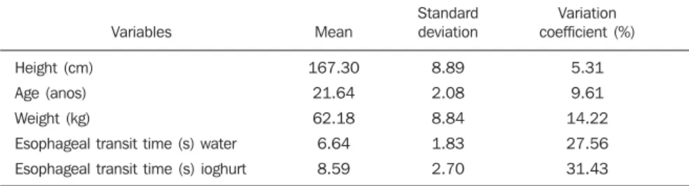
The mean esophageal transit time for water was 6.64 ± 1.83 seconds, and for yo-ghurt, 8.59 ± 2.70 seconds (Table 1).
The analysis of the difference (yoghurt-water) between esophageal transit times has indicated that the mean esophageal transit time with water ingestion was sta-tistically lower as compared with yoghurt ingestion (t = 2.9905, gl = 21, p = 0.0039).
DISCUSSION
In the present experiment, the volun-teers had no difficulty keeping water or yoghurt in the oral cavity. All the steps of the investigation could be accomplished in a calm environment for all the individuals inside the examination room. The intra-abdominal esophagus could be easily ac-cessed and identified at US, allowing the visualization of the progression of the wa-ter/yoghurt bolus through this region.
The esophageal transit time could be de-termined with the aid of the stopwatch in all the volunteers, by measuring the time interval between the moment where the
device was activated once the deglutition of water or yoghurt was initiated, up to the visualization of the bolus transit through the intra-abdominal esophagus. All the examinations could be easily and simply performed, with the patients comfortably positioned and taking a short time to be completed.
The method could differentiate the esophageal transit time for water that was statistically shorter than for yoghurt (p <
0.0039), demonstrating that both sub-stances present a defined time for passing through the esophagus, and that both can be utilized in an initial screening for con-ditions affecting the peristalsis of this or-gan and, consequently, the delay in the bolus transit through the esophagus.
The evaluation of the thoracic esopha-gus with external approach by ultrasonog-raphy is impaired by the access difficulty, both through the anterior and the posterior mediastinum because of the presence of air in the lungs and bone tissue in the dorsal spine through which sound waves cannot be transmitted, with the exception of the segment posterior to the aorta and the heart(14). The cervical esophagus can be evaluated by ultrasonography, considering its lateral location in relation to the trachea, proximity with the surface and interposition of soft tissue such as the left thyroid lobe(15). Given its posterior positioning in relation to the left hepatic lobe, the intra-abdomi-nal segment of the esophagus is easily ac-cessible by ultrasonography, acting as an access window for ultrasound imaging (a relatively homogeneous organ that dis-places the bowel loops with gas)(16–19).
With the advent of new technologies, new method have been introduced for evaluating the esophagus such as nuclear medicine, optic fiber endoscopy, manom-etry with pH-mmanom-etry, and endoscopic
ultra-sonography. All these methods demonstrate the location and anatomical and functional aspects of the esophagus(20–22), but are in-vasive or utilize ionizing radiation with highly expensive equipment.
Therefore, the method evaluated in the present study has allowed a less invasive, a cost-effective and probably more easily available technique for the patients as com-pared with esophageal radiography or scin-tigraphy, considering that this method re-quires only a conventional ultrasonography unit and a stopwatch to be reproduced.
CONCLUSION
The new experimental method for evaluating the esophageal transit time with external approach by ultrasonography is a simple, non-invasive technique that pro-vides significant information without re-quiring ionizing radiation. This method gives us information on the differences between the esophageal transit time for liq-uid and pasty food. It is widely available and can be simply performed, with no con-traindication and playing a significant role in the initial evaluation of conditions that may affect the esophagus. It is a safe, reli-able and discriminative method including bedside capability (suitable for utilization in residences, hospitals and infirmaries), allowing the obtention of fast results with a good cost-benefit ratio.
REFERENCES
1. Ramos JH, Santos MB, Tavares-Neto J. Estudos dos critérios clínicos para requisição de radiogra-fias de tórax em um hospital universitário (Sal-vador, Bahia). Radiol Bras. 1999;32:243–6.
2. Song KS, Song HH, Park SH, et al. Impact of clinical history on film interpretation. Yonsei Med J. 1992;33:168–72.
3. Valadares CP, Silva RAP, Tavares Jr WC, et al. Apresentação da técnica de estudo do tempo de esvaziamento gástrico por meio da ultra-sonogra-fia. Radiol Bras. 2006;39:15–8.
Table 1 Mean, standard deviation and variation coefficient in percentage of the variables height, age, weight and esophageal transit times with water and yoghurt.
Variables
Height (cm)
Age (anos)
Weight (kg)
Esophageal transit time (s) water
Esophageal transit time (s) ioghurt
312
Sakate M et al.
Radiol Bras. 2008 Set/Out;41(5):309–312 4. Meschan I. Oropharynx, laryngopharynx, and
esophagus. In: Meschan I, editor. Roentgen signs in diagnostic imaging. Volume 1 – abdomen. Philadelphia: WB Saunders; 1984. p. 487–560. 5. Parkman HP, Maurer AH, Caroline DF, et al. Op-timal evaluation of patients with nonobstructive esophageal dysphagia. Manometry, scintigraphy, or videoesophagography? Dig Dis Sci. 1996;41: 1355–68.
6. Henry MA, Harbermann MC, Rocha OM. Esoph-ageal motor disturbances in progressive systemic sclerosis. Dis Esophagus. 1999;12:51–3.
7. Machado MM, Rosa ACF, Barros N, et al. Ultra-sonografia endoscópica (USE) do esôfago, estô-mago, cólons e reto. Radiol Bras. 2002;35:219– 23.
8. Ling TC, Johnston BT. Esophageal investigations in connective tissue disease: which test are most appropriate? J Clin Gastroenterol. 2001;32:33– 6.
9. Nakajima K, Kawano M, Kinuya K, et al. The di-agnostic value of oesophageal transit scintigraphy for evaluating the severity of oesophageal
com-plications in systemic sclerosis. Nucl Med Commun. 2004;25:375–81.
10. Nassif MS, Jauregui GF, Rocha VB, et al. Análise crítica da seriografia do esôfago, estômago e duo-deno em um serviço de radiologia de um hospi-tal geral. Radiol Bras. 2004;37:425–9. 11. Penas ME. Motilidade esofagiana: ensaio
icono-gráfico sobre cintilografia dinâmica do esôfago. Radiol Bras. 2007;40:423–7.
12. Costa MMB, Canevaro LV, Azevedo ACP, et al. Valores típicos do “produto dose-área” obtidos durante o estudo videofluoroscópico da degluti-ção. Radiol Bras. 2003;36:17–20.
13. Zar JH. Bioestatistical analysis. New Jersey: Prentice-Hall, 1996.
14. Westra SJ, Derkx HHF, Taminiau JAJM. Symp-tomatic gastroesophageal reflux: diagnosis with ultrasound. J Pediatr Gastroenterol Nutr. 1994;19: 58–64.
15. Esposito F, Lombardi R, Grasso AC, et al. Trans-abdominal sonography of the normal gastroe-sophageal junction in children. J Clin Ultrasound. 2001;29:326–31.
16. Johnson MC. The esophagus. Gastroenterology. 2001;28:459–85.
17. Costa CD, Zomignan HP, Rocha JIP, et al. Re-fluxo gastroesofágico em pediatria: estudo radio-lógico. Pediatria (S. Paulo). 1986;8:136–40.
18. Defagó M, Kuschnir E, Ninci CM, et al. Evalua-tion of methods for the study of gastroesophageal reflux. Rev Fac Cienc Med Univ Nac Córdoba. 1985;43:9–13.
19. Foley LC, Slovis TL, Campbell JB, et al. Evalu-ation of the vomiting infant. Am J Dis Child. 1989;143:660–1.
20. Cerri GG. Vísceras ocas. In: Rocha DC, Cerri GG, Prando A, et al. Ultra-sonografia abdominal. São Paulo: Sarvier; 1988; p. 169–86.
21. Naik DR, Bolia A, Moore DJ. Comparison of barium swallow and ultrasound in diagnosis of gastro-oesophageal reflux in children. Br Med J (Clin Res Ed). 1985;290:1943–5.