The modulatory effect of estradiol
benzoate on superoxide dismutase
activity in the developing rat brain
1Laboratory for Molecular Biology and Endocrinology,
Vin a Institute of Nuclear Sciences, Belgrade, Yugoslavia
2Institute of Zoology, Faculty of Biology, University of Belgrade,
Belgrade, Yugoslavia S. Peji1,
J. Kasapovi1,
D. Cvetkovi2 and
S.B. Pajovi1
Abstract
The sensitivity of copper,zinc (CuZn)- and manganese (Mn)-superoxide dismutase (SOD) to exogenous estradiol benzoate (EB) was investigated in Wistar rats during postnatal brain development. Enzyme activities were measured in samples prepared from brains of rats of both sexes and various ages between 0 and 75 days, treated sc with 0.5 µg EB/100 g body
weight in 0.1 ml olive oil/100 g body weight, 48 and 24 h before sacrifice. In females, EB treatment stimulated MnSOD activity on days 0 (66.1%), 8 (72.7%) and 15 (81.7%). In males, the stimulatory effect of EB on MnSOD activity on day 0 (113.6%) disappeared on day 8 and on days 15 and 45 it became inhibitory (40.3 and 30.5%, respectively). EB had no effect on the other age groups. The stimulatory effect of EB on CuZnSOD activity in newborn females (51.8%) changed to an inhibitory effect on day 8 (38.4%) and disappeared by day 45 when inhibition was detected again (48.7%). In males, the inhibitory effect on this enzyme was observed on days 0 (45.0%) and 15 (28.9%), and then disappeared until day 60 when a stimulatory effect was observed (38.4%). EB treatment had no effect on the other age groups. The sensitivity of MnSOD to estradiol differed significantly between sexes during the neonatal and prepubertal period, whereas it followed a similar pattern thereafter. The sensitivity of CuZnSOD to estradiol differed significantly between sexes during most of the study period. Regression analysis showed that the sensitivity of MnSOD to this estrogen tended to decrease similarly in both sexes, whereas the sensitivity of CuZnSOD showed a significantly different opposite tendency in female and male rats. These are the first reports indicating hormonal modulation of antioxidant enzyme activities related to the developmental process.
Correspondence
S.B. Pajovi
Laboratory for Molecular Biology and Endocrinology
Vin a Institute for Nuclear Sciences P.O. Box 522
11001 Belgrade Yugoslavia
Fax: +38-1-11-455-561 E-mail: [email protected]
Received June 27, 2002 Accepted January 15, 2003
Key words
·Brain
·Brain development ·Estradiol
·Superoxide dismutase
Introduction
Antioxidant enzymes such as superoxide dismutase (SOD), catalase (CAT), and gluta-thione peroxidase (GPx) reduce levels of superoxide radicals (O2·-) and H
2O2, thus
protecting cells from oxygen toxicity. Data suggest that estradiol contributes to the
ovulatory cycle in humans (4), whereas GPx activity in erythrocytes is positively corre-lated with plasma estradiol levels during the menstrual cycle (5). It has been reported that estradiol influences the expression of SOD in the corpus luteum (6) and significantly alters the activity of CAT (3) and the produc-tion of O2·- and H
2O2 (7) in rat macrophages.
The regulatory role of estradiol on anti-oxidant enzymes is more intriguing in light of the hypothesis that reactive oxygen spe-cies can act as physiological signaling mol-ecules (8). Namely, there is growing evi-dence suggesting that O2·- and H
2O2 play a
number of significant, diverse roles in repro-duction (9) and brain development (10). Fi-nally, estrogens function as radical scaven-gers and inhibit neuronal peroxidation both
in vivo and in vitro (11). The marked antioxi-dant activity of estradiol makes this steroid a candidate for the therapy of neurodegen-erative diseases associated with aging (12). It is well known that estradiol exerts mul-tiple effects on morphogenesis, neuroendo-crine function, sex differentiation and neuro-transmission during brain development. Also, the importance of antioxidant enzymes in neu-roprotection during brain development and aging has been well documented (13-15). Yet, we found no data concerning hormonal modu-lation of antioxidant enzyme activities during developmental processes. Our previous stud-ies have shown that estradiol benzoate (EB) and progesterone modulate the activities of SOD, GPx, CAT, and glutathione reductase in brain of adult rats (16-18). The inhibition of brain mitochondrial manganese-SOD (MnSOD) activity by EB without affecting cytosol copper,zinc-SOD (CuZnSOD) activ-ity was a response of rats of both sexes.
In the present study we determined whether exogenous EB affects SOD activity during postnatal brain development in rats of both sexes. We postulated that the regulatory role of estradiol as well as its antioxidant activity in developmental processes could be expressed, at least in part, by modulation of
SOD activity and consequently of the levels of O2·- and H
2O2.
Material and Methods
Wistar rats of both sexes and various ages (0, 8, 15, 30, 45, 60 and 75 days) were housed in open colony cages under controlled condi-tions of temperature (23 ± 2ºC) and illumina-tion (lights on from 5:00 to 17:00 h), with free access to tap water and laboratory chow. Ani-mals were treated 48 and 24 h prior to sacrifice with a sc injection of 0.5 µg EB/100 g body weight (ß-estradiol-3-benzoate; Sigma, St. Louis, MO, USA) suspended in 0.1 ml olive oil/100 g body weight. To obtain the experi-mental group of newborn rats (0 days), the pregnant females received the same dose of EB 48 and 24 h before delivery (on the 19th and 20th day of pregnancy). Two groups of control animals were either sham-injected or injected sc with 0.1 ml olive oil/100 g body weight by the same schedule. Animals were sacrificed by decapitation with a guillotine (Harvard Apparatus Inc., Edenbridge, Kent, UK) and the brains were removed immedi-ately for homogenate preparation.
SOD activity was determined by the method of Misra and Fridovich (21). The auto-oxidation of adrenaline to adrenochrome was carried out in 3 ml of 50 mM Na2CO3 and 100 µM EDTA, pH 10.2, at 26ºC. The inhibition of auto-oxidation was monitored at 480 nm. After assaying total SOD activity the samples were treated with 4 mM KCN in order to inhibit cytosol SOD (22) and sub-jected again to the enzyme assay as described above. The values thus obtained were sidered to be due to MnSOD. Protein con-centration in the cytosol was determined by the method of Lowry et al. (23).
Data were analyzed using the statistical packages OriginPro 6.1 and Statistica 5.0. The differences in enzyme activity (U/mg protein) between EB-treated and control groups were determined by the t-test. In further analysis, relative values for enzyme activity were used, expressed as percent of the enzyme activity in controls of the same
age. Departures of relative values from nor-mal distribution were determined by the Lilliefors and Shapiro-Wilks test. Since the observed variables did not show significant departures from normal distribution, no data transformation was employed. The effects of EB treatment on enzyme activity during development were tested by ANOVA and the Tukey honest significant difference test. A linear regression model with replication was employed in the analyses of the devel-opmental pattern of the response of SOD activity to EB. The slopes of the regression lines for females and males were compared to determine intergender differences in the response of SOD activity.
Results
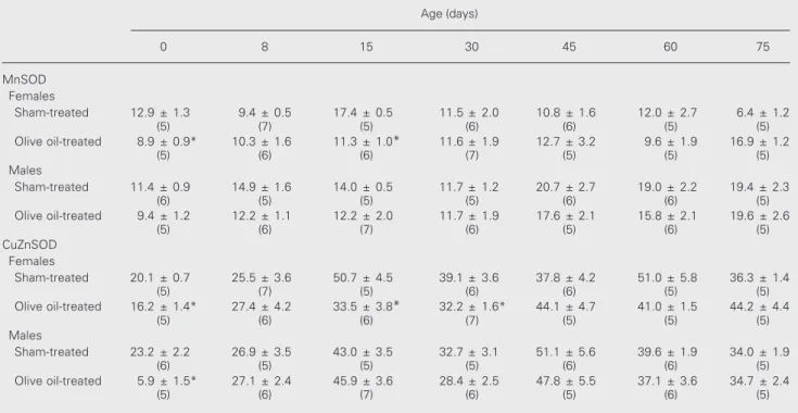
Table 1 shows that olive oil treatment significantly inhibited the activity of MnSOD and CuZnSOD on days 0 and 15 in female
Table 1. The activity of manganese (MnSOD)- and copper,zinc-superoxide dismutase (CuZnSOD) in sham-treated control and olive oil-treated control rats of different ages.
Age (days)
0 8 15 30 45 60 75
MnSOD Females
Sham-treated 12.9 ± 1.3 9.4 ± 0.5 17.4 ± 0.5 11.5 ± 2.0 10.8 ± 1.6 12.0 ± 2.7 6.4 ± 1.2
(5) (7) (5) (6) (6) (5) (5)
Olive oil-treated 8.9 ± 0.9* 10.3 ± 1.6 11.3 ± 1.0* 11.6 ± 1.9 12.7 ± 3.2 9.6 ± 1.9 16.9 ± 1.2
(5) (6) (6) (7) (5) (5) (5)
Males
Sham-treated 11.4 ± 0.9 14.9 ± 1.6 14.0 ± 0.5 11.7 ± 1.2 20.7 ± 2.7 19.0 ± 2.2 19.4 ± 2.3
(6) (5) (5) (5) (6) (6) (5)
Olive oil-treated 9.4 ± 1.2 12.2 ± 1.1 12.2 ± 2.0 11.7 ± 1.9 17.6 ± 2.1 15.8 ± 2.1 19.6 ± 2.6
(5) (6) (7) (6) (5) (6) (5)
CuZnSOD Females
Sham-treated 20.1 ± 0.7 25.5 ± 3.6 50.7 ± 4.5 39.1 ± 3.6 37.8 ± 4.2 51.0 ± 5.8 36.3 ± 1.4
(5) (7) (5) (6) (6) (5) (5)
Olive oil-treated 16.2 ± 1.4* 27.4 ± 4.2 33.5 ± 3.8* 32.2 ± 1.6* 44.1 ± 4.7 41.0 ± 1.5 44.2 ± 4.4
(5) (6) (6) (7) (5) (5) (5)
Males
Sham-treated 23.2 ± 2.2 26.9 ± 3.5 43.0 ± 3.5 32.7 ± 3.1 51.1 ± 5.6 39.6 ± 1.9 34.0 ± 1.9
(6) (5) (5) (5) (6) (6) (5)
Olive oil-treated 5.9 ± 1.5* 27.1 ± 2.4 45.9 ± 3.6 28.4 ± 2.5 47.8 ± 5.5 37.1 ± 3.6 34.7 ± 2.4
(5) (6) (7) (6) (5) (6) (5)
rats, and the activity of CuZnSOD on day 30 when compared to sham-treated controls. In males, olive oil had no effect on MnSOD activity and inhibited the activity of CuZnSOD on day 0. To investigate the modu-latory role of EB on SOD activities during brain development and to facilitate the com-parison between sexes, we report enzyme activity as percent of the values for olive oil-treated controls of the same age.
MnSOD activity in the brain of female and male rats (Figure 1)
In the brain of female rats EB treatment stimulated MnSOD activity on days 0
(P<0.001, t-test), 8 and 15 (P<0.01), but had no effect on the activity of the enzyme dur-ing further development (P>0.05, t-test). EB prominently stimulated brain MnSOD activ-ity immediately after birth both in males and in females (P<0.001, t-test). This effect dis-appeared on day 8 (P>0.05, t-test) and on day 15 of postnatal development it became inhibitory (P<0.05) and opposite compared to females. Inhibition was detected again on day 45 (P<0.05, t-test), while EB seemed to have no effect on the other age groups (P>0.05). Time variation in the effect of EB on MnSOD activity in females (tested by ANOVA and the Tukey test) was statistically significant between days 0, 8 and 15, and days 30 and 45 of postnatal development, with the most sig-nificant effect occurring between days 15 and 30 (P<0.001, Tukey test), when the stimula-tory effect of this hormone disappeared. In males, the effect of EB on MnSOD activity showed a highly significant difference be-tween day 0 and all the other time points examined (P<0.001, Tukey test). The effect of the hormone significantly changed between day 8 and day 15 (P<0.05, Tukey test) when it became inhibitory.
As shown in Figure 1, the sensitivity of MnSOD to estradiol administration differed significantly between sexes during the neona-tal and prepuberneona-tal period (females vs males for 0, 8, 15 days, P<0.01, t-test). During further development this sensitivity followed a simi-lar pattern in female and male rats (P>0.05). Regardless of the sex differences found for the above mentioned time points during early de-velopment, regression analysis (Figure 1B) showed a similar decreasing tendency of MnSOD sensitivity to EB in both sexes during the entire period examined (comparison of regression slopes: t = -1.06, P>0.05).
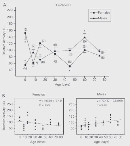
CuZnSOD activity in the brain of female and male rats (Figure 2)
The stimulatory effect of EB on CuZnSOD activity in newborn females
Relative activity (%)
220
200
180
160
140
120
100
80
60
40
250
200
150
100
50
Relative activity (%)
0 10 20 30 40 50 60 70 80
Age (days)
Females Males
Females
Males
0 10 20 30 40 50 60 70 80 0 10 20 30 40 50 60 70 80
Age (days) Age (days)
y = 168.59 + -1.121x R = -0.54
y = 123.90 + -0.6790x R = -0.34
250
200
150
100
50 (5)
(7)
(7) (5)
(7)
(6)
(6) (6)
(5)
(5)
(5)
(5)
(5) (6) *
* * *
* *
MnSOD
Figure 1. A, Effects of estradiol benzoate on manganese-superoxide dismutase (MnSOD) activity in female and male rats of different ages. Data are reported as means ± SEM as percent of olive oil control values for the number of rats given in parentheses (*P<0.05, t-test).
B, Scatter-plot of MnSOD activity. P - significance of slope for females (P<0.001) and males (P<0.001), ANOVA.
A
(P<0.05, t-test) changed to a markedly inhib-itory effect on day 8 of postnatal develop-ment (P<0.01), and disappeared by day 45 when strong inhibition of enzyme activity was detected again (P<0.001). At the age of 60 and 75 days estradiol had no effect on this enzyme (P>0.05, t-test). In the brain of males, the inhibitory effect of EB on CuZnSOD activity was observed during the early post-natal development, i.e., on days 0 (P<0.01, t -test) and 15 (P<0.05), and then disappeared until day 60 when a stimulatory effect was observed (P<0.05). At the age of 75 days EB treatment had no effect on CuZnSOD activ-ity (P>0.05, t-test).
The variation with time of the effects of EB on CuZnSOD activity in females showed highly significant differences in inhibition or activation between days 0 and 8 (P<0.001, Tukey test), 8 and 15 (P<0.01), and 15 and 45 (P<0.001), when the effect of the hor-mone was inverted, disappeared and was reestablished again. In males, the effect of EB on the same enzyme was significantly different between days 0, 8 and 15, and day 60 (P<0.001, P<0.05, P<0.001, respectively, Tukey test).
Contrary to MnSOD, for which both in-hibition and activation were restricted to the period of early postnatal development, these diverse effects of EB seemed to dominate during the entire period examined in the case of CuZnSOD activity (females vs males for 0, 8, 15, 45, 60 days, P<0.02, t-test). Oppo-site and significantly different tendencies in the response of this enzyme activity to EB between sexes were also shown by regres-sion analysis (Figure 2B, comparison of re-gression slopes: t = -3.69, P<0.001).
Discussion
We used exogenous EB to investigate different developmental periods when modu-lation of SOD activity by this hormone might be relevant to processes of estradiol-regu-lated brain differentiation.
We observed that olive oil alone, used as the conventional solvent of EB, reduced SOD activity (24) at certain times during early development (Table 1). This reduction may be related to growing evidence demonstrat-ing the powerful antioxidant properties of olive oil phenolics both in vitro and in vivo
(25). In healthy men, ingestion of virgin olive oil increases the incorporation of oleic acid, phenols and vitamin E into low-density lipoprotein (LDL), which increases LDL re-sistance to oxidation (26). Olive oil phenol hydroxytyrosol reduces H2O2 generation, H2O2-induced DNA damage and mRNA lev-els of GPx in oxidative stress-sensitive hu-man prostate cells (27). It has also been
Figure 2. A, Effects of estradiol benzoate on copper,zinc-superoxide dismutase (CuZnSOD) activity in female and male rats of different ages. Data are reported as means ± SEM as percent of olive oil control values for the number of rats given in parentheses (*P<0.05, t -test). B, Scatter-plot of CuZnSOD activity. P - significance of slope for females (P<0.01) and males (P<0.001), ANOVA.
Relative activity (%)
220
200
180
160
140
120
100
80
60
40
250
200
150
100
50
Relative activity (%)
0 10 20 30 40 50 60 70 80
Age (days)
Females Males
Females
Males
0 10 20 30 40 50 60 70 80 0 10 20 30 40 50 60 70 80
Age (days) Age (days)
y = 107.08 + -0.46x R = -0.29
y = 72.427 + 0.63103x R = 0.53
250
200
150
100
50 (6)
(7) (5)
(7)
(6) (6)
(5)
(5) (5)
(5) (5)
(6)
*
* *
CuZnSOD
* *
* (5)
(7)
A
shown that polyphenol mixtures prepared from olive oil decrease the production of O2·- in cultured human promonocyte (28)
and that olive oil-fed rats have lower liver CuZnSOD, CAT and GPx activities com-pared to fish oil-fed rats (29). The reduction of SOD activity by olive oil stopped after day 30 of development and was not observed in adult rat brain (30). This could be due to the marked perturbations in oxidative metabo-lism and lipid composition during differen-tiation. Namely, the increased degree of li-pid unsaturation demonstrated early during development has been suggested to provide antioxidative protection at the time when antioxidant enzyme defenses are low (31).
When examining the sensitivity of brain SOD to exogenous EB during postnatal de-velopment we applied the hormone dose used in studies of its feedback effect on neonatal and infantile rats of both sexes (32,33). EB treatment of the pregnant female rat immediately before delivery increased the activity of both SOD in the brain of female neonates, particularly MnSOD. In the brain of male neonates, strong stimula-tion of MnSOD activity significantly ex-ceeded the reduction of CuZnSOD activity, representing the most intensive SOD response to EB during the examined developmental period. This indicates that the stimulating effects of EB on brain SOD activities in newborn pups coincided with the high se-rum estradiol levels found in the neonates (aged 0 and 1 days) of both sexes, which were significantly higher than the maximal values observed in adult rats (34). Since the increased SOD activity in the brain and other tissues of rats during the perinatal period is often related to the transition of newborns from a relatively hypoxic to a hyperoxic environment (10,35), these data suggest a possible influence of estradiol on the SOD response to antioxidant stress induced by delivery.
Although there was a difference in the effects of EB on MnSOD and CuZnSOD
activities at the same age, the intragender developmental profile of both SOD responses showed similar increases, stagnations and falls, with the exception of day 8 in females and day 0 in males. This indicates a different mechanism of mitochondrial and cytosol enzyme regulation by exogenous EB during most of the developmental period examined, with a certain degree of time synchroniza-tion in their regulasynchroniza-tion. Previous findings also indicated that during the postnatal de-velopment of rats, MnSOD and CuZnSOD activities often do not correlate with one another or with activities of other antioxi-dant enzymes, and do not synchronously follow the increase of aerobic brain metabo-lism (13,14,36). Mavelli et al. (13) concluded that two types of SOD respond with a dis-tinct time constant to brain differentiation, pointing to a distinct mechanism of regula-tion.
ERa (37) and possibly ERß forms in the
brain of female and male rats during devel-opment, as well as the possibility of differen-tial transcription activity of ERa and ERß
through the same regulatory site (38) could be the cause of the observed sex difference in SOD response to EB. On the other hand, different neuroprotective effects of pharma-cological and physiological doses of estra-diol on H2O2, glutamate and ß-amyloid pep-tide toxicity (mediated by H2O2) operate through ER-independent mechanisms (12). It is possible that estradiol by the same mechanisms influences not only the neuro-toxic, but also the physiological levels of H2O2, thus modulating SOD activities by changing the enzyme product level. Still, we found no data concerning sex dependence in the ER-independent mechanisms of the neu-roprotective and antioxidant effects of estra-diol.
Some of the activities of antioxidant en-zyme are synergistically coordinated in their response to increased aerobic metabolism during brain development, while others are independently regulated (36). Similar find-ings, together with the observation of in-creased transcriptional activity of NF-kB by increased H2O2 levels, led de Haan et al. (10) to postulate a role of reactive oxygen species in normal development. Namely, nonsyn-chronization of antioxidant enzyme activi-ties during development could be related to the tissues need to produce reactive oxygen species for the selective gene transcription via activation of transcription factors, rather than solely for its antioxidant defense
re-quirements. New data on the role of reactive oxygen species in intracellular signaling of transcription, cell proliferation and growth (8) support this assumption. In the light of this, the intergender difference in develop-mental sensitivity of brain SOD to exog-enous EB indicates that estradiol could have a differential influence on females and males not only in O2·- detoxification, but also in
fine regulation of nontoxic levels of O2·- and
H2O2 as secondary messengers in the mito-chondrial and cytosol compartment during development.
Considering the pleiotropic influences of estradiol on brain development in females and in males as a metabolite of testosterone (39), sex-specific modulation of SOD by this hormone during the infantile and prepuber-tal period could contribute to establishing sex-related brain morphology and function. This sex specificity decreased during pu-berty and adult development, possibly due to the more similar later physiological role of EB modulation of SOD in the differentiation of the female and male brain.
The developmental intra- and intergender influences of EB on SOD activity shown in the present study suggest the contribution of this estrogen to protection against oxidative stress and to the production of signals selec-tively needed for different periods of brain development. Since the hormonal modula-tion (40) and the sex specificity of antioxi-dant enzyme activity (2) were related only to aging, these are the first data indicating hor-monal modulation of antioxidant enzymes related to development.
References
1. Guemouri L, Artur Y, Herbeth B, Jeandel C, Cuny G & Siest G (1991). Biological variability of superoxide dismutase, glutathione peroxi-dase and catalase in blood. Clinical Chemistry,37: 1932-1937. 2. Rikans LE, Moore DR & Snowden CD (1991). Sex dependent
differ-ences in the effects of aging on antioxidant defense mechanisms of rat liver. Biochimica et Biophysica Acta, 1074: 195-200.
3. Azevedo RB, Lacava ZGM, Miyasaka CK, Chavez SB & Curi R (2001). Regulation of antioxidant enzyme activities in male and female rat
macrophages by sex steroids. Brazilian Journal of Medical and Bio-logical Research, 34: 683-687.
4. Shiotani M, Noda Y, Narimoto K, Imai K, Mori T, Fujimoto K & Ogawa K (1991). Immunohistochemical localization of superoxide dismu-tase in the human ovary. Human Reproduction,6: 1349-1353. 5. Massafra C, Gioia D, De Felice C, Picciolini E, De Leo V, Bonifazi M &
peroxidase activities during the menstrual cycle. Journal of Endocri-nology,167: 447-452.
6. Sugino N, Hirosawa-Takamori M, Zhong L, Telleria CM, Shiota K & Gibori G (1998). Hormonal regulation of copper-zinc superoxide dis-mutase and manganese superoxide disdis-mutase messenger ribo-nucleic acid in the rat corpus luteum: Induction by prolactin and placental lactogens. Biology of Reproduction,59: 599-605. 7. Chao T, Van Alten PV & Valter RJ (1994). Steroid sex hormones and
macrophage function: modulation of reactive oxygen intermediates and nitrite release. American Journal of Reproductive Immunology, 32: 43-52.
8. Finkel T (1998). Oxygen radicals and signaling. Current Opinion in Cell Biology, 10: 248-253.
9. Behrman HR, Riley JCM & Aten RF (1993). Reactive oxygen species and ovarian function. In: Adashi EY & Leung PCK (Editors), The Ovary. Raven Press Ltd., New York, NY, USA, 455-471.
10. de Haan JB, Tymms MJ, Cristiano F & Kola I (1994). Expression of copper/zinc superoxide dismutase and glutathione peroxidase in organs of developing mouse embryos, fetuses, and neonates. Pedi-atric Research,35: 188-196.
11. Brinton RD & Yamazaki RS (1998). Advances and challenges in the prevention and treatment of Alzheimer’s disease. Pharmacological Research, 15: 386-398.
12. Simpkins JW, Rabbani O, Shi J, Panickar KS, Green PS & Day AL (1998). A system for the brain-enhanced delivery of estradiol: An assessment of its potential for the treatment of Alzheimer’s disease and stroke. Pharmazie,53: 505-511.
13. Mavelli I, Rigo A, Federico R, Ciriolo MR & Rotilio G (1982). Super-oxide dismutase, glutathione peroxidase and catalase in developing rat brain. Biochemical Journal,204: 535-540.
14. Del Maestro R & McDonald W (1987). Distribution of superoxide dismutase, glutathione peroxidase and catalase in developing rat brain. Mechanisms of Ageing and Development,41: 29-38. 15. Mariucci G, Ambrosini MV, Colarieti L & Bruschelli G (1990).
Differ-ential changes in Cu,Zn and Mn superoxide dismutase activity in developing rat brain and liver. Experientia,46: 753-755.
16. Pajovi S, Nikezi G & Martinovi JV (1993). Effects of ovarian
steroids on superoxide dismutase activity in the rat brain. Experientia, 49: 73-75.
17. Pajovi S, Sai i ZS, Spasi MB, Petrovi VM & Martinovi JV (1996). Effects of progesterone and estradiol benzoate on superox-ide dismutase activity in the brain of male rats. Experientia, 52: 221-224.
18. Pajovi SB, Sai i ZS, Spasi MB, Petrovi VM & Martinovi JV (1999). Effects of progesterone and estradiol benzoate on glutathi-one dependent antioxidant enzyme activities in the brain of female rats. General Physiology and Biophysics,18: 35-44.
19. Rossi MA, Cecchini G & Dianzani MU (1983). Glutathione peroxi-dase, glutathione reductase and glutathione transferase in two dif-ferent hepatomas and in normal liver. International Research Com-munications System. Medical Science, 11: 805.
20. de Waziers I & Albrecht R (1987). The effects of vitamin A nutritional status on glutathione, glutathione transferase and glutathione per-oxidase activities in rat intestine. Experientia, 43: 394-395. 21. Misra HP & Fridovich I (1972). The role of superoxide anion in the
autooxidation of epinephrine and a simple assay for superoxide dismutase. Journal of Biological Chemistry, 247: 3170-3175. 22. Geller BL & Winge DR (1983). A method for distinguishing
Cu,Zn-and Mn-containing superoxide dismutases. Analytical Biochemistry, 128: 86-92.
23. Lowry OH, Rosebrough NJ, Farr AL & Randall RJJ (1951). Protein measurement with the Folin phenol reagent. Journal of Biological Chemistry, 193: 265-275.
24. Peji S, Kasapovi J & Pajovi SB (1999). Effects of olive oil on superoxide dismutase activity in brain of newborn and young female rats. Physiological Research, 48: 297-301.
25. Visioli F, Poli A & Galli C (2002). Antioxidant and other biological activities of phenols from olives and olive oil. Medicinal Research Reviews, 22: 65-75.
26. Gimeno E, Fito M, Lamuela-Raventos RM, Castellote AI, Covas M, Farre M, de La Torre-Boronat MC & Lopez-Sabater MC (2002). Effect of ingestion of virgin olive oil on human low-density lipopro-tein composition. European Journal of Clinical Nutrition, 56: 114-120.
27. Quiles JL, Farquharson AJ, Simpson DK, Grant I & Wahle KW (2002). Olive oil phenolics: effects on DNA oxidation and redox enzyme mRNA in prostate cells. British Journal of Nutrition, 88: 225-234. 28. Leger CL, Kadiri-Hassani N & Descomps B (2000). Decreased
super-oxide anion production in cultured human promonocyte cells (THP-1) due to polyphenol mixtures from olive oil processing wastewa-ters. Journal of Agricultural and Food Chemistry, 48: 5061-5067. 29. Ruiz-Gutierrez V, Perez-Espinosa A, Vazquez CM & Santa-Maria C
(1999). Effects of dietary fats (fish, olive and high-oleic-acid sun-flower oils) on lipid composition and antioxidant enzymes in rat liver.
British Journal of Nutrition, 82: 233-241.
30. Pajovi SB, Kasapovi J & Martinovi J (1997). Superoxide dismu-tase activities in different tissues of female rats treated with olive oil. Physiological Research, 46: 381-384.
31. Allen RG & Balin AK (1989). Oxidative influence on development and differentiation: an overview of a free radical theory of development.
Free Radical Biology and Medicine, 6: 631-661.
32. Goldman BD & Gorski RA (1971). Effects of gonadal steroids on the secretion of LH and FSH in neonatal rats. Endocrinology, 89: 112-115.
33. Rodriguez-Sierra JF & Clough RW (1987). Sexual dimorphism in the synaptogenic effect of estradiol in prepubertal rats. Synapse, 1: 258-264.
34. Dohler KD & Wuttke W (1975). Changes with age in levels of serum gonadotropins, prolactin, and gonadal steroids in prepubertal male and female rats. Endocrinology, 97: 898-907.
35. Tanswell AK & Freeman BA (1984). Pulmonary antioxidant enzyme maturation in the fetal and neonatal rat. I. Developmental profiles.
Pediatric Research,18: 584-587.
36. Ninfali P, Aluigi G & Pompella A (1998). Postnatal expression of glucose-6-phosphate dehydrogenase in different brain areas. Neuro-chemical Research,23: 1197-1204.
37. Yokosuka M, Okamura H & Hayashi S (1997). Postnatal develop-ment and sex difference in neurons containing estrogen receptor-alpha immunoreactivity in the preoptic brain, the diencephalon, and the amygdala in the rat. Journal of Comparative Neurology, 389: 81-93.
38. Paech K, Webb P, Kuiper GGJM, Nilsson S, Gustafsson J-A, Kushner PJ & Scanian TS (1997). Differential ligand activation of estrogen receptors ERalpha and ERbeta at AP1 sites. Science, 277: 1508-1510.
39. MacLusky NJ & Naftolin F (1981). Sexual differentiation of the central nervous system. Science,211: 1294-1311.
40. Bolzan AD, Brown OA, Goya RG & Bianchi MS (1995). Hormonal modulation of antioxidant enzyme activities in young and old rats.