ISSN 0100-879X
BIOMEDICAL SCIENCES
AND
CLINICAL INVESTIGATION
www.bjournal.com.br
www.bjournal.com.br
Volume 43 (11) 1010-1134 November 2010
Institutional Sponsors
The Brazilian Journal of Medical and Biological Research is partially financed by
Hotsite of proteomics metabolomics developped by:
Braz J Med Biol Res, November 2010, Volume 43(11) 1095-1101
Effect of hormone replacement on exercise cardiopulmonary
reserve and recovery performance in subclinical hypothyroidism
Effect of hormone replacement on exercise
cardiopulmonary reserve and recovery
performance in subclinical hypothyroidism
M.R.M. Mainenti
1,2, P.F.S. Teixeira
1, F.P. Oliveira
2and M. Vaisman
11Serviço de Endocrinologia, Hospital Universitário Clementino Fraga Filho, Departamento de Clínica Médica, Faculdade de Medicina, 2Laboratório de Fisiologia do Exercício, Escola de Educação Física e Desportos, Universidade Federal do Rio de Janeiro, Rio de Janeiro, RJ, Brasil
Abstract
Subclinical hypothyroidism (SH) patients present cardiopulmonary, vascular and muscle dysfunction, but there is no consensus
about the benefits of levothyroxine (L-T4) intervention on cardiopulmonary performance during exercise. The aim of the present study was to investigate the effects of L-T4 on cardiopulmonary exercise reserve and recovery in SH patients. Twenty-three SH women, 44 (40-50) years old, were submitted to two ergospirometry tests, with an interval of 6 months of normalization of thyroid-stimulating hormone (TSH) levels (L-T4 replacement group) or simple observation (TSH = 6.90 µIU/mL; L-T4 = 1.02 ng/ dL). Patients with TSH >10 µIU/mL were excluded from the study to assure that they would receive treatment in this later stage of SH. Twenty 30- to 57-year-old women with no thyroid dysfunction (TSH = 1.38 µIU/mL; L-T4 = 1.18 ng/dL) were also evaluated. At baseline, lower values of gas exchange ratio reserve (0.24 vs 0.30; P < 0.05) were found for SH patients. The treated group presented greater variation than the untreated group for pulmonary ventilation reserve (20.45 to 21.60 L/min; median variation = 5.2 vs 25.09 to 22.45 L/min; median variation = -4.75, respectively) and for gas exchange ratio reserve (0.19 to 0.27; median variation = 0.06 vs 0.28 to 0.18; median variation = -0.08, respectively). There were no relevant differences in cardiopulmonary recovery for either group at baseline or after follow-up. In the sample studied, L-T4 replacement improved exercise
cardiopul-monary reserve, but no modification was found in recovery performance after exercise during this period of analysis.
Key words: Subclinical hypothyroidism; Thyroid dysfunction; Levothyroxine; Ergospirometry; Exercise physiology
Introduction
Correspondence: M.R.M. Mainenti, Estrada do Bananal, 981, Bloco 1, Apto. 703, 22750-012 Rio de Janeiro, RJ, Brasil. E-mail: [email protected]
Received February 20, 2010. Accepted October 7, 2010. Available online November12, 2010. Published November 12, 2010. Subclinical hypothyroidism (SH) is a condition
character-ized by elevated serum thyroid-stimulating hormone (TSH) concentrations, whereas free thyroxin (T4) levels are in the reference range (1). The prevalence of such dysfunction is gender and age dependent, and detected more often in women and elderly people (2). Studies from the United States analyzing both males and females show a prevalence of 4.3% (2) and 9% (3), respectively. In Rio de Janeiro, Brazil, an epidemiologic study detected a prevalence of 7.27% of SH among women older than 35 years (4).
Since heart, vasculature, lungs, and muscles are target organs for thyroid hormones, a reduction of their performance in SH is expected, and has already been documented (5-8). However, there are only few studies analyzing exercise performance in SH and the effects of hormone replacement with levothyroxine (L-T4). In 1985,
Forfar et al. (9) showed a greater left ventricular ejection fraction after L-T4 treatment. Following this study, others
showed either little improvement (10,11) or significantly
better performance (6,12,13) after hormone replacement. There are many ways to evaluate exercise performance. Ergospirometry (also called spiroergometry or cardiopulmo-nary test) is a noninvasive, direct and reliable method that permits the evaluation of cardiovascular and respiratory functions through gas exchange analysis during exercise. Measurements of minute ventilation and of oxygen and carbon dioxide inspired and expired fractions allow the computation of a large number of parameters related to
car-diopulmonary fitness (14). The effort level most commonly
1096 M.R.M. Mainenti et al.
methods have been applied (computation of increments in cardiopulmonary parameters and signal kinetics analysis), to better evaluate subjects that do not reach high absolute intensities during the ergospirometry test.
The papers concerning SH and cardiopulmonary exer-cise performance cited here have important limitations such as the absence of a control group in most of them (6,9,10,12). Furthermore, only three studies used ergospirometry to quantify cardiopulmonary capacity during exercise (6,11,13). In a previous study by our group, SH patients showed im-provements in cardiopulmonary parameters at submaximal intensity after L-T4 replacement (13), but there was no
investigation about reserve capacity or recovery. On this basis, the aim of the present study was to investigate the effects of L-T4 replacement on cardiopulmonary exercise
reserve and recovery in SH patients.
Patients and Methods
Study design
Initially, a sectional study was performed, in which the parameters studied in SH women were compared to those investigated in a similar group regarding age, menopausal status and sedentary life style, with no thyroid dysfunc-tion.
Next, SH patients were included in a prospective con-trolled study without placebo to investigate the effect of L-T4 replacement on cardiopulmonary exercise reserve
and recovery.
Patients
Twenty-three women aged 30 to 56 years with spontane-ous autoimmune SH (anti-thyroid peroxidase antibodies >35 IU/mL) were recruited from the outpatient Endocrine Clinic of the University Hospital Clementino Fraga Filho (Federal University of Rio de Janeiro). In addition, 21 women aged 30 to 57 years with no thyroid dysfunction (healthy women) were also evaluated in order to compare their performance to the baseline results for patients. The inclusion criteria for SH were: women aged 30 to 60 years, the presence of elevated serum TSH levels, free thyroxin (FT4) values within
the reference range and positive anti-thyroid peroxidase antibodies (TPO-Ab) (Diagnostic Products Corporation Kit criteria: TSH >4.0 µIU/mL, FT4 between 0.8 and 1.9 ng/dL
and TPO-Ab >35 IU/mL) in two blood samples obtained with at least a 4-week interval between them. Patients with TSH >10 µIU/mL were excluded from the study to assure they would receive treatment in this higher stage of SH. The healthy group inclusion criteria were: women aged 30 to 60 years, TSH and FT4 levels within the reference
range and negative anti-thyroid peroxidase antibody titers. The excludingcriteria for all groups were: use of drugs that
could influence thyroid function, heart rate (HR) and blood
pressure; diagnosed cardiac diseases or systemic arterial hypertension; presence of pain or other physical problems
that could interfere with walking. The protocol was approved by the University Hospital Clementino Fraga FilhoEthics Committee and all subjects gave written informed consent to participate.
Intervention study protocol
Patients were randomly divided into two groups accord-ing to the proposed intervention in a blind manner: L-T4
-treated patients (N = 11) and un-treated patients (N = 12). Each treated patient received an accurate count of tablets containing L-T4. The initial dosage was 0.75 µg·kg-1·day-1,
which was progressively titrated to achieve serum TSH normalization. No patient used more than 125 µg/day to reach TSH reference values (checked every 2 months during outpatient visits). Patients were instructed to take the medication once a day in the morning, at least 30 min before breakfast. Untreated patients did not receive any medication or intervention.
After the first procedures at the Endocrine Clinic (symp
-tom investigation, protocol explanations, written consent), patients were taken to the Exercise Physiology Laboratory of the Physical Education and Sports School at the same University, where the cardiopulmonary test was performed (baseline) and 6 months after TSH normalization (treated patients) or observation (untreated patients).
Ergospirometry
Before the cardiopulmonary test, all subjects were instructed to avoid physical exercise, drinking alcohol, taking caffeine (for one day), and smoking (for at least 4 h before the exam).
The cardiopulmonary test was performed with a treadmill
EG 700.2 (Ecafix, Brazil) and the modified Balke protocol
was used (constant velocity of 4.8 km/h and a 3% grade increment every 2 min). Patients were monitored during the test with a 12-lead electrocardiogram (EKG) Cardio
Perfect (Ecafix). Respiratory gas exchange was sampled from a mouthpiece connected to a medium flow meter and
a gas analyzer VO2000 (Medical Graphics, USA), that was calibrated before each test with gas standards of known concentrations. Oxygen (O2) and carbon dioxide (CO2)
expired fractions, and the ventilation flow were measured
breath-by-breath, allowing the calculation of important cardiopulmonary parameters (cited below). A nose clip was used to avoid gas escape. Blood pressure was determined by the auscultatory method (Narcosul, 1400-C, Brazil) during
exercise and in the recovery phase (first and third minutes).
All participants were informed about the test interruption criteria: chest pains, systolic blood pressure (SBP) >220 mmHg, diastolic blood pressure (DBP) >115 mmHg, a blood pressure decrease with increase load, dizziness, physical manifestations of extreme fatigue, EKG changes, and subjects’ request to stop (18). The person conducting the ergospirometry exam was not aware of group allocation.
the difference between the variable’s maximal value (at the end of the test) and the lowest value (at the beginning of the test). The parameters analyzed were: oxygen uptake (V
.
O2, mL·kg-1·min-1); HR; bpm; minute ventilation (V .
E; L/min), and gas exchange ratio (R = V
. O2/V
. CO2).
The recovery from exercise was determined by the fol-lowing equation: Recovery (%) = [(PV - MinV) / PV] x 100; where PV = the peak value of the parameter analyzed; MinV = the value of the parameter analyzed at the considered minute of recovery (1st or 3rd). HR, SBP and DBP, V
. E and
V
.
O2 were measured during the recovery investigation.
Statistical analysis
Data are reported as median (interquartile range) and were analyzed by nonparametric statistical tests, since the numerical variables did not have a normal distribution by the Kolmogorov-Sminorv test.
The Mann-Whitney test was applied in order to identify differences between SH women and those with no thyroid dysfunction for cardiopulmonary reserve and exercise recovery, and variation (6 months-baseline) of cardiopul-monary reserve in treated and untreated patients. This test was also used to assure similar characteristics (regarding numerical variables) after randomization.
The Wilcoxon test was performed to analyze treated and untreated patients before and after TSH normalization or observation.
Proportions of categorical variables were compared
by the chi-square (Χ2) test. Statistical significance was
considered when P ≤ 0.05. SPSS 13.0 for Windows (SPSS
Inc., USA) was used for statistical analysis.
Results
Baseline results
Hormone levels. The hormone levels of SH patients were 6.90 (5.40-9.10) µIU/mL TSH and 1.02 (0.82-1.19) ng/dL FT4. Healthy women presented normal TSH [1.38
(0.96-1.89) µIU/mL] and FT4 [1.18 (1.04-1.32) ng/dL] values.
Exercise cardiopulmonary reserve. All cardiopulmonary reserve values were higher in women with no thyroid
dys-function than in SH patients, with a statistically significant
difference for gas exchange ratio (R; Table 1).
It is important to note that patient and healthy women
groups were similar regarding variables that influence
cardiopulmonary performance such as age, body mass, smoking, sedentary lifestyle, and menopause (Table 1).
Recovery from exercise. There was no difference in V
. E, V
.
O2, HR, or blood pressure percent recovery
be-tween patients and healthy women at any recovery minute (P always >0.15; Table 2).
Results of intervention
Hormone levels. Randomization into treated and un-treated assured comparable groups in terms of confounding
variables such as smoking and menopause (P > 0.05, Χ2
test). Sedentary lifestyle, measured by regular exercise self-report, was also similar for the two groups (treated = 83%;
untreated = 73%, P > 0.05, Χ2 test). Body mass and age,
that could also confound the results, did not differ between the two groups of patients (treated = 65.90 kg; untreated = 66.10 kg, median values, P > 0.05, Mann-Whitney test).
TSH values were significantly reduced in patients treated
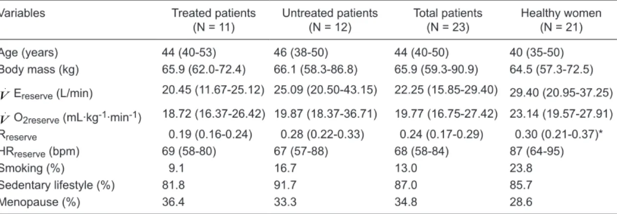
Table 1. Baseline characteristics and exercise cardiopulmonary variable reserve of subclinical hypothyroidism patients and healthy women.
Variables Treated patients (N = 11)
Untreated patients (N = 12)
Total patients (N = 23)
Healthy women (N = 21)
Age (years) 44 (40-53) 46 (38-50) 44 (40-50) 40 (35-50) Body mass (kg) 65.9 (62.0-72.4) 66.1 (58.3-86.8) 65.9 (59.3-90.9) 64.5 (57.3-72.5)
V
.
Ereserve (L/min) 20.45 (11.67-25.12) 25.09 (20.50-43.15) 22.25 (15.85-29.40) 29.40 (20.95-37.25)
V
.
O2reserve (mL·kg-1·min-1) 18.72 (16.37-26.42) 19.87 (18.37-36.71) 19.77 (16.75-27.42) 23.14 (19.57-27.91) Rreserve 0.19 (0.16-0.24) 0.28 (0.22-0.33) 0.24 (0.17-0.29) 0.30 (0.21-0.37)* HRreserve (bpm) 69 (58-80) 67 (57-88) 68 (58-84) 87 (64-95)
Smoking (%) 9.1 16.7 13.0 23.8
Sedentary lifestyle (%) 81.8 91.7 87.0 85.7
Menopause (%) 36.4 33.3 34.8 28.6
Data are reported as median (interquartile range), except for smoking, sedentary lifestyle and menopause (%). Treated patients with subclinical hypothyroidism received 0.75 µg·kg-1·day-1 levothyroxine orally for 6 months. When necessary, the dose was adjusted to reach normal thyroid-stimulating hormone levels. V
.
E = minute ventilation; V
.
O2 = oxygen
1098 M.R.M. Mainenti et al.
with hormone replacement (medians = 6.46 vs 2.94 µIU/mL, P = 0.008), but not in untreated patients (medians = 7.24 vs 5.65 µIU/mL, P > 0.05). FT4
val-ues were significantly increased in treated patients
(medians = 1.05 vs 1.26 ng/dL). As expected, this
significant rise was not detected in untreated patients
(medians: 0.90 vs 1.12 ng/dL, P > 0.05). The time interval between randomization and evaluation after 6 months of euthyroidism was 11 ± 2.72 months for the treated group. In the untreated group, the time between the two exams was 9.83 ± 2.69 months.
Exercise cardiopulmonary reserve. Treated patients presented increased V
.
E and Rreserve
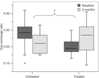
after 6 months of TSH normalization, whereas the cardiopulmonary reserve for the cited parameters decreased in untreated patients after 6 months of observation (Table 3). Comparisons of the reserve variation (6 months-baseline) showed a statistically
significant difference between treated and untreated
patients for V .
Ereserve (P = 0.01) and Rreserve (P =
0.04; Figures 1 and 2), which did not occur for HR or V
.
O2reserve. It is important to note that treated
and untreated patients were comparable regarding exercise cardiopulmonary reserve variables before treatment or observation (P > 0.05), presenting no
fitness level difference at baseline. Reserve data
presentation can be found in Table 3.
Recovery from exercise. HR recovery was slightly better in the second evaluation for both treated and
un-Table 2. Baseline results for cardiopulmonary variables during recovery from exercise.
Variables Treated patients (N = 11)
Untreated patients (N = 12)
Total patients (N = 23)
Healthy women (N = 21)
V
.
E recovery
1st min (%) 18.70 (14.77-33.42) 21.53 (12.35-24.36) 20.25 (14.69-29.27) 21.50 (15.17-29.51) 3rd min (%) 41.02 (36.35-46.05) 44.03 (36.41-51.41) 41.09 (37.14-49.68) 46.09 (33.11-52.90)
V
.
O2 recovery
1st min (%) 22.02 (18.98-35.67) 20.08 (16.79-24.13) 21.75 (18.25-27.76) 26.51 (17.15-34.20) 3rd min (%) 44.83 (37.99-51.30) 47.73 (38.42-54.16) 44.83 (38.06-53.72) 52.87 (43.93-59.16) HR recovery
1st min (%) 16.40 (11.55-20.68) 14.26 (11.14-18.82) 14.93 (11.65-19.91) 15.44 (10.90-17.69) 3rd min (%) 25.20 (23.49-27.13) 27.93 (22.89-29.96) 26.72 (23.49-28.98) 28.76 (23.47-30.92) SBP recovery
1st min (%) 6.06 (2.94-10.00) 6.25 (0.00-9.52) 6.15 (0.00-10) 5.88 (0.00-10.57) 3rd min (%) 13.89 (10.00-17.24) 10.71 (6.90-23.68) 12.30 (9.77-21.00) 15.63 (11.76-23.53) DBP recovery
1st min (%) 6.25 (0.00-11.76) 5.56 (5.00-10.00) 5.72 (0.00-11.76) 5.00 (0.00-11.11) 3rd min (%) 6.25 (0.00-10.53) 10.00 (5.26-15.00) 6.69 (3.94-11.94) 11.11 (5.56-16.45)
Data are reported as median (interquartile range). Treatment of patients with subclinical hypothyroidism with levothyroxine is described in the legend of Table 1. V
.
E = minute ventilation; V
.
O2 = oxygen uptake; HR = heart rate; SBP = systolic blood pressure; DBP = diastolic blood pressure. All P values were >0.05 (total patients vs healthy women,Mann-Whitney test).
Table 3. Exercise cardiopulmonary reserve of treated and untreated patients with subclinical hypothyroidism.
Variables Treated group (N = 11)
Untreated group (N = 12)
V
.
Ereserve (L/min)
Baseline 20.45 (11.67-25.12) 25.09 (20.50-43.15) 6 months 21.60 (19.40-28.40) 22.45 (16.52-29.40) Variation 5.20 (-2.95-6.97) -4.75 (-11.02-1.14)*
V
.
O2reserve (mL·kg-1·min-1)
Baseline 18.72 (16.37-26.42) 19.87 (18.37-36.71) 6 months 17.99 (13.81-19.66) 20.40 (17.36-23.44) Variation -4.60 (-7.44-1.30) 1.29 (-10.89-2.72) Rreserve
Baseline 0.19 (0.16-0.24) 0.28 (0.22-0.33) 6 months 0.27 (0.18-0.32) 0.18 (0.15-0.27) Variation 0.06 (-0.07-0.15) -0.08 (-0.14-0.06)* HRreserve (bpm)
Baseline 69 (58-80) 67 (57-88) 6 months 67 (56-81) 62 (51-76) Variation -2 (-23-3) -6 (-18-3)
Data are reported as median (interquartile interval). Treatment of pa-tients with subclinical hypothyroidism with levothyroxine is described in the legend of Table 1. V
.
E = minute ventilation; V
.
O2 = oxygen
up-take; R = gas exchange ratio; HR = heart rate. *P ≤ 0.05 (untreated vs
treated patients at all investigated minutes, with no statistical
significance compared to baseline tests (mean increase of
2%). Systolic and diastolic blood pressure recovery showed no changes with treatment or only observation. V
. E and
V
.
O2 recovery either increased, decreased or remained
the same, with no statistical or clinical significance.
Discussion
The findings of the present study suggest that SH
patients have some impairment in their exercise cardio-pulmonary reserve, while the recovery of hemodynamic
and ventilatory parameters does not show any significant
alteration. The normalization of serum TSH for 6 months through L-T4 replacement allowed an improvement in
exer-cise cardiopulmonary reserve, but no significant alterations
were evident in the recovery phase (probably because no important impairment was shown in the baseline condition). It is also important to note that untreated patients showed a worse reserve performance after 6 months without hormone replacement intervention. The use of variation (6 months-baseline) to analyze the treated and untreated groups was important because it represents the general behavior and the magnitude of follow-up. The limitation to the present study is the absence of a placebo group (only an observation, untreated group was assessed). There was
no financial support for this purpose.
Some other studies have evaluated exercise perfor-mance of SH patients before and after hormone replace-ment. One study, carried out in Scotland with 10 SH patients (9), examined the ejection fraction at rest and during exercise. The authors detected higher ejection frac-tion values at high intensity exercise when TSH level was normalized. Nevertheless, no control group was examined to determine if this improvement was due to hormone level normalization or to a possible exercise retest bias. Ten years later, researchers from the United States (10) repeated the same study but waited 3 months in the normalized TSH state before reevaluating the patients. They analyzed the structure and function of the heart and found only modest improvements for left ventricular diastolic dimension and pre-ejection period at exercise.
The first study that evaluated the effect of L-T4
replace-ment in SH patients by means of ergospirometry was carried out in Germany (6). Twenty SH patients before and after restoration of euthyroidism were included. All patients were treated with L-T4, and the results showed greater increases
of V .
E, V
.
O2 and oxygen pulse, as also shown by the
pres-ent findings regarding exercise cardiopulmonary reserve,
since the results of the cited paper were calculated in the same way as the reserve in the present study. However, a recent double-blind, randomized, placebo-controlled study from Italy (11), also using ergospirometry, did not demonstrate any differences in maximal V
.
O2 or maximal
power output after 6 months of stable euthyroidism. It is
important to note that the current data analysis was different, considering not only the maximal point, but to what extent one subject could increase from the rest (reserve), a fact that may explain the difference between results. Previous
Figure 1. Box plots (median, 1st and 3rd quartiles, minimum and maximum) of minute ventilation (V
.
E) reserve for untreated and treated patients at baseline and after 6 months. *P = 0.01 com-paring the variation (6 months-baseline) between the treated and untreated groups (Mann-Whitney test). P values were >0.05 for all other comparisons (Mann-Whitney and Wilcoxon tests). Treat-ment of patients with subclinical hypothyroidism with levothyrox-ine is described in the legend of Table 1.
1100 M.R.M. Mainenti et al.
data from our group have shown improvements in V .
E, V
.
O2 and HR at submaximal intensity of exercise (5th
min-ute of exercise; velocity = 4.8 km/h; gradient = 6%) for treated patients and no alterations in untreated women (13).
The baseline impairment of exercise cardiopulmonary reserve found in this study (considering the gas exchange ratio reserve) in SH patients can be explained by the re-duced thyroid hormone (TH) action. Although they were in the reference range, FT4 values were lower in SH patients
compared to healthy women. Some expected TH actions
that could influence cardiopulmonary exercise performance
are: myocardial contraction, vascular dilatation, enzyme activation (mainly inside mitochondria, involved in energy production), and ventilation capacity of the lungs. Once these actions are impaired (due to the slight reduction of
TH levels), the exercise performance will also be influenced.
When hormone levels are restored (as in the present study, by L-T4 replacement), those specific actions will occur in a
more efficient manner and the exercise performance will
be improved. One good example of this enhancement was published by Kahaly (6), who showed that increased vital capacity is obtained after TSH normalization in treated SH patients. If this parameter (vital capacity) is restored with L-T4 replacement, the minute ventilation (V
.
E = tidal volume,
part of vital capacity, * respiratory rate) reserve (increment peak exercise-rest analyzed in the present study) must also be restored, as observed in the treated patients evaluated in the present study.
It is well established that HR recovery after exercise is a predictor of mortality (19-22). The results of the recovery of exercise revealed that the alterations in TH levels were
not sufficient to influence the hemodynamic and ventilatory
parameters measured during this phase, probably due to
the mild hormone deficiency characteristic of the present
sample.
In the present study, levothyroxine replacement proved exercise cardiopulmonary reserve, which was im-paired in subclinical hypothyroid patients (when compared to healthy women). The recovery after exercise was not affected by this level of thyroid dysfunction and intervention
did not lead to an enhanced performance. These findings
support the consistency of the literature about mild thyroid dysfunction management, which can help physicians in treatment decisions about each patient with subclinical hypothyroidism. More studies are needed to determine which exercise cardiopulmonary parameters will constantly
benefit from L-T4 replacement.
References
1. Surks MI, Ortiz E, Daniels GH, Sawin CT, Col NF, Cobin
RH, et al. Subclinical thyroid disease: scientific review and
guidelines for diagnosis and management. JAMA 2004; 291: 228-238.
2. Hollowell JG, Staehling NW, Flanders WD, Hannon WH, Gunter EW, Spencer CA, et al. Serum TSH, T(4), and thyroid antibodies in the United States population (1988 to 1994): National Health and Nutrition Examination Survey (NHANES III). J Clin Endocrinol Metab 2002; 87: 489-499.
3. Canaris GJ, Manowitz NR, Mayor G, Ridgway EC. The Colorado thyroid disease prevalence study. Arch Intern Med
2000; 160: 526-534.
4. Sichieri R, Baima J, Marante T, de Vasconcellos MT, Moura AS, Vaisman M. Low prevalence of hypothyroidism among Black and Mulatto people in a population-based study of Brazilian women. Clin Endocrinol 2007; 66: 803-807. 5. Duyff RF, Van den Bosch J, Laman DM, van Loon BJ,
Lins-sen WH. Neuromuscular findings in thyroid dysfunction: a
prospective clinical and electrodiagnostic study. J Neurol Neurosurg Psychiatry 2000; 68: 750-755.
6. Kahaly GJ. Cardiovascular and atherogenic aspects of sub-clinical hypothyroidism. Thyroid 2000; 10: 665-679. 7. Biondi B, Palmieri EA, Lombardi G, Fazio S. Effects of
subclinical thyroid dysfunction on the heart. Ann Intern Med
2002; 137: 904-914.
8. Fazio S, Palmieri EA, Lombardi G, Biondi B. Effects of thy-roid hormone on the cardiovascular system. Recent Prog Horm Res 2004; 59: 31-50.
9. Forfar JC, Wathen CG, Todd WT, Bell GM, Hannan WJ, Muir AL, et al. Left ventricular performance in subclinical
hypothyroidism. Q J Med 1985; 57: 857-865.
10. Arem R, Rokey R, Kiefe C, Escalante DA, Rodriguez A. Cardiac systolic and diastolic function at rest and exercise in subclinical hypothyroidism: effect of thyroid hormone therapy. Thyroid 1996; 6: 397-402.
11. Caraccio N, Natali A, Sironi A, Baldi S, Frascerra S, Dardano A, et al. Muscle metabolism and exercise tolerance in sub-clinical hypothyroidism: a controlled trial of levothyroxine. J Clin Endocrinol Metab 2005; 90: 4057-4062.
12. Brenta G, Mutti LA, Schnitman M, Fretes O, Perrone A, Matute ML. Assessment of left ventricular diastolic function by radionuclide ventriculography at rest and exercise in subclinical hypothyroidism, and its response to L-thyroxine therapy. Am J Cardiol 2003; 91: 1327-1330.
13. Mainenti MR, Vigario PS, Teixeira PF, Maia MD, Oliveira FP, Vaisman M. Effect of levothyroxine replacement on exercise performance in subclinical hypothyroidism. J Endocrinol Invest 2009; 32: 470-473.
14. Wasserman K, Hansen JE, Sue DY, Whipp BJ. Principles of exercise testing and interpretation. Philadelphia: Lea & Febiger; 1987.
15. Calbet JA, Chavarren J, Dorado C. Running economy and delayed onset muscle soreness. J Sports Med Phys Fitness
2001; 41: 18-26.
16. Ziemba AW, Chwalbinska-Moneta J, Kaciuba-Uscilko H, Kruk B, Krzeminski K, Cybulski G, et al. Early effects of short-term aerobic training. Physiological responses to graded exercise. J Sports Med Phys Fitness 2003; 43: 57-63.
course of blood pressure changes immediately after maximal exercise. J Sports Med Phys Fitness 2006; 46: 605-610.
18. Fletcher GF, Balady GJ, Amsterdam EA, Chaitman B, Eckel R, Fleg J, et al. Exercise standards for testing and training: a statement for healthcare professionals from the American Heart Association. Circulation 2001; 104: 1694-1740. 19. Cole CR, Blackstone EH, Pashkow FJ, Snader CE, Lauer
MS. Heart-rate recovery immediately after exercise as a predictor of mortality. N Engl J Med 1999; 341: 1351-1357. 20. Nishime EO, Cole CR, Blackstone EH, Pashkow FJ, Lauer
MS. Heart rate recovery and treadmill exercise score as predictors of mortality in patients referred for exercise ECG.
JAMA 2000; 284: 1392-1398.
21. Cole CR, Foody JM, Blackstone EH, Lauer MS. Heart rate recovery after submaximal exercise testing as a predictor of mortality in a cardiovascularly healthy cohort. Ann Intern Med 2000; 132: 552-555.