CLINICAL SCIENCE
Serum levels of hypothalamic-pituitary-testicular axis
hormones in men with or without prostate cancer or
atypical small acinar proliferation
Caio da Silva Schmitt, Ernani Luis Rhoden, Gilberto Laurino Almeida
Federal University of Health Sciences of Porto Alegre – Urology, Porto Alegre, RS, Brazil.
INTRODUCTION: Substantial controversy exists regarding the association between testosterone serum levels and prostate cancer.
OBJECTIVE: To evaluate the levels of hypothalamic-pituitary-testicular axis hormones in the sera of men with prostate cancer and atypical small acinar proliferation as well as those with normal biopsies.
METHODS: A study cohort of 186 men with suspected prostate cancer who had undergone transrectal prostate biopsies was used in this study. The patients were divided into the following three groups based on the histology of the biopsy samples: no neoplasia, atypical small acinar proliferation or prostate cancer. Demographic data were also collected. Levels of total testosterone, follicle-stimulating hormone, luteinizing hormone, prolactin, estradiol, and serum prostate-specific antigen were measured in blood samples.
RESULTS:Initially, 123 men were found to be without neoplasia, 26 with atypical small acinar proliferation and 37 with prostate cancer. After a second biopsy was taken from the men diagnosed with atypical small acinar proliferation, the diagnoses were revised: 18 were diagnosed with atypical small acinar proliferation and 45 with prostate cancer. No significant differences between the groups were identified regarding age, smoking history, chronic diseases, body mass index or PSA levels (P.0.05). The mean serum levels of testosterone, follicle-stimulating
hormone, luteinizing hormone, prolactin and estradiol were similar in all of the groups (P.0.05). Furthermore, in individuals with prostate cancer, the Gleason scores and prevalence of hypogonadism were not significantly different (P.0.05).
CONCLUSION: The present study revealed no difference in the serum levels of testosterone, follicle-stimulating hormone, luteinizing hormone, prolactin or estradiol in men without neoplasia compared with those with atypical small acinar proliferation or prostate cancer.
KEYWORDS: Testosterone; Luteinizing hormone; Follicle-stimulating hormone; Estradiol; Prolactin.
Schmitt CS, Rhoden EL, Almeida GL. Serum levels of hypothalamic-pituitary-testicular axis hormones in men with or without prostate cancer or atypical small acinar proliferation. Clinics. 2011;66(2):183-187.
Received for publication onSeptember 14, 2010;First review completed onOctober 4, 2010;Accepted for publication onOctober 24, 2010 E-mail: [email protected]
Tel.: 55 51 3378-9995
INTRODUCTION
Prostate cancer is the second most frequent malign neoplasia in the world, as well as the most frequent tumor affecting men except non-melanoma skin tumors.1,2 The etiology of prostate neoplasia is unknown, although some risk factors have been determined like familiar history, age and race. The androgens, specifically testosterone, have a fundamental role in the growth of the prostate gland.3In the 1940s, Huggins and Hodges4 demonstrated that, in men with advanced prostate cancer, androgenic deprivation
caused tumor regression. This observation causes some concern regarding the possibility of the potential role of androgens as an etiologic factor in the development of prostate cancer. However, most epidemiologic studies were unable to establish a consistent relationship between higher androgen serum levels as a risk factor for prostate cancer. Interestingly, the Prostate Cancer Prevention Trial (PCPT)5 revealed that finasteride, an 5a-redutase inhibitor,
admin-istration reduced prostate cancer diagnoses by 25% after seven years of treatment. Although, in this same trial, the occurrence of higher grades of prostate cancer in the finasteride group, when compared to the control group, was unexpected and intriguing.
Furthermore, other publications have demonstrated that low testosterone serum levels was associated with undiffer-entiated tumors,6 advanced tumor stage7 and unfavorable prognosis after radical prostate surgery.8 All of these
Copyrightß2011CLINICS– This is an Open Access article distributed under
observations revealed that the relationship between prostate cancer and testosterone is far from a complete under-standing requiring further investigation.
Some prostate biopsy characteristics such as, prostate intra-epithelial neoplasia and, more recently, atypical small acinar proliferation (ASAP) have been implicated as prostate cancer risk factors during follow-up of those patients.9 The role of serum hormones, including the androgens, in patients with ASAP or prostate cancer, has not been explored yet, but might be helpful to improve our understanding of the relationship between the hormones from hypothalamic-pituitary-testicular axis in men with or without prostate cancer.
Based on the discussion above, the objective of the current study is to evaluate serum levels of the hypothalamic-pituitary-testicular axis hormones in men submitted to prostate biopsies and histological characteristics of ASAP, prostate cancer and negative results.
MATERIALS AND METHODS
Study Population
All men visiting the Urology Division of Santa Casa Hospital of Porto Alegre, Brazil, between March 2006 and December 2007 with indication of prostate biopsy were invited to participate in this study. A prostate biopsy was indicated if the serum prostate specific antigen (PSA) was over 2.5 ng/ml or had increased by more than 0.75 ng/ml/ year, and/or suspicious results from a digital rectal exam of the prostate gland (prostatic lobe asymmetry, nodules or altered consistency). Exclusion criteria included previous or current use of testosterone replacement therapy, previous prostate surgery, urinary infection or other inflammatory or infectious condition affecting the genito-urinary tract, current use of a vesical catheter and chemotherapy or radiotherapy for treatment of pelvic neoplasia.
Of the 437 men evaluated during this period, all signed an informed consent form. The study was approved by the Research Ethics Committee of the Institution.
Men fulfilling the inclusion criteria were interviewed approximately 30 days before the prostate biopsy by two of the authors (CSS and GLA) to collect the data. Weight and height were checked using a scale calibrated previously (FilizolaH, Sao Paulo, Brazil) and the body mass index (BMI) was calculated by dividing the weight (Kg) by the square of the height (m2). The individuals themselves classified their race as Caucasian or not Caucasian. Also during this interview, individual data about medical history were collected, such as previous surgery, acute and chronic diseases, medications and smoking. After the interview, the patients were sent to the laboratory to have a blood sample drawn and then to their scheduled prostate biopsy date.
Biochemical Analysis
After four hours fasting,10 a venous blood sample was obtained between 7 and 11 am, to assess serum levels of total testosterone (TT), follicle-stimulating hormone (FSH), luteinizing hormone (LH), prolactin and estradiol. Serum levels of TT (normal range: 241 to 827 ng/dL), FSH (normal range: 1.4 to 18.1 mIU/mL), LH (normal range: 1.5 to 9.3 mIU/mL) and prolactin (normal range: 2.1 to 17.7 ng/ mL) were determined by electrochemiluminescence using the ADVIA Centaur 2000 system (SiemensH, Sao Paulo, Brazil), while estradiol (normal range: up to 56.0 pg/mL)
and total PSA (normal range: up to 2.5 ng/mL) were determined by the Immulite immunometric chemilumines-cent method (Diagnostic Products Corp, Los Angeles, CA, USA).
Prostate biopsy
An ultrasound-guided transrectal biopsy was performed by standard methods using a Tosbee 4 endocavity trans-ductor (ToshibaH, New York, USA) under local anesthesia (20 mL of 1% lidocain injected bilaterally around the neurovascular bundle).11 A mean of twelve biopsy cores were removed from each prostate gland, but this number was exceeded when indicated by the presence of a nodule or other suspect image on ultrasound. The tissue samples were stored in bottles containing 10% of formalin.
Histological examination of the prostate
Microscopically, all samples isolated by biopsy were analyzed, after staining with hematoxylin and eosin using standard laboratory methods, by a single expert pathologist of our Institution.
The individuals were divided into three groups according to the histological results: without neoplasia (including benign prostate hyperplasia, acinar atrophy and high grade prostate intra-epithelial neoplasia), ASAP and prostate cancer. Men diagnosed with ASAP by biopsy were subjected to a second biopsy. These results were used to newly diagnose men with prostate cancer, while the others remained in the ASAP group.
Complicating factors
Potential complicating factors that could interfere with serum TT levels were measured to avoid false results, including age, obesity, smoking, chronic diseases such as hypertension, diabetes, peripheral vascular disease, ischemic heart disease, dyslipidemia and hyperthyroidism, and serum levels of other hormones of the hypothalamic-pituitary-testicular axis like FSH, LH, prolactin and estra-diol. We did not include free testosterone serum levels in the current study by protocol and laboratory difficulties.
Statistical analysis
Data like smoking, race and chronic disease were analyzed with the Chi-square test. The same test was applied when a stratified analysis was done using the hypogonadism criteria for patients with serum TT under 300 ng/mL. To evaluate the effect of age, BMI and some of the hormones (TT, prolactin and estradiol), ANOVA was used, and for the total PSA, FSH and LH we used a non-parametric test (Kruskal-Wallis). Results were considered statistically significant when thePvalue was less than 0.05. All analyses were done using the SPSS statistical program, version 14.0 for Windows (SPSS, Chicago, IL, USA).
RESULTS
After screening, 186 men who had indication for prostate biopsy, based on current protocol, were eligible for analyses. The mean age of the study population was 64.54¡6.7 years
as no neoplasia, 26 (14%) with ASAP and 37 (19.9%) with prostate cancer. Overall, 57 (30.6%) of the individuals presented at least one associated medical condition repre-sented by dyslipidemia (30.6%), hypertension (24.7%), diabetes mellitus (17.7%), peripheral vascular disease (10.2%), ischemic heart disease (8.6%) and hyperthyroidism (1.6%). Other data are described in Table 1.
Subsequent analyses divided the patients according to the histological results: without neoplasia, ASAP and prostate cancer. Men with ASAP on the first biopsy but prostate
cancer on the second biopsy, 8 (31%) cases, were re-allocated by the second result. The groups do not differ in relation to age (P= 0.37), BMI (P= 0.66), race (P= 0.36), smoking (P= 0.79) or the presence of a chronic disease (P= 0.27), as well as the median total PSA serum level (6.05 vs. 5.63 vs. 6.63 ng/mL, respectively;P= 0.22; Table 2).
The mean TT serum level were respectively 414.4
¡ 147.33 ng/dL for patients without neoplasia, 421.17¡
149.24 ng/dL for patients with ASAP, and 408.40 ¡
129.05 ng/dL for patients with prostate cancer (P= 0.95). The other hormones (FSH, LH, prolactin and estradiol) were similar among the different studied groups (P.0.05; Table 3). Analysis of the prostate cancer group revealed no correlation between stratified Gleason score (#6, 7 and$
8) and TT (P= 0.59), FSH (P= 0.91), LH (P= 0.49), prolactin (P= 0.59) or estradiol (P= 0.42), however the median total PSA serum level was higher in the group of men with the highest Gleason score (P= 0.01; Table 4).
When men were differentiated between low TT (less than 300 ng/mL) and those with normal TT (more than 300 ng/ dL), we did not find a statistically difference regarding the histological groups. The prevalence of men with levels of TT less than 300 ng/mL was 24.4% in the group without neoplasia, 33.3% in the ASAP group, and 22.2% in the prostate cancer group (P= 0.65).
DISCUSSION
The present study included a homogeneous group of men with similar characteristics that can influence androgen levels such as age, BMI, history of smoking, and chronic diseases. The median total PSA serum level did not differ
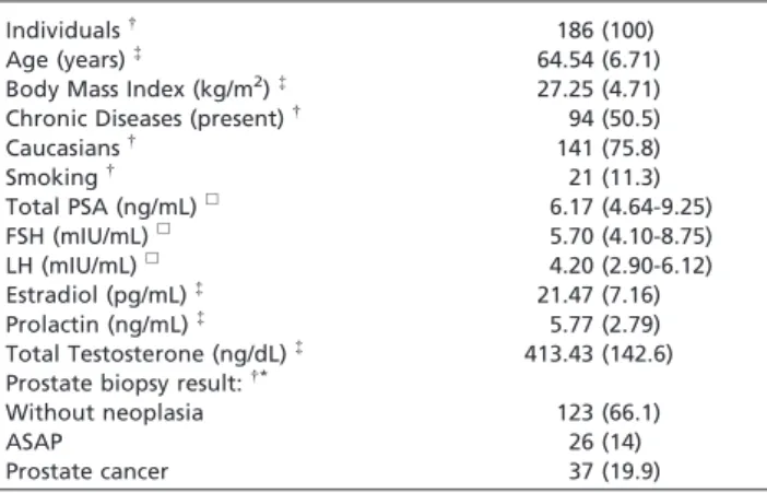
Table 1 -General characteristics of the study population.
Individuals{
186 (100)
Age (years){ 64.54 (6.71)
Body Mass Index (kg/m2){
27.25 (4.71)
Chronic Diseases (present){ 94 (50.5)
Caucasians{
141 (75.8)
Smoking{ 21 (11.3)
Total PSA (ng/mL)h 6.17 (4.64-9.25)
FSH (mIU/mL)h 5.70 (4.10-8.75)
LH (mIU/mL)h 4.20 (2.90-6.12)
Estradiol (pg/mL){
21.47 (7.16) Prolactin (ng/mL){
5.77 (2.79) Total Testosterone (ng/dL){
413.43 (142.6) Prostate biopsy result:{*
Without neoplasia 123 (66.1)
ASAP 26 (14)
Prostate cancer 37 (19.9)
{
: N and %;
{
: Mean and standard deviation;
h: Median (percentile 25-75)
*: Results before the second biopsy of the first ASAP
ASAP: atypical small acinar proliferation
Table 2 -Men categorized according to the prostate biopsy result: without neoplasia, ASAP and prostate cancer.
Prostate biopsy
Without neoplasia ASAP Prostate cancer Pvalue
Individuals{ 123 (66.1) 18 (9.7) 45 (24.2)
Age (years){
64.3¡6.6 66.2¡5.5 64.0¡7.6 0.37
Caucasians{ 94 (77.7) 11 (69.2) 36 (78.4) 0.36
BMI (kg/m2){
27.2¡4.2 26.6¡6.2 27.7¡4.9 0.66
Chronic disease{(present) 67 (54.5) 7 (38.5) 20 (45.9) 0.27
Smoking{
15 (12.2) 2 (11.5) 4 (8.1) 0.79
Total PSA (ng/mL)h 6.05 (4.58-9.06) 5.63 (4.36-7.42) 6.63 (5.10-10.95) 0.22
{: N (%); {
: Mean¡standard deviation;
h: Median (percentile 25-75)
ASAP: atypical small acinar proliferation
Table 3 -Serum total testosterone (TT), follicle stimulant hormone (FSH), luteinizing hormone (LH), prolactin and estradiol levels in men diagnosed by prostate biopsy as without neoplasia, or ASAP or prostate cancer.
Prostate biopsy
Without neoplasia (N = 123) ASAP (N = 18) Prostate cancer (N = 45) Pvalue
TT (ng/mL){ 414.14 (387.84-440.44) 421.17 (346.95-495.38) 408.40 (369.63-447.17) 0.95
FSH (mIU/mL)h 6.00 (3.90-8.50) 5.60 (4.30-12.07) 5.20 (4.20-8.65) 0.86
LH (mIU/mL)h 4.10 (2.70-6.00) 4.65 (2.72-5.45) 4.50 (3.35-6.00) 0.58
Prolactin (ng/mL){
5.62 (5.21-6.04) 6.04 (3.72-8.37) 6.07 (5.17-6.98) 0.59
Estradiol (pg/mL){
21.06 (19.83-22.28) 20.79 (16.36-25.22) 22.86 (20.69-25.03) 0.32
{
: Mean¡standard deviation;
h: Median (percentile 25-75)
significantly among men without prostate cancer, ASAP or with prostate cancer. Importantly the mean TT serum level in men with negative prostate biopsy was similar to those individuals diagnosed with ASAP or prostate cancer. Furthermore, the other hormones evaluated as FSH, LH, prolactin and estradiol, also did not differ between the three studied groups. In relation to the Gleason score of the prostate cancer group, no significant differences could be established regarding the mean serum levels of the hormones of hypothalamic-pituitary-testicular axis, includ-ing total testosterone. In addition, there was no statistically significant difference in the prevalence of hypogonadal serum levels of TT (considered as less than 300 ng/dL) between the study groups.
Recently, several authors12-14have directed their attention trying to understand the relationship between total testoster-one serum levels and prostate cancer. Epidemiological studies have presented controversial results, with some showing a positive relationship between testosterone levels and a higher risk of prostate cancer,15but most demonstrat-ing that this relationship could not be verified conclusively or was negative.16 More recently, a pooled database covering 18 prospective studies,17 published by the Endogenous Hormones and Prostate Collaborative Group, which enrolled 3886 men with prostate cancer and 6438 controls, was unable to demonstrate a significant relationship between serum TT and free testosterone levels, or its metabolites, or even serum levels of other androgens, like dehydroepiandrosterone sulfate and androstenedione, and a higher risk of prostate cancer, even after they were grouped by quintiles of serum concentrations. In this same study, the Gleason score of prostate cancer cases as well as tumor stage did not correlate with either low or elevated serum levels of androgens or their metabolites. It is interesting that this pooled database included 95% of the available data in the world regarding this topic. Based in this context, the current study data suggest, even with a modest number of men, an inconsistent relationship between serum testosterone levels and prostate cancer diagnosed in a clinical setting. The broad variation in TT serum levels could partially explain the lack of difference observed in our study. Although, even when men were classified as having normal or subnormal TT level using a standard cutoff point (300 ng/dL) from the literature,10the histological findings were similar between groups.
However, recent new studies18-20 have demonstrated a positive correlation between men with more undifferen-tiated prostate cancer with subnormal TT serum levels, the
results of the current analysis are consistent with data from other studies,21,22which demonstrates that this correlation is not established and most likely depends on the patients characteristics, as well as the technique used to measure serum TT, the type of analysis performed and whether or not potential complicating factors were eliminated from the analysis. Also, it is interesting to observe that the majority of these studies are based in retrospective analyses which has several methodological limitations.
The relationship between hypothalamic-pituitary-testicular axis hormonal levels and prostate cancer was initially suggested by Miller and colleagues23 and, subse-quently, by Madersbacher and colleagues.24Hypothetically, the tumor or a tumor cell product could inhibit androgen production, thereby suppressing the release of hypothala-mic-pituitary hormones and, consequently, testicular ster-oidogenesis. Though clinical studies have demonstrated that removing prostate cancer results in an elevation of FSH, LH and testosterone, the mechanism of this response remains to be clarified.23,24 In the current study, we did not find that hypothalamic-pituitary-testicular axis hormo-nal levels were different independent of the diagnosis, even with ASAP or prostate cancer.
Perhaps the most important contribution of the present study is the inclusion of men with the diagnosis of ASAP, a condition frequently correlated to a concomitant or a sub-sequent prostate cancer diagnosis.9It is well established that ASAP is a histological prostate alteration that not fill out the characteristics for the diagnosis of prostate cancer. A study in which men diagnosed with ASAP had a radical prostatectomy revealed that all 25 (100%) patients had a final histological diagnosis of prostate cancer from the surgical specimen.25For researchers investigating whether prostate cancer is related to higher levels of total testosterone, this is the best population to study since this condition is increasingly considered as a risk factor for prostate cancer. Although, in the current study, serum levels of all hormones of the hypothalamic-pituitary-testicular axis were similar. It is important to point out that there is a lack of data in the literature regarding androgen levels in men with ASAP, which limits discussions or comparisons based in the results of other studies.
Certainly the relationship between prostate cancer and testosterone is complex and requires further investigation. The hypothesis that higher serum levels of testosterone increases prostate cancer risk was originated in the observa-tion that established prostate cancer regresses after surgical or medical castration.4 However, this occurs in advanced
Table 4 -Serum total prostatic specific antigen (PSA) and hypothalamic-pituitary-testicular axis hormones levels in men with prostate cancer, categorized by Gleason score.
Gleason score
#6 7 $8 Pvalue
N (%) 18 (40) 19 (42.3) 8 (17.7)
Total PSA (ng/mL)h 5.78 (4.61-6.47) 7.35 (6.16-16.85) 15.32* (10.00-21.90) 0.01
TT (ng/mL){
393.56 (333.36-453.75) 431.74 (358.39-505.08) 386.38 (316.61-456.14) 0.59
FSH (mIU/mL)h 8.33 (3.52-13.14) 7.82 (4.89-10.75) 9.23 (1.84-16.62) 0.91
LH (mIU/mL)h 8.66 (0.59-16.74) 5.18 (3.87-6.49) 4.30 (3.29-5.30) 0.49
Estradiol (pg/mL){ 21.61 (18.36-24.87) 22.84 (19.71-25.98) 25.70 (17.31-34.10) 0.42
Prolactin (ng/mL){
6.06 (4.92-7.20) 6.47 (4.54-8.40) 5.15 (3.94-6.36) 0.59
{
: Mean¡standard deviation;
tumors and may not correspond to lower stage prostate tumors, especially when studying the etiology of the prostate cancer. Recently, in an elegant publication, Morgentaler and Traish26suggested The Saturation Theory as explanation for some of the controversial data regarding the issue of prostate and testosterone levels. Based in this theory, testosterone serum levels just over the levels of a castrated condition would have the maximum possible androgen effect, since all the prostate cell receptors would be occupied by androgens or their metabolites. Also, this theory would differentiate the effects of testosterone based on the individual androgen status. In the clinical field, this theory also could be a reasonable explanation for the negative results of the current data as well as for the majority epidemiological studies, especially those with negative results regarding testosterone and risk of prostate cancer.26
Maybe one of the most important complicating factor of all studies published in literature which tried to establish some relationship regarding androgens and prostate cancer, includ-ing the current study, is the fact that TT serum levels do not necessarily reflect the androgenic activity inside the prostate gland. Certainly, the most important factor related to this observation is the fact that dihydrotestosterone, the active metabolite of the testosterone, is converted in the prostate tissue, and not in the bloodstream. The direct observation of this metabolic aspect is the fact that serum levels of androgens may not correlate with the active hormonal environment inside the prostate gland. Furthermore, natural variations in the production of hormones on a daily basis, as well as variation in serum hormone level measurement techniques between laboratories, should be also considered.
Other limitations of the present study need to be considered. The most relevant limitations are the number of patients and the single measurement of the different hormones. A single dosage of serum testosterone may not represent properly the hormone exposure of that person during his life.27However, well-defined inclusion criteria, as well as selection of cases based on the usually recommended indication of prostate biopsy and the prospective nature of the study as well as including individuals with normal biopsies, should reduce potential selection bias. Other authors18,21included free or bioavailable testosterone levels in their studies regarding this issue. Unfortunately, in the current study these analyses were not included as explained initially. Although, based in the Endogenous Hormones and Prostate Collaborative Group publication17and also in the observation that the study population presented similar characteristics regarding factors that may influence testoster-one levels we would not expect significant different results.
In conclusion, our study did not demonstrate different serum levels of TT, FSH, LH, prolactin and estradiol in men with prostate biopsy without neoplasia, ASAP or prostate cancer.
REFERENCES
1. Gronberg H. Prostate cancer epidemiology. Lancet. 2003;361:859-64, doi: 10.1016/S0140-6736(03)12713-4.
2. National Institute of Cancer – Health Department/Brazil. Available from: URL: http://www.inca.gov.br/estimativa/2005
3. Reiter RE, deKernion JB. Epidemiology, etiology, and prevention of prostate cancer. In: Walsh PC, editor. Campbell’s Urology. 8th ed. Philadelphia: Saunders; 2002. p. 3003.
4. Huggins C, Hodges CV. Studies on prostate cancer. I. The effect of castration, of estrogen and androgen injection on serum phosphatases in metastatic carcinoma of the prostate. Cancer Res. 1941;1:293-7.
5. Thompson IM, Goodman PJ, Tangen CM, Lucia MS, Miller GJ, Ford LG, et al. The influence of finasteride on the development of prostate cancer. N Engl J Med. 2003;349:215-24, doi: 10.1056/NEJMoa030660.
6. Massengill JC, Sun L, Moul JW, Wu H, McLeod DG, Amling C, et al. Pretreatment total testosterone level predicts pathological stage in patients with localized prostate cancer treated with radical prostatect-omy. J Urol. 2003;169:1670-6, doi: 10.1097/01.ju.0000062674.43964.d0. 7. Morgentaler A, Bruning CO 3rd, DeWolf WC. Occult prostate cancer in
men with low serum testosterone levels. JAMA. 1996;276:1904-6, doi: 10. 1001/jama.276.23.1904.
8. Yamamoto S, Yonese J, Kawakami S, Ohkubo Y, Tatokoro M, et al. Preoperative serum testosterone levels as an independent predictor of treatment failure following radical prostatectomy. Eur Urol. 2007;52:696-701, doi: 10.1016/j.eururo.2007.03.052.
9. Leite KR, Mitteldorf CA, Camara-Lopes LH. Repeat prostate biopsies following diagnoses of prostate intraepithelial neoplasia and atypical small gland proliferation. Int Braz J Urol. mar-apr 2005;31:131-6, doi: 10. 1590/S1677-55382005000200007.
10. Nieschag E, Swerdloff R, Behre HM, Gooren LJ, Kaufman JM, Legros JJ, et al. International Society of Andrology (ISA), International Society for the study of the aging male (ISSAM) European Association of Urology (EAU). Investigation, Treatment And Monitoring Of Late-Onset Hypogonadism In Males ISA, ISSAM, And EAU Recommendations. Eur Urol. 2005;48:1-4, doi: 10.1016/j.eururo.2005.04.027.
11. Carter HB, Partin AW. Diagnosis and Staging of Prostate Cancer. In: Walsh PC, editor. Campbell’s Urology. 8th ed. Philadelphia: Saunders; 2002. p. 3055.
12. Barret-Connor E, Garland C, McPhillips JB, Khaw KT, Wingard DL. A prospective, population based study of androstenedione, estrogens and prostate cancer. Cancer Res. 1990;50:169.
13. Isom-Batz G, Bianco FJ Jr, Kattan MW, Mulhall JP, Lilja H, Eastham JA. Testosterone as a predictor of pathological stage in clinically localized prostate cancer. J Urol. 2005;173:1935-7, doi: 10.1097/01.ju.0000158040. 33531.e7.
14. Chodak GW, Vogelzang NJ, Caplan RJ, Soloway M, Smith JA. Independent prognostic factors in patients with metastatic (stage D2) prostate cancer. The Zoladex Study Group. JAMA. 1991;265:618, doi: 10. 1001/jama.265.5.618.
15. Gann PH, Hennekens CH, Ma J, Longcope C, Stampfer MJ. Prospective study of sex hormone levels and risk of prostate cancer. J Natl Cancer Inst. 1996;88:1118-26, doi: 10.1093/jnci/88.16.1118.
16. Prehn RT. On the prevention and therapy of prostatic cancer by androgen administration. Cancer Res. 1999;59:4161.
17. Endogenous Hormones, , Prostate Cancer Collaborative Group, Roddam AW, Allen NE, Appleby P, Key TJ. Endogenous sex hormones and prostate cancer: A collaborative analysis of 18 prospective studies. JNCI. 2008;100:170-83.
18. San Francisco IF, Regan MM, DeWolf WC, Olumi AF. Low age adjusted free testosterone levels correlate with poorly differentiated prostate cancer. J Urol. 2006;175:1341-5, doi: 10.1016/S0022-5347(05)00680-4. 19. Lane BR, Stephenson AJ, Magi-Gallazzi C, Lakin MM, Klein EA. Low
testosterone and risk of biochemical recurrence and poorly differentiated prostate cancer at radical prostatectomy. Urology. 2008;72:1240-5, doi: 10. 1016/j.urology.2008.06.001.
20. Schatzl G, Madersbacher S, Thu¨rridl T, Waldmu¨ller J, Kramer G, Haitel A, et al. High-grade prostate cancer is associated with low serum testosterone levels. Prostate. 2001;47:52-8, doi: 10.1002/pros.1046. 21. Morgentaler A, Rhoden EL. Prevalence of prostate cancer among
hypogonadal men with prostate-specific antigen levels of 4.0 ng/dL or less. Urology. 2006;68:1263-7, doi: 10.1016/j.urology.2006.08.1058. 22. Curran MJ, Bihrle W, 3rd. Dramatic rise in prostate-specific antigen after
androgen replacement in a hypogonadal man with occult adenocarci-noma of the prostate. Urology. 1999;53:423-4, doi: 10.1016/S0090-4295 (98)00348-3.
23. Miller LR, Partin AW, Chan DW, Bruzek DJ, Dobs AS, Epstein JI, et al. Influence of radical prostatectomy on serum hormone levels. J Urol. 1998;160:449-53, doi: 10.1016/S0022-5347(01)62922-7.
24. Madersbacher S, Schatzl G, Bieglmayer C, Reiter WJ, Gassner C, Berger P, et al. Impact of radical prostatectomy and TURP on the hypothalamic-pituitary-gonadal hormone axis. Urology. 2002;60:869-74, doi: 10.1016/ S0090-4295(02)01893-9.
25. Brausi M, Castagnetti G, Dotti A, De Luca G, Olmi R, Cesinaro AM. Immediate radical prostatectomy in patients with atypical small acinar proliferation. Over treatment? J Urol. 2004;172:906-9.
26. Morgentaler A, Traish AM. Shifting the paradigm of testosterone and prostate cancer: the saturation model and the limits of androgen-dependent growth. Eur Urol. 2009;55:310-20, doi: 10.1016/j.eururo.2008. 09.024.