Dual effect of silymarin on experimental non-alcoholic steatohepatitis
induced by irinotecan
Eudmar Marcolino Assis-Júnior
a, Anielle Torres Melo
a, Venúcia Bruna Magalhães Pereira
a,
Deysi Viviana Tenazoa Wong
b, Nathalia Ribeiro Pinho Sousa
a, Christiane Mendes Gonçalves Oliveira
a,
Lara Raissa Cavalcante Malveira
a, Leonardo Silva Moreira
a, Marcellus Henrique Loiola Ponte Souza
c,
Paulo Roberto Carvalho Almeida
b, Roberto César Pereira Lima-Júnior
a,⁎
aDepartment of Physiology and Pharmacology, Faculty of Medicine, Federal University of Ceará, Brazil bDepartment of Pathology and Forensic Medicine, Faculty of Medicine, Federal University of Ceará, Brazil cDepartment of Clinical Medicine, Faculty of Medicine, Federal University of Ceará, Brazil
a b s t r a c t
a r t i c l e
i n f o
Article history:
Received 23 January 2017 Revised 20 April 2017 Accepted 23 April 2017 Available online 26 April 2017
Irinotecan-based regimens are commonly used for treatment of colorectal cancer, which is limited by mucositis and non-alcoholic steatohepatitis (NASH). Silymarin (SIL) prevents fatty liver disease in the clinical setting and in models of liver damage induced chemically. This study investigated the possible effect of SIL on irinotecan (IRI)-induced NASH. Swiss female mice were injected with saline (SAL 5 ml/kg i.p.), IRI (50 mg/kg i.p.), SIL (150 mg/kg p.o.) or IRI (50 mg/kg i.p.) + (SIL 1.5, 15 or 150 mg/kg p.o.) thrice/week/7 weeks. On the seventh week, blood samples were collected for transaminases assay and livers were collected for histopathology, measurement of the total lipids, malondyadehyde (MDA), non-protein sulfhydryl groups (NPSH), cytokines (IL-1β, IL 6 and IL-10), 3-nitrotyrosine (N-Tyr) and toll-like receptor 4 (TLR4) immunoexpression, quantification of NF-kB,α -smooth muscle actin (α-SMA), andEscherichia coli16S rRNA gene (RRS) expression. IRI increased liver transam-inases, neutrophil infiltration, lipid accumulation, MDA, IL-1βand IL-6 levels, N-Tyr and TLR4 immunostaining, NF-kB,α-SMA expression and RRS versus the SAL group (pb0.05). Additionally, SIL (1.5 mg/kg) improved these parameters (pb0.05), except neutrophil infiltration and RSS versus the IRI group. Furthermore, the SIL (15 mg/kg) only improved the inflammatory parameters, the expression ofα-SMA and RRS versus the IRI group (pb0.05). The higher dose of SIL (150 mg/kg) was even more deleterious than the intermediate dose. Therefore, silymarin showed a dual effect on liver damage induced by IRI. Hepatoprotection seems to involve the inhibition of oxidative stress and protein nitrosylation, preventing activation of hepaticfibrosis mechanisms. © 2017 Elsevier Inc. All rights reserved. Keywords:
Irinotecan Steatohepatitis Silymarin
Cancer chemotherapy toxicity Inflammation
Liver
1. Introduction
Colorectal Cancer (CRC) is the third most common type of cancer among men (10% of total cases) and the second among women (9.2% of total cases). Although the majority of cases (55%) occur in developed regions, the highest mortality rates occur in less developed regions (52% of deaths) (WHO-World Health Organization, 2004).
The main site of CRC metastasis is the liver in about 30% of patients (Kuvshinov and Fong, 2007). Clinical management of metastatic CRC in-volves tumor resection (Costa et al., 2014). Non-resectable tumors can become resectable by the use of conversion therapy, which is mainly based on irinotecan or oxaliplatin combined with 5-Fluorouracil
(Bismuth et al., 1996; Adam et al., 2004; Pozzo et al., 2004). However, irinotecan-based regimens are related to limiting side effects such as in-testinal mucositis (IM) (Ikuno et al., 1995; Freitas, 2007). In addition, about 1 in 12 patients undergoing irinotecan treatment develop non-al-coholic steatohepatitis (NASH), representing a 3.45-fold increased risk for those under chemotherapy treatment (Robinson et al., 2012), which might limit metastasectomy. Histopathological damage criteria were established byKleiner et al. (2005), who consider steatosis, lobular inflammation and vacuolization as important parameters for NASH di-agnosis, whilefibrosis and portal infiltration are considered indicators of severe disease (Kleiner et al., 2005). Costa and co-workers developed an experimental model of irinotecan-related NASH characterized by ox-idative stress, inflammatory infiltrate, lipid accumulation,fibrosis and hepatic failure (Costa et al., 2014).
Silymarin is a naturally-occuring, polyphenol-rich antioxidant, which acts as a free radical scavenger (Fraschini et al., 2002). It is report-ed that silymarin reversibly inhibits P-450 cytochrome enzymes ⁎ Corresponding author at: Departamento de Fisiologia e Farmacologia, Faculdade de
Medicina, Universidade Federal do Ceará, Rua Cel Nunes de Melo, 1127, Rodolfo Teófilo, 60430-270 Fortaleza, Ceará, Brazil.
E-mail address:robertocesar@ufc.br(R.C.P. Lima-Júnior).
http://dx.doi.org/10.1016/j.taap.2017.04.023
0041-008X/© 2017 Elsevier Inc. All rights reserved.
Contents lists available atScienceDirect
Toxicology and Applied Pharmacology
(Fraschini et al., 2002), stabilizes mitochondrial (Bindoli et al., 1997) and microsomal membranes (Trouillas et al., 2008). Polyak et al. (2007)andKim et al. (2013)reported anti-inflammatory and immuno-modulatory effects of silymarin with the involvement of NF-κB inhibi-tion (Lee et al., 2013) and over-expression of immunomodulators such as IL-10 and IFN-γ(Wilasrusmee et al., 2002). In addition,Täger et al.
(2001)reported that silymarin treatment over-expressed the endoge-nous IL-1βinhibitor (IL-1ra). However, the effects of silymarin vary ac-cording to the dose and route of administration.
Silymarin hepatic protection has been widely demonstrated in ani-mal models and in the clinical setting (Cacciapuoti et al., 2013; Zhang et al., 2013; Trouillas et al., 2008; Chen et al., 2012). It is effective in the treatment of obesity-related NASH (Cacciapuoti et al., 2013), pre-vents the establishment of alcoholic steatohepatitis (Zhang et al., 2013), and protects against chemically-induced liver damage andfi bro-sis by carbon tetrachloride (Trouillas et al., 2008) and thioacetamide (Chen et al., 2012).
In the present study we aimed to evaluate the effects of silymarin on irinotecan-induced non-alcoholic steatohepatitis.
2. Materials and methods
2.1. Chemicals and antibodies
Irinotecan hydrochloride (Evoterin®, Evolabis, São Paulo, Brazil, vial of 100 mg); Silymarin S0292 (Sigma-Aldrich, MO, USA); Monoclonal primary antibody rabbit anti-nitrotyrosine (Life Technology, NY, USA); Monoclonal primary antibody rabbit anti TLR-4 (Life Technology, NY, USA); Polyclonal primary antibody anti-NF-kB NLS rabbit (Santa Cruz Biotechnology, CA, USA); Polyclonal primary antibody Rabbit Anti-Actinβ(Santa Cruz Biotechnology, CA, USA); Primary Antibody Mono-clonal Goat Anti-α-SMA (Sigma-Aldrich, MO, USA); Secondary antibody linked to alkaline phosphatase anti-rabbit IgG (Sigma Aldrich, MO, USA); Secondary antibody linked to alkaline phosphatase anti-goat IgG (Sigma-Aldrich, MO, USA); ImunoCruz - Anti-rabbit ABC kit (Santa Cruz Biotechnology, CA, USA).
2.2. Animals
Swiss mice, weighing between 20 and 25 g, and obtained from the animal facility at Federal University of Ceará were used. The animals had free access to drinking water and food and were kept in a controlled temperature (22 ± 2 °C) environment with a light-dark cycle (12 h/12 h). All animal care and experimental procedures complied with the laboratory animal care and use principles outlined by the Na-tional Institutes of Health (NIH publication no. 85–23, revised 1985) and were approved by the local Ethics in Animal Research Committee (CEPA) of the Federal University of Ceará (Protocol number 21/12).
2.3. Experimental design
The animals were divided into six experimental groups (n = 8–10): group 1 SAL: Saline 5 ml/kg, p.o. 1 h before saline 5 ml/kg i.p; group 2 -SIL: Silymarin 150 mg/kg, p.o. 1 h before saline 5 ml/kg i.p.; group 3 - IRI: Saline 5 ml/kg, p.o. 1 h before irinotecan 50 mg/kg, i.p.; group 4 - S1.5: Silymarin 1.5 mg/kg p.o. 1 h before irinotecan 50 mg/kg i.p.; group 5 -S15: Silymarin 15 mg/kg p.o. 1 h before irinotecan 50 mg/kg i.p.; group 6 - S150: Silymarin 150 mg/kg p.o. 1 h before irinotecan 50 mg/kg i.p. The animals were injected three times a week on alternate days. On the sixth day of each week the animals received silymarin or saline reinforcement. This schedule of treatment was continued for seven consecutive weeks. The survival rate (%) of the animals was recorded.
2.4. Serum concentration of liver transaminases
The mice were anaesthetised using 2.5% tribromo-ethanol solution (10 ml/kg, i.p.) for blood sample collection at the retro orbital plexus. After centrifugation (at 100 ×gfor 10 min), the plasma was obtained for alanine and aspartate aminotransferase (ALT and AST) biochemical analysis following the manufacture's guidebook (Labtest Diagnostica commercial kits, Minas Gerais, Brazil).
2.5. Histopathological analysis
Liver samples were fixed in 10% formalin buffered solution, dehydrated and embedded in paraffin. Microtome sections (5 mm) were stained using hematoxylin-eosin (H&E) and mounted on 24 mm2coverslips, to be examined by optical microscopy (×100). The slides were analysed by an experienced pathologist who was unaware of treatment and group sorting. NASH diagnosis and graduation were made using NASH Activity Score (NAS), as proposed byKleiner et al. (2005). The concomitant presence of the following three histological parameters is necessary to diagnose NASH: steatosis (b5% score 0; 5– 33% score 1; 33–36% score 2;N66% score 3); lobular inflammation (no infiltrated byfield score 0, mild infiltration score 1; moderate infi ltra-tion score 2; intense infiltration score 3) and hepatocyte ballooning (Score 0 = none, score 1 = mild ballooning, score 2 = severe ballooning).
In addition, neutrophil infiltration sites in all histopathologic slides were counted blind by two independent collaborators (10fields). The firstfield choice was random; the followingfields were contiguous to the previousfield.
2.6. Liver lipid contents
Lipid content was assayed using theBligh and Dyer (1959)method. Briefly, samples were homogenized in chloroform:methanol (1:2), 1.9 ml per 100 mg tissue sample, before adding more chloroform (0.63 ml) and stirring. Distilled water (0.63 ml) was added to reach a final proportion of 2:2:1 chloroform:methanol:water. The mixture was centrifuged at room temperature, 200 ×gfor 2 min, obtaining a two-phase system (polar above, nonpolar below). The lower phase was placed into a tare tube and evaporated at 100 °C. Total lipid was expressed in mg of lipids/g of liver.
2.7. Malondialdehyde assay (MDA)
Liver samples were homogenized in 1.15% KCl solution (10% homog-enate). A homogenate aliquot (500μl), placed in a test tube, was mixed with 3 ml 1% H3PO4 and 0.6 ml 1% thiobarbituric acid (TBA). This mix-ture was placed in a water bath (45 min) and cooled in ice, n-butanol (4 ml) was added, and the mixture was vortexed (1 min) and centri-fuged at 1200gfor 15 min. The butanolic fraction was placed in a cuvette and read in spectrophotometer at 520 and 535 mm and the difference between the two readings was used to calculate the MDA, based on the molar extinction coefficient (13,700 M/cm) (Mihara and Uchiyama, 1978).
2.8. Non-protein sulfhydryl groups (NP-SH)
using linear regression and is expressed in NP-SH/tissue (mg/500 mg) (Boyd et al., 1979).
2.9. Detection of cytokines IL-1β, IL-6 and IL-10 by ELISA
IL-1β, IL-6 and IL-10 levels were assayed by ELISA (Cunha et al.,
1993). The liver samples were homogenized (Safieh-Garabedian et al., 1995). A 96-well plate was coated with an antibody against the desired interleukin (4μg/ml, DuoSet ELISA Development kit R&D Systems) overnight at 4 °C. After blocking unspecific binds with BSA, the sample and standard were loaded in duplicate and incubated at 4 °C for 2 h (Adam et al., 2004). The plates were washed 3 times with PBS-Tween 20, 0.1% v/v (washing step). After washing, specific detection biotinylat-ed antibody (dilutbiotinylat-ed 1:1000 with assay buffer 1% BSA, R&D System, USA) was added to the wells, followed by another 2 h incubation at 4 °C. The plates were washed as before, and 100μl of streptavidin-HRP di-luted 1:200 were added, followed by 100μl of substrate solution (1:1 mixture of H2O2and tetramethylbenzidine; R&D System, USA), the plate was then incubated in the dark at room temperature for 20 min. The reaction was stopped by addition of 2 N H2SO4, and the absorbance was measured at 450 nm. The results are expressed in pg/g of tissue and reported as the mean ± SEM.
2.10. Immunohistochemistry
Immunohistochemical analyses of the liver samples for iNOS, N-Tyr and TLR4 were performed as follows: Histological specimens were deparaffinised and then hydrated. After hydration, the slides were sub-merged in citrate buffer (pH 6.0), and heated at 100 °C for 15 min for an-tigen recovery. After cooling to room temperature, the slides were washed in PBS three times. The slides were submerged in 3% hydrogen peroxide to block endogenous peroxidases and washed again. The pri-mary antibody was incubated for 12 h (rabbit anti-iNOS antibodySanta Cruz Biotechnology, antibodySanta Cruz, CA, antiNTyr and AntiTLR4 -Life Technology, -Life®, NY), and diluted in either PBS - BSA 5% ratio 1: 400 (iNOS and TLR4) or 1: 800 (N-Tyr). The slides were washed again, before incubation of the biotinylated secondary antibody (30 min, room temperature), anti-rabbit IgG diluted in PBS-5% BSA at a ratio of 1:200. Following further washing the slides were incubated with ABC complex (30 min) (horseradish peroxidase Standard). The slides were finally washed as before, and DAB/peroxide (2 min) was used to colour the proteins, the DAB/peroxide stain confreres a characteristic brown colour to the positive cells. Harry's hematoxylin was used as a contrast stain, before rinsing the slides in distilled water and dehydrating with a graded series of alcohol and xylene, and mounting. Quantitative reac-tion analysis, following Yeoh et al. (2005) parameters, was carried out by a pathologist using a light microscope. TLR4 positive cells were counted in ten microscopefields in each sample, similarly, N-tyr posi-tive areas were quantified in tenfields of each sample using the image processing software Fiji (Schindelin et al., 2012).
2.11. Quantitative real-time PCR (qPCR)
To assay enteric bacteria translocation, liver samples were collected in sterile conditions and homogenized in Trisol buffer for DNA extrac-tion using the Qiagen kit according to the manufacturer's specifications. The polymerase chain reaction was performed as described below:
E. coli16S rRNA gene (RRS), associated withEscherichia colidetection, was detected using IQ5 Real-Time PCR Detection System (Bio-Rad Labora-tories, USA) (Quetz et al., 2012). The housekeeping gene used was PPIA peptidylprolyl isomerase A (Tatsumi et al., 2008). The primer pairs for mouse were as follows: 16SrRNA (Forward −5′ -GTGGAAAAACCTCGTCCAGA-3′; Reverse: 5′ -GCTCGGCTTCCAGTAFFGAG-3′) and PPIA (Forward: 5′-GACATGGAGAAGATCTGGCA-3′; Reverse: 5′ -GGTCTTTACGGATGTCAACG-3′). The conditions used were: for 16SrRNA, 95 °C-20″, 63 °C-20″and 72 °C-45″and for PPIA, 95 °C-20″58 °C-20″
and 72 °C-45″. iQ SYBR Green Supermix (10 uL) (Bio-Rad, San Diego, CA, USA), each primer (2μl) (0.2μM), cDNA samples (3μl) and nu-clease-free water to afinal volume of 20μl, were used in the reaction. All amplifications resulted in a melting curve, used to ensure the am-plification specificity and to detect primer-dimer formation or other non-specific product. Tested genes' quantification cycles (Cq) were exported to Microsoft Excel (Microsoft, USA) and the RSS relative gene expression levels were calculated according to 2-ΔΔCT methodology, whereΔΔCT = (CT Target Gene - CT housekeeping gene in the treated group)/(target gene CT - CT of the housekeeping gene control), with PPIA as the housekeeping gene (Livak and Schmittgen, 2001).
2.12. Western blot
Animals' livers were homogenized in RIPA buffer (50 mM Tris; 150 mM NaCl; 1 mM EDTA; 1% Triton) and centrifuged at 1500 g for 10 min, before their supernatants were separated into appropriately la-belled Eppendorf tubes. The total protein in each sample was assayed using a BioRad® Kit, following the manufacturer's instructions. Equal amounts of total protein were loaded onto a SDS-PAGE gel and run at 150 V. The proteins were then transferred to a nitrocellulose membrane using the BioRad system. The membrane was incubated in 5% skimmed milk (NESTLÉ®) in TBS-T buffer (Tris-NaCl with 1% Tween 20) for 2 h. Afterwards, the membranes were washed 3 times (10 min each) with TBS-T buffer. Next, they were incubated with the respective primary an-tibodies (anti-NF-kB NLS rabbit (1:600); Rabbit Anti-Actinβ(1:5000) -Santa Cruz Biotechnology, CA, USA; Goat Anti-α-SMA (1:600) - Sigma-Aldrich, MO, USA), and diluted in 5% BSA-TBS buffer (Tris-NaCl). The in-cubation was carried out over-night at 4 °C with gentle agitation. The membranes were then washed as described above and incubated with secondary antibodies (alkaline phosphatase anti-rabbit IgG (1:1000); alkaline phosphatase anti-goat IgG (1:1000) - Sigma-Aldrich, MO, USA), diluted in 5% skimmed milk-TBS-T for 2 h. Later, the membranes were washed again and developed using the alkaline-phosphatase sub-tract BCIP®/NBT (Sigma Aldrich).
2.13. Statistical analysis
Data are expressed as the mean ± standard error of the mean (SEM), except for the histopathological scores which are reported as median values (minimum–maximum). Data were analysed using one-way ANOVAs followed by the Newman-Keuls's test or the Kruskal–Wallis test, followed by Dunn's test, as appropriate. The survival curve was expressed as a percentage of surviving animals and the log-rank (Man-tel–Cox) test was used to determine differences between survival curves. The level of statistical significance was set at 5% (pb0.05).
3. Results
3.1. Clinical-like aspects of irinotecan-induced-NASH and liver functional parameters
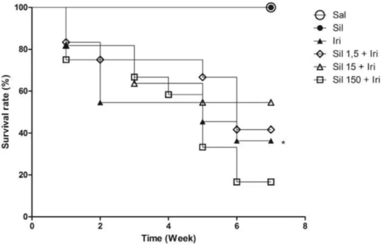
The treatment with irinotecan caused higher mortality versus saline group (Fig. 1). While the intermediate and low doses of silymarin did not interfere with mortality, the highest dose of silymarin increased the death ratio to nearly 100% and therefore this dose was not used for subsequent assays.
Irinotecan significantly (pb0.05) increased serum transaminase
The lower dose of silymarin significantly improved almost all liver function markers (Fig. 2A, B and D), except the neutrophil infiltrate (Fig. 2C). In this group, the liver parenchyma was unharmed, with ob-servable hepatocyte cord arrangement and sinusoids with preserved ar-chitecture (Fig. 3). The intermediate dose presented large and diffused histopathologic damage, with necrotic areas and compromised architecture (Fig. 3). This intermediate dose promoted higher serum concentration of transaminases (Fig. 2A) and greater expression ofα
-SMA (Fig. 2D), but reduced the neutrophil infiltration count (Fig. 2C) and lipid accumulation (Fig. 2B). Thesefindings were significantly dif-ferent from irinotecan group (pb0.05).
3.2. Oxidative stress plays a role in pathophysiology but its control is not sufficient to prevent disease progression
To investigate the role of oxidative stress in IRI-related NASH, sam-ples of each group were used in MDA, NPSH and nitrotyrosine (N-Tyr) assays. MDA presence indicates oxidative stress, since this aldehyde is the main the sub-product of the membrane's polyunsaturated fatty acid peroxidation (Zhu et al., 2014). Its presence aggravates the oxida-tion by causing denaturaoxida-tion and membrane peroxidaoxida-tion (Zhang et al., 2013).
Fig. 1.Irinotecan increased mice mortality, and silymarin was not able to attenuate this parameter. The graph represents the weekly increased mice death ratio (Kaplan Meier test). Mantel-Cox log-rank for survival curves were used for comparison. *pb0.05 vs.
saline group (negative control).
Fig. 2.Irinotecan altered all the liver functions markers evaluated: Transaminases levels (A), lipid accumulation (B), neutrophil infiltration (C) andα-SMA expression (D). Silymarin prevented the increase on transaminases levels, lipid accumulation andα-SMA expression only in the 1.5 mg/kg dose, while the 15 mg/kg dose was able to prevent only the
neutrophil infiltration. Data are expressed as means ± SEM (n = 6 per group) and analysed with one-way ANOVA and Newman-Keuls post hoc test. *pb0.05 vs. saline group.#pb
0.05 vs. irinotecan group.
Table 1
Histopathological changes ranked by Kleiner’score. Irinotecan significantly increased total Kliener’score versus saline group. Low doses of silymarin was capable of preventing these
changes. The results are reported as medians ± SEM (n = 5 per group) and analysed using Kruskal-Wallis and Dunn's test.
Groups
Kleiner's score
Lobular inflammation Steatosis Ballonization Total Saline 1 (0–1) 0 (0–1) 0 (0–1) 1 (0–2) Silymarin 0,5 (0–1) 0 (0–1) 0 (0–0) 0,5 (0–2) Irinotecan 1,5 (1–2) 0 (0–1) 1,5 (1–2)⁎ 3 (3–4)⁎
Sil 1,5 1,5 (1–2) 0 (0–1) 0 (0–0)# 2 (1–2)#
Sil 15 1 (1–3) 0 (0–0) 0 (0–2) 1 (1–5)
⁎ pb0.05 vs. saline #p
Another important oxidative stress parameter is the staining for N-Tyr. When peroxynitrite reacts with normal tissue proteins, it modifies tyrosine residues to generate nitrotyrosine. Then, peroxynitrite action can be indirectly detected by N-Tyr staining in the liver tissue (Huie and Padmaja, 1993).
Irinotecan increased (pb0.05) the levels of MDA (Fig. 4A) and N-Tyr
(Fig. 4C), while decreased NP-SH (Fig. 4B) versus the saline injected group. Lower doses of silymarin reduced the MDA accumulation (Fig. 4A) and the N-Tyr staining (Fig. 4C), but increased the total NPSH avail-able (Fig. 4B) in comparison with the irinotecan group. The intermediate doses had similar effects. The only parameter that was not significantly different than the saline group was MDA accumulation (pN0.05).
Repre-sentative images from the immunostaining for N-Tyr show strong stain-ing in the irinotecan group compared to saline-treated group, which was markedly attenuated by all doses of silymarin (Fig. 4D).
3.3. Control of bacteria accumulation in the liver depends on the dose of silymarin and adequate anti-inflammatory and immune-modulatory response
Irinotecan treatment increased the expression of inflammatory markers in the liver when compared to saline-treated animals (pb
0.05).Fig. 5shows that IL-6, IL-1βand NFκB are increased in irinotecan injected mice's livers (Fig. 5A, B and D). Silymarin treatment showed a reduced NFκB (Fig. 5D) and IL-6 (Fig. 5A) expression, but only 1.5 mg/ml doses reduced the expression of IL-1β(Fig. 5B). IL-10 did not change on irinotecan injection, but intermediate doses of silymarin significantly overexpressed this cytokine (Fig. 5C).
3.4. Bacteria seem to contribute to the pathophysiology of irinotecan-relat-ed NASH
RSS-16S gene expression showed the presence of bacteria in the liver of animals treated with irinotecan versus saline injected animals (Fig. 6A). Animals that received a low dose of silymarin, but not those treated with intermediate doses, showed a significantly reduced num-ber of RSS-16S copies compared to the irinotecan group (Fig. 6A). Im-munostaining for TLR4 confirmed our PCR data, which showed that irinotecan enhanced the staining for TLR4 (Fig. 6B) versus the saline group, and that lower doses of silymarin notably reduced it (Fig. 6B). Representative photomicrographs from the immunostaining for TLR4 show a higher number of positively stained cells in the irinotecan group, and the low dose silymarin-treated animals displayed reduced immunoexpression (Fig. 6C).
4. Discussion
This study describes the effect of silymarin on Irinotecan-related NASH. Our results showed that drug has a dual effect, showing preven-tive characteristics in low doses and enhancing liver damage in higher doses. Silymarin alone did not cause toxicity, even in high doses.
The same damage pattern associated with irinotecan injection as de-scribed previously (Costa et al., 2014) was reproduced in this study. Irinotecan injections resulted in higher lipid content and inflammation in the liver, as similarly shown byCosta et al. (2014). According to Costa et al. (2014), irinotecan causes intestinal damage, allowing trans-location of Gram negative bacteria to the liver. Translocating bacteria cause tissue damage associated with inflammation and massive neutro-phil infiltration, oxidative burst with high levels of ROS, and mitochon-dria damage culminating in lipid accumulation and lipid peroxidation, which in turn enhances free radical levels and exacerbates local damage in a vicious cycle. Silymarin pre-treatment reduced the accumulation of fat in the liver of the mice that received irinotecan. The consensus on fatty liver appearance is that lipid accumulation can involve either one, or a combination of three causes: a higher blood concentration of fatty acids due to a fat rich diet or adipocyte break down; greater endog-enous synthesis of fat, or a mitochondrial dysfunction (Chitturi and Farrell, 2001). As found here, there was no difference in dietary condi-tions between groups, or any reason for higher fatty acids synthesis. It is therefore possible that lipid accumulation in the liver occurred due to mitochondrial dysfunction. Previous studies have demonstrated that silymarin prevents mitochondrial dysfunction by stabilising the mi-tochondrial membrane, and improvingfluidity and membrane potential state (Zhu et al., 2014; Trouillas et al., 2008). However, there are no data concerning a direct role of silymarin in lipid metabolism. This supports the assumption that lipid accumulation occurs due to mitochondrial dysfunction.
Low doses of silymarin prevented the appearance of clinical signs highly correlated to NASH (Kleiner et al., 2005; Kleiner and Brunt, 2012) such as lobular inflammation, steatosis and vaccuolation, and also prevented the expression ofα-SMA, a knownfibrosis marker asso-ciated with advanced NASH. On the other hand, higher doses promoted Fig. 3.Silymarin improved microscopic injury in liver during irinotecan induced-NASH.
liver histopathological damage, with increased levels of serum hepatic transaminases and higher expression ofα-SMA.
Both low and high doses of silymarin showed low staining for N-tyr and high levels of NP-SH. Higher doses showed lower neutrophil infi l-trate and lower expression of IL-6, suggesting damage caused directly by bacteria; action on the liver cells. The similarities and differences be-tween groups highlight the importance of a basal oxidation level, as low doses of silymarin prevented oxidative stress, but did not interfered with some inflammatory functions, such as neutrophil accumulation, which is essential to control infections. It is possible to speculate that in-termediate doses of silymarin reduced the neutrophils' oxidative capac-ity and infiltration ability. Thus bacterial infection was not properly handled, resulting in significant liver damage.
Local inflammation is also likely involved in lipid accumulation. Costa et al. (2014)previously showed that irinotecan induces several in-flammatory and oxidative events which precede lipid accumulation in irinotecan-related NASH. In addition, several reports show that infl am-mation inhibits the lipoprotein lipase in hepatocytes (Butterwith and Griffin, 1989). Inflammation can also lead to excessive production of ROS (Abbas and Lichtman, 2011), damaging mitochondrial membranes. This indicates a potential beneficial use for anti-inflammatory drugsfin treating NASH.
Silymarin prevented some inflammatory events, agreeing with liter-ature data that report itsanti-inflammatory and immunomodulatory ef-fects (Fraschini et al., 2002). It was demonstrated that silymarin over-expresses IL-1ra and down-regulates IL-6 (Täger et al., 2001). The groups treated with silymarin showed reduced levels of IL-6 and
NF-κB in comparison to irinotecan. However, the intermediate dose group failed to down-regulate IL-1β. A proper inflammatory response is essen-tial for adequate bacterial clearance, which may explain the liver dam-age observed in the group that received higher doses of silymarin. Additionally, IL-6 shows a role in liver regeneration, so its downregula-tion could compromise damage recovery. Accordingly, IL-6 knockout mice fail to regenerate the liver after hepatectomy due to lack of DNA synthesis (Cressman et al., 1996). IL-6 downregulation could therefore have contributed to the extensive damage observed in animals treated with higher silymarin doses.
Lower IL-6 expression could either be due to reduced nuclear local-ization of NF-κB, (Barnes and Karin, 1997), or negative modulation by anti-inflammatory cytokines such as IL-10.Knolle et al. (1997)showed that exogenous IL-10 interferes with IL-6 expression in a dose-depen-dent manner. Although IL-10 expression is also enhanced by transcrip-tion factor NF-κB, it can be alternatively regulated by cAMP (Opal et al.,
1998). It is well-known that IL-10 is released at later stages of the in-flammatory process as a natural mechanism against exacerbated pro-inflammatory stimuli (Howard et al., 1993). Silymarin is reported to in-crease IL-10 expression (Wilasrusmee et al., 2002), therefore similar mechanisms may be operating in this study.
decreasing the diapedesis rate to the liver (Menezes et al., 2009). This may explain the lower neutrophil infiltration observed in the groups injected with high doses of silymarin.
Oxidative burst inhibition could also explain the liver damage ob-served in the intermediate dose group.Costa et al. (2014)showed that Escherichia coli translocates from the gut to the liver during Fig. 5.Irinotecan injection caused overexpression of IL-6, IL-1βand NF-κB (A, B and D). Lower doses of silymarin reduced all those mediators expression. The middle dose of silymarin was able to reduce IL-6 expression and NF-κB activity, but not IL-1βexpression. The middle dose also increased IL-10 expression (C). Data are expressed as means ± SEM (n = 6 per group) and analysed with one-way ANOVA and Newman-Keuls post hoc test. *pb0.05 vs. saline group.#pb0.05 vs. irinotecan group.
Fig. 6.Irinotecan increased bacterial infection in mice's livers. RSS-16S copy numbers were higher in the irinotecan group when compared to saline. The low dose significantly reduced the copies of this gene (A). Conversely, the number of TLR4+ cell in the irinotecan injected mice and middle dose were higher when compared to saline, the low dose was able to also reduce the number of TLR4+ cell when compared with irinotecan group. In‘C’is possible to see the representative pictures from TLR4 immuno-staining quantified in‘B’, the TLR4 positive cells are
stained in brown. Data are expressed as means ± SEM (n = 6 per group) and analysed with one-way ANOVA and Newman-Keuls post hoc test. *pb0.05 vs. saline group.#pb0.05 vs.
steatohepatitis. Bacterial LPS activates inflammatory response in a TLR4-dependent pathway (Raetz et al., 1991). This pathway acts as thefirst line of defence against infection. However other types of cellu-lar damage are also associated withE. coliinfection.E. coliuses the Type Three Secretion System (T3SSs) to import effectors into the cytoplasm of eukaryotic cells. Those effectors are recognised by mitochondrial as-sociated protein (Map) or by the EspF effectors phosphorylating N-WAS and WAVE-2, leading to cytoskeletal rearrangement, mitochondri-al dysfunction, apoptosis and necrosis (Coburn et al., 2007). Then, bac-terial interaction with liver cells and secretory mechanisms might explain the damage observed in the Sil 15 + Iri group.
The discrepancy between IL-1βbeing reduced at low doses, but not at intermediate doses, could be explained by the involvement of cell processing. IL-1βis produced physiologically as a precursor, pro-IL-1β, which must be cleaved to become an active molecule and be secreted, which explains its relatively fast release following a trigger event, such as bacterial infection (Dinarello, 1998). As it is found pre-formed, the transcription activity of NF-κB would not be essential to IL-1βrelease. That does not exclude the possibility of an alternative pathway through MAP3K/ERK JNK/AP-1, for example (Samavati et al., 2009). In addition, silymarin is reported to supress the IL-1βsignal, but not its expression (Täger et al., 2001).
In the clinical setting, silymarin daily doses range from 280 to 800 mg. Taking into account the difference between the protective ef-fects of low doses of silymarin in oppose to increased risk of liver dam-age with its intermediate and high doses, clinical trials are highly recommended in order to assess the optimal dose to be used in patients to prevent irinotecan-related hepatotoxicity. Then, based on present re-sults, it is reasonable to consider that the therapeutic index of silymarin might be shortened when administered together with irinotecan. How-ever, with the current knowledge of the animal doses in this co-admin-istration scenario (1.5 mg/kg as therapeutic dose and 15 mg/kg as Lowest Observed Adverse Effect Level - LOAEL - in mice), it is impossible to determine a starting dose for the clinical trial with co-administration, based on standard risk management practise. Taken into account the ten-fold uncertainty factor for interspecies extrapolation and ten-fold uncertainty factor for LOAEL to NOAEL (No Observed Adverse Effect Level) extrapolation, the NOAEL in human would be 0.15 mg/kg, much lower than the therapeutic dose of 1.5 mg/kg. According to Blanchard and Smoliga (2015), interspecies dose scaling must be scien-tifically justified using a broader physiologic, pharmacokinetic, and tox-icology data. Then, it might be unethical to dose human at 1.5 mg/kg in co-administration scenario based on the current knowledge.
In summary, the lowest dose of silymarin protected the liver tissue against the damage induced by irinotecan. Silymarin-related protection seems to be mediated by the control of oxidative stress without compromising the innate and adaptive immune functions. Here, we showed the effectiveness of a clinically used drug in an atypical applica-tion. In addition to better understanding the pathophysiology of NASH in-duced by irinotecan, the present study provides new insights on how to modulate the balance of pro- and anti- inflammatory cytokines and oxidative stress to control infection in that context. However an investiga-tion of the impact of antibiotics in this animal model is still necessary.
Conflicts of interest
The authors indicate that they have no potential conflicts of interest.
Acknowledgments
We are grateful to Maria Silvandira Freire for her technical assistance. This study was supported by CNPq (Conselho Nacional de Desenvolvimento Científico e Tecnológico, grant number: 307143/2014-7) and FUNCAP (Fundação Cearense de Apoio ao Desenvolvimento Cien-tífico, grant number: PR2-0101-00054.01.00/15). RCPLJ is a CNPq fellow-ship holder.
References
Abbas, A.K., Lichtman, A.H., 2011.Basic Immunology: Functions and Disorders of the Im-mune System. Saunders/Elsevier, Philadelphia, PA.; London.
Adam, R., Delvart, V., Pascal, G., Valeanu, A., Castaing, D., Azoulay, D., Giacchetti, S., Paule, B., Kunstlinger, F., Ghémard, O., Levi, F., Bismuth, H., 2004.Rescue surgery for unresectable colorectal liver metastasisdownstaged by chemotherapy: a model to predict long-term survival. Ann. Surg. 240 (4), 644–657.
Barnes, P.J., Karin, M., 1997.Nuclear factor-kappaB: a pivotal transcription factor in chron-ic inflammatory diseases. N. Engl. J. Med. 336 (15), 1066–1071.
Bindoli, A., Cavallini, L., Siliprandi, N., 1997.Inhibitory action of silymarin of lipid peroxide formation in rat liver mitochondria and microsomes. Biochem. Pharmacol. (26), 2405–2409.
Bismuth, H., Adam, R., Lévi, F., Farabos, C., Waechter, F., Castaing, D., Majno, P. e Engerran, L., 1996.Resection of nonresectable liver metastases from colorectal cancer after neo-adjuvant chemotherapy. Ann. Surg. 224 (4), 509–522.
Blanchard, O.L., Smoliga, J.M., 2015.Translating dosages from animal models to human clinical trials—revisiting body surface area scaling. FASEB J. 29 (5), 1629–1634.
Bligh, E.G., Dyer, W.J., 1959.A rapid method of total lipid extraction and purification. Can. J. Biochem. Physiol. 37 (8), 911–917.
Boyd, S.C., Sasame, H.A., Boyd, M.K., 1979.High concentration of glutathione in glandular stomach: possible implications for carcinogenesis. Science 205 (1979), 1010–1112.
Butterwith, S.C., Griffin, H.D., 1989.The effects of macrophage-derived cytokines on lipid metabolism in chicken (Gallus domesticus) hepatocytes and adipocytes. Comp. Biochem. Physiol. A Comp. Physiol. 94 (4), 721–724.
Cacciapuoti, F., Scognamiglio, A., Palumbo, R., Forte, R., Cacciapuoti, F., 2013.Silymarin in non alcoholic fatty liver disease. World J. Hepatol. 5 (3), 109–113.
Chen, I.S., Chen, Y.C., Chou, C.H., Chuang, R.F., Sheen, L.Y., Chiu, C.H., 2012.
Hepatoprotection of silymarin against thioacetamide-induced chronic liverfibrosis. J. Sci. Food Agric. 92 (7), 1441–1447.
Chitturi, S., Farrell, G.C., 2001.Etiopathogenesis of nonalcoholic steatohepatitis. Semin. Liver Dis. 21 (1), 27–41.
Coburn, B., Sekirov, I., Finlay, B.B., 2007.Type III secretion systems and disease. Clin. Microbiol. Rev. 20 (4), 535–549.
Costa, M.L., Lima-Júnior, R.C., Aragão, K.S., Medeiros, R.P., Marques-Neto, R.D., de Sá Grassi, L., Leite, L.L., Nunes, L.G., de Mesquita Neto, J.W., de Castro Brito, G.A., de Souza, M.H., de Almeida, P.R., Ribeiro, R.A., 2014.Chemotherapy-associated steatohepatitis in-duced by irinotecan: a novel animal model. Cancer Chemother. Pharmacol. 74 (2).
Cressman, D.E., Greenbaum, L.E., DeAngelis, R.A., Ciliberto, G., Furth, E.E., Poli, V., Taub, R., 1996.Liver failure and defective hepatocyte regeneration in interleukin-6-deficient mice. Science (New York, N.Y.) 274 (5291), 1379–1383.
Cunha, F.Q., Boukili, M.A., Motta, J.I.B., Vargaftig, B.B., Ferreira, S.H., 1993.Blockade by fenspiride of endotoxin-induced neutrophil migration in the rat. Eur. J. Pharmacol. 238, 47–52.
Dinarello, C.A., 1998.Interleukin-1β, interleukin-18, and the interleukin-1βconverting enzymea. Ann. N. Y. Acad. Sci. 856 (1), 1–11.
Fraschini, D.F., Demartini, G., Esposti, D., 2002.Pharmacology of Silymarin. Clin. Drug In-vest. 22 (1), 51–65.
Freitas, H.C., 2007.Envolvimento da interleucina-18 (IL-18) na patogênese da mucosite gastrintestinal induzida pelo cloridrato de irinotecano (CPT-11). 104f. Dissertação (Mestrado em Farmacologia). Faculdade de Medicina, Universidade Federal do Ceará, Fortaleza.
Howard, M., Muchamuel, T., Andrade, S., Menon, S., 1993.Interleukin 10 protects mice from lethal endotoxemia. J. Exp. Med. 177 (4), 1205–1208.
Huie, R.E., Padmaja, S., 1993.The reaction of no with superoxide. Free Radic. Res. Commun. 18 (4), 195–199.
Ikuno, N., Soda, H., Watanabe, M., Oka, M., 1995.Irinotecan (CPT-11) and characteristic mucosal changes in the mouse ileum and cecum. J. Natl. Cancer Inst. 87 (24), 1876–1883.
Kim, B.-R., Seo, H.-S., Ku, J.-M., Kim, G.-J., Jeon, C.Y., Park, J.H., Jang, B.-H., Park, S.-J., Shin, Y.-C., Ko, S.-G., 2013.Silibinin inhibits the production of pro-inflammatory cytokines through inhibition of NF-κB signaling pathway in HMC-1 human mast cells. Inflamm. Res. 62, 941–950.
Kleiner, D.E., Brunt, E.M., 2012.Nonalcoholic fatty liver disease: pathologic patterns and biopsy evaluation in clinical research. Semin. Liver Dis. 32 (1), 3–13.
Kleiner, D.E., Brunt, E.M., Van Natta, M., Behling, C., Contos, M.J., Cummings, O.W., Ferrell, L.D., Liu, Y.C., Torbenson, M.S., 2005.Design and validation of a histological scoring system for nonalcoholic fatty liver disease. Hepatology 41 (6), 1313–1321.
Knolle, P.A., Loser, E., Protzer, U., Duchmann, R., Schmitt, E., Zum Buschenfelde, K.-H.M., Rose-John, S., Gerken, G., 1997.Regulation of endotoxin-induced IL-6 production in liver sinusoidal endothelial cells and Kupffer cells by IL-10. Clin. Exp. Immunol. 107 (3), 555–561.
Kuvshinov, B., Fong, Y., 2007.Surgical Therapy of Liver Metastasis. Semin Oncologia 34 (3), 177–185.
Lee, M.-H., Huang, Z., Kim, D.J., Kim, S.-H., Kim, M.O., Lee, S.-Y., Xie, H., Park, S.J., Kim, J.Y., Kundu, J.K., Bode, A.M., Surh, Y.-J., Dong, Z., 2013.Direct targeting of MEK1/2 and RSK2 by silybin induces cell-cycle arrest and inhibits melanoma cell growth. Cancer Prev. Res. (Phila.) 6 (5), 455–465.
Livak, K.J., Schmittgen, T.D., 2001.Analysis of relative gene expression data using real-time quantitative PCR and the 2(-Delta Delta C(T)) Method. Methods 25 (4), 402–408.
Menezes, G.B., Lee, W.-Y., Zhou, H., Waterhouse, C.C.M., Cara, D.C., Kubes, P., 2009. Selec-tive down-regulation of neutrophil Mac-1 in endotoxemic hepatic microcirculation via IL-10. J. Immunol. 183 (11), 7557–7568 Baltimore, Md.: 1950.
Opal, S.M., Wherry, J.C., Grint, P., 1998.Interleukin-10: potential benefits and possible risks in. Clin. Infect. Dis. 27 (6), 1497–1507.
Polyak, S.J., Morishima, C., Shuhart, M.C., Wang, C.C., Liu, Y., Lee, D.Y.-W., 2007.Inhibition of T-cell inflammatory cytokines, hepatocyte NF-kappaB signaling, and HCV infection by standardized Silymarin. Gastroenterology 132 (5), 1925–1936.
Pozzo, C., Basso, M., Cassano, A., Quirino, M., Schinzari, G., Trigila, N., Vellone, M., Giuliante, F., Nuzzo, G. e Barone, C., 2004.Neoadjuvant treatment of unrectable liver disease with irinotecan and 5-fluorouracil plus folinic acid in colorectal câncer patients. Ann. Oncol. 15 (6), 933–939.
Quetz, J., da, Lima, S.I.F., Havt, A., Prata, M.M., Cavalcante, P.A., Medeiros, P.H., Cid, D.A., Moraes, M.L., Rey, L.C., Soares, A.M., Mota, R.M., Weigl, B.H., Guerrant, R.L., Lima, A.A., 2012.Campylobacter jejuni infection and virulence-associated genes in children with moderate to severe diarrhoea admitted to emergency rooms in northeastern Brazil. J. Med. Microbiol. 61 (Pt 4), 507–513.
Raetz, C.R., Ulevitch, R.J., Wright, S.D., Sibley, C.H., Ding, A., Nathan, C.F., 1991. Gram-neg-ative endotoxin: an extraordinary lipid with profound effects on eukaryotic signal transduction. FASEB J. 5 (12), 2652–2660.
Robinson, S.M., Wilson, C.H., Burt, A.D., Manas, D.M., White, S.A., 2012. Chemotherapy-as-sociated liver injury in patients with colorectal liver metastases: a systematic review and meta-analysis. Ann. Surg. Oncol. 19 (13), 4287–4299.
Safieh-Garabedian, B., Poole, S., Allchorne, A., Winter, J., Woolf, C.J., 1995.Contribution of interleukin-1 beta to the inflammationinduced increase in nerve growth factor levels and inflammatory hyperalgesia. Br. J. Pharmacol. 115, 1265–1275.
Samavati, L., Rastogi, R., Du, W., Hüttemann, M., Fite, A., Franchi, L., 2009.STAT3 tyrosine phosphorylation is critical for interleukin 1 beta and interleukin-6 production in re-sponse to lipopolysaccharide and live bacteria. Mol. Immunol. 46 (8–9), 1867–1877.
Schindelin, J., Arganda-Carreras, I., Frise, E., Kaynig, V., Longair, M., Pietzsch, T., Preibisch, S., Rueden, C., Saalfeld, S., Schmid, B., Tinevez, J.Y., White, D.J., Hartenstein, V., Eliceiri, K., Tomancak, P., Cardona, A., 2012.Fiji: an open-source platform for biolog-ical-image analysis. Nat. Methods 9 (7), 676–682.
Täger, M., Dietzmann, J., Thiel, U., Neumann, K.H., Ansorge, S., 2001.Restoration of the cel-lular thiol status of peritoneal macrophages from CAPD patients by theflavonoids silibinin and silymarin. Free Radic. Res. 34 (2), 137–151.
Tatsumi, K., Ohashi, K., Taminishi, S., Okano, T., Yoshioka, A., Shima, M., 2008.Reference gene selection for real-time RT-PCR in regenerating mouse livers. Biochem. Biophys. Res. Commun. 374 (1), 106–110.
Trouillas, P., Marsal, P., Svobodová, A., Vostálová, J., Gazák, R., Hrbác, J., Sedmera, P., Kren, V., Lazzaroni, R., Duroux, J.L., Walterová, D., 2008.Mechanism of the antioxidant ac-tion of silybin and 2,3-dehydrosilybinflavonolignans: a joint experimental and theo-retical study. J. Phys. Chem. A 112 (5), 1054–1063.
WHO-World Health Organization, 2004. Disponível em.http://www.who.int/cancer/en/
(Accessed in: 14/08/2014).
Wilasrusmee, C., Kittur, S., Shah, G., Siddiqui, J., Bruch, D., Wilasrusmee, S., Kittur, D.S., 2002.Immunostimulatory effect ofSilybum marianum(milk thistle) extract. Med. Sci. Monit. 8 (11), BR439–BR443.
Zhang, W., Hong, R., Tian, T., 2013.Silymarin's protective effects and possible mechanisms on alcoholic fatty liver for rats. Biomol. Ther. 21 (4), 264–269.