Iranian Journal of Basic Medical Sciences
ijbms.mums.ac.ir
Protective effect of silymarin against chemical-induced
cardiotoxicity
Bibi Marjan Razavi
1, Gholamreza Karimi
2*
1Targeted Drug Delivery Research Center, Department of Pharmacodynamy and Toxicology, School of Pharmacy, Mashhad University of
Medical Sciences, Mashhad, Iran
2 Pharmaceutical Research Center, School of Pharmacy, Mashhad University of Medical Sciences, Mashhad, Iran
A R T I C L E I N F O A B S T R A C T
Article type: Review article
Cardiac disorders remain one of the most important causes of death in the world. Oxidative stress has been suggested as one of the molecular mechanisms involved in drug-induced cardiac toxicity. Recently, several natural products have been utilized in different studies with the aim to protect the progression of oxidative stress-induced cardiac disorders. There is a large body of evidence that administration of antioxidants may be useful in ameliorating cardiac toxicity. Silymarin, a polyphenolic flavonoid has been shown to have utility in several cardiovascular disorders. In this review, various studies in scientific databases regarding the preventive effects of silymarin against cardiotoxicity induced by chemicals were introduced. Although there are many studies representing the valuable effects of silymarin in different diseases, the number of researches relating to the possible cardiac protective effects of silymarin against drugs induced toxicity is rather limited. Results of these studies show that silymarin has a broad spectrum of cardiac protective activity against toxicity induced by some chemicals including metals, environmental pollutants, oxidative agents and anticancer drugs. Further studies are needed to establish the utility of silymarin in protection against cardiac toxicity. Article history:
Received: Feb 21, 2016 Accepted: Jun 30, 2016
Keywords: Cardiotoxicity Metals Oxidative stress Silybum marianum Silymarin
►Please cite this article as:
Razavi BM, Karimi G. Protective effect of silymarin against chemical-induced cardiotoxicity. Iran J Basic Med Sci 2016; 19:916-923.
Introduction
Milk thistle (Silybum marianum) is the most ancient and broadly used therapeutic plant for its useful effects on liver and other organs (1). This plant is native to the Mediterranean and grows throughout Europe and North America.It is also cultivated in China, India, South America, Africa, Iran and Australia (2).
Silymarin, a polyphenolic flavonoid, is isolated from the milk thistle (3). Silymarin is a combination of some bioflavonoids found in the fruit, seeds and leaves of this plant including silybin, isosilybin, silydianin and silychristin (4).
Silybin, the most common constituent and the main active phytochemical of the silymarin complex (50-60% of silymarin) is responsible for the beneficial properties of the silymarin (5). It is orally absorbed, but due to its low water solubility, poor bioavailability has been observed (5). Besides the antioxidant effect (6), silymarin indicates effective anti-inflammatory (7), antifibrotic (4), antineoplastic (8), immunomodulating (9) and membrane stabilizing (10) properties in different animal and human studies. Furthermore, according to the literature, protective effects of silymarin in different tissues including brain (11), heart (9), liver (12), kidney (13, 14), lung (15), pancreas (16)
and skin (17) have been reported against some toxic materials and different disorders. It is established that silymarin has been utilized medicinally to cure liver diseases including viral hepatitis, cirrhosis and alcoholic liver disorders (18). Inhibition of hepatotoxin binding to receptor sites on the hepatocyte membrane; increase in the level of reduced glutathione in the liver, stimulatory effect on ribosomal RNA polymerase and finally protein synthesis leading to increased hepatocyte regeneration, are considered as its hepatoprotective mechanisms of action (5). In addition to hepatoprotective activity, the other organ protective effect could be related to its antioxidant activitiy via free radical scavenging and increasing endogenous antioxidant defense such as reduced glutathione (19).
Based on pharmacological studies, silymarin has been established as a safe herbal product. Animal studies indicated that silymarin is nontoxic. It has been
shown that silymarin is not teratogen andhad no
post-mortem toxicity (4). Some adverse effects including gastroenteritis, headache and dermatological sym-ptoms have been reported. Among them gastroin-testinal symptoms are the most prevalent (20).
Heart disease remains one of the most important causes of death in the world (21), and the quest for
new treatment options has recently directed the attention to herbal therapy because of the safety, efficacy and cultural acceptability (22). According to several important documents, administration of antioxidants may be useful in ameliorating drug-induced cardiac toxicity (4, 6, 9, 10, 23, 24).
Silymarin, as a potent antioxidant, has been
shown to have utility in several heart disorders (9, 21). Hence, in this review, different in vivo and
in vitro studies in scientific databases regarding the
protective effect of silymarin against cardiotoxicity induced by chemicals were discussed. According to the literature these chemicals include metals, environmental pollutants, oxidative agents and anticancer drugs.
Metals Iron
Tissue iron deposition may induce organ dysfunction. Cardiac iron deposition is the leading cause of death in the patients with sickle cell disease and thalassemia, possibly due to cell apoptosis (25, 26). It is reported that silybin has an important role in the chelation therapy of chronic iron overload, as occurs in the treatment of Cooley's anemia (Beta
thalassemia major). The polyphenol structure of
silymarin allows both the scavenging of free radicals, with concomitant formation of fairly stable aroxyl radicals and the chelation of transition metals, including iron (27, 28).
The protective effect of silymarin on iron overload-induced hepatotoxicity has been shown (29). In another study, the effect of deferoxamine (a synthetic iron chelator) and silymarin against heart iron deposition in an iron overload rat model (100 mg/kg every other day for two weeks) were investigated. Results showed that the serum levels of ferritin and malondialdehyde (MDA) in silymarin or deferoxamine group were less than those that received combination of these agents. Furthermore, co-administration of silymarin and deferoxamine did not attenuate the intensity of iron deposition in heart probably due to the pharmacokinetic interaction.
The results confirmed that silymarin and
deferoxamine may reduce the level of iron and oxidative stress in the subjects (30).
Although silymarin is well known as antioxidant, it may paradoxically affect cells by inducing intracellular oxidative stress. Silymarin reduce Fe (III) to Fe (II) before making iron complexes through Fentontype reactions with production of hydroxyl radicals, or Haber–Weiss reactions with superoxide anions. It can exert prooxidant effects, by generation of oxygen radicals in the presence of metal ions and inducing lipid peroxidation, protein modification and DNA damage (26, 27). It seems that treatment of iron overload by silymarin is challenge to physicians that
exposes them to great dilemma regarding handling of problem.
Arsenic (As)
Inorganic arsenic is a naturally occurring toxic metalloid. The important sources of arsenic exposure
are contaminated drinking water and food.
Approximately 100 million people in the world exposed
to arsenic at levels above 5 μg/l (31). Chronic arsenic
exposure induces ROS mediated oxidative stress and plays an important role in the pathogenesis of cardiac toxicity, which is associated with myocardial injury, cardiac arrhythmias and cardiomyopathy (32, 33).
arsenic-induced ROS generation causes lipid
peroxidation, enzymes inactivation and DNA damage in the heart tissue (34).
It has been demonstrated that silibinin (75 mg/kg/day, 4 week, orally) attenuated
arsenic-induced cardiotoxicity and dyslipidemia in rats. Muthumani and Milton Prabu (2014) showed that consumption of sodium arsenite (5 mg/kg, orally) for 4 weeks could induce cardiotoxicity as evidenced by increasing the activity of serum cardiac markers, such
as creatine kinase-MB (CK-MB) and lactate
dehydrogenase (LDH). Moreover, mitochondrial
enzymes activities such as isocitrate dehydrogenase (ICDH), succinate dehydrogenase (SDH), malate dehydrogenase (MDH), a-ketoglutarate dehydrogenase (a-KDH) and NADH dehydrogenase were decreased in arsenic-intoxicated rats. Reduction in cardiac SOD activity and glutathione content in arsenic exposed animals were also observed. Arsenic disturbed the mitochondrial phospholipid bilayer which leads to an elevation in lipid peroxidation. Arsenic decreased the level of cardiolipin via the increased level of lipid peroxidation in heart mitochondria. Plasma total cholesterol (TC), triglycerides (TG), low density lipoprotein (LDL) and very low density lipoprotein (VLDL) significantly increased in the arsenic treated rats followed by a significant decrease in the high
density lipoprotein (HDL). These alterations in lipid profile are indication of arsenic-induced
hyperlipidemia. A significant decrease in the activity of Na+/K+-ATPase and Mg2+ ATPase and a significant increase in the activity of Ca2+ATPase in the heart were observed in arsenic-treated rats. Besides, arsenic up-regulated myocardial NADPH (NOX) oxidase sub units such as NOX2 and NOX4 and down-regulated of Nrf2 and HO-1(heme oxygenase 1) protein expressions. Nrf2 is found to have an important role in protecting the cell against oxidative stress. Silibinin, activates Nrf2 resulting in the activation of antioxidant response elements within the cells such as HO-1 (heme oxygenase 1) and SOD (superoxide dismutase). Therefore, Nrf2 activators such as silibinin are useful in protecting oxidative
induced histopatological changes in the cardiac tissue such as necrosis, mononuclear inflammatory cell infiltration, myofibrillar derangement and hemo-rrhage (10). Arsenic-intoxicated rats demonstrated swelling of heart mitochondria together with loss of cristae, irregular shape and size. Silibinin remarkably improved all these altered markers and abnormal-lities (10). As a result, silibinin could protect the Arsenic-induced free radicals in mitochondria and increased the amount of NADPH due to its free radical scavenging activity of hydroxyl and methoxy groups and facilitates the dismutation of arsenic-induced free radicals because of the presence of C=O in silibinin structure. The up-regulation of Nrf2 expression and the prevention of lipid peroxidation are also involved in the mechanisms of silibinin protection against arsenic-induced cardiotoxicity.
Oxidative agents Copper-ascorbate
Copper-ascorbate produces reactive oxygen species (ROS) via the following reaction:
Cu+2+ascorbic acid Cu+2+dehydroascorbic acid + H2O2 (hydrogen peroxide)
Cu+2+O2 Cu+2+ O20–
Cu+2 + H2O2 Cu+2+0OH+OH-
According to a study by Dutta et al, (2014), silymarin was used as a protective agent in mitochondrial oxidative stress due to presence of copper-ascorbate. In this study, copper -ascorbate was acted as an inducer of oxidative stress in goat heart mitochondria. It was indicated that incubation of isolated goat heart mitochondrial with
copper-ascorbate (0.2 mM Cu2+ and 1 mM ascorbic acid for 1
hr) increased the levels of MDA and protein carbonylation of the mitochondrial membrane, reduced the content of mitochondrial glutathione and altered the status of antioxidant enzymes. Moreover, copper-ascorbate reduced the activity of
Kreb’s cycle enzymes and disturbed mitochondrial
morphology. Co-incubation with silymarin (0.05-0.50 mg/ml) ameliorated all cardiac mitochondria damages induced by cupper-ascorbate. It seems that silymarin may protect against copper-ascorbate induced mitochondrial oxidative stress through different mechanisms including improvement the alteration in mitochondrial TCA cycles and respire-tory chain enzyme activities, reduction of mitochon-drial swelling and mitochonmitochon-drial NO concentration as well as mitochondrial dityrosine level as a marker of protein oxidation (35).
Sodium fluoride
Fluoride (F) is usually distributed in the environment in various forms. Fluoride anions naturally exist in water sources and drinking water. F is released from the runoff of F containing rocks
drinking water is the largest supplier to daily F intake. Moreover, F is found in various insecticide
formulations, fluoridated food stuffs, dentifrices,
drugs and vapors emitted from industries using
F containing compounds (36). Increased F
concentration accumulates in different soft tissues and organs, such as the heart. It was indicated that
experimental fluorosis causes changes in
electrocardiogram such as P-Q interval prolongation and sinus bradycardia due to hypofunction of the thyroid gland. Also, mean duration and amplitude of the T wave were higher and longer in chronic fluorosis as a result of a decrease in the level of blood potassium. Moreover, chronic fluorosis induces severe pathological damages in the heart of animals including degeneration and disruption of cardiac myofibers, wide cardiac fibrillar spaces, cardiac inflammation and marked necrosis (37, 38). ROS play an important role in the pathogenesis of myocardial tissue damage induced by F (23, 39). Furthermore, it has been reported that some cardiac abnormalities including atherosclerosis and hypertension are related to the chronic exposure to F. It was reported that expression of some inflammatory genes including vascular cell adhesion molecule-1(VCAM-1), P-selectin, monocyte chemotactic protein-1 (MCP-1), IL-8, and IL-6 at the RNA and protein levels were increased following chronic F exposure (40). As long term F exposure produces oxidative stress in cardiac
tissue; therefore antioxidants could protect
myocardial damage induced by F (39). It was indicated that silymarin can improve damages induced by sodium fluoride in rat cardiac tissue via its antioxidant activity. Silymarin (10 and 20 mg/kg /day), one week prior to sodium fluoride (600 ppm through drinking water, 1 week) could decrease MDA level, increase SOD and catalase activities and reduce GSH content in heart tissues of rats treated by sodium fluoride. Vitamin C (10 mg/kg/day) was used as a positive control. The effect of silymarin (20 mg/Kg) was similar to that of vitamin C (39). According to the results of this study, silymarin-rich foods can prevent cardiac tissues from fluoride-induced oxidative stress which needs further studies to understand the exact mechanism of action.
H2O2-Phenylephrine
Recent in vitro and in vivo studies indicate that reactive oxygen species involve in the hypertrophic
response. Increase of heart mass (cardiac
mechanisms that are associated with cardiac
hypertrophy include stimulation via -adrenergic
agonists, endothelin-1 (ET-1) and angiotensin II (AngII), as well as peptidic growth factors (epidermal growth factor, insulin growth factor, fibroblast growth factor) (41, 43).
Recently, several lines of evidence suggested that oxidative stress plays an important role in pathologic cardiac hypertrophy. So, inhibition of the increased ROS generation could be considered as a promising therapeutic approach for treating of cardiovascular
diseases including cardiac hypertrophy (44).
Phenylephrine is an -adrenergic drug which can
induce cardiac hypertrophy. It is well known that activation of the several signaling pathways including ERK/MAPK and the PI3K/Akt are involved in the regulation and progression of cardiac hypertrophy. One study showed that silibinin is able to inhibit the development of cardiac hypertrophy in embryonic rat heart-derived H9c2 cells. Results showed that phenylephrine phosphorylated ERK1/2 after 15 and 30-min of incubation times, whereas silibinin co-incubation inhibited ERK1/2 phosphorylation. This study also revealed that although slight elevation in the Akt phosphorylation was observed in H9c2 cells
following phenylephrine treatment, however,
incubation with silibinin completely reversed this modest activation of Akt by phenylephrine in H9c2 cells (45). Therefore, Akt phosphorylation has a minor role in cardiac hypertrophy induced by phenylephrine.
As a result, silibinin could protect cardiac against hypertrophy induced by phenylephrine probably due to the inhibition of ERK1/2 and Akt phosphorylation (45).
H2O2, as a physiologically appropriate form of oxidative stress (46), stimulates H9c2 cells death as verified by decrease in the cell viability. Results indicated that increased cell viability was observed following preincubation with silibinin. Furthermore, it was demonstrated that silibinin pretreatment alleviated significantly apoptosis in H9c2 cells induced by H2O2. Results showed that silibinin prevented H9c2 cells from H2O2-induced oxidative stress and apoptotic cell death (45).
Isoproterenol
Isoproterenol is a beta-adrenergic agonist.
Evidences have shown that beta-adrenergicagonists
can induce apoptosis in cultured neonatal cardiac myocytes (47). So, it is suggested that myocardial cell injury in heart failure might be induced by this factor in animal studies. Several studies showed that silymarin could protect cardiomyocytes against isoproterenol induced cytotoxicity. According to a study by zhou et al, (2006), silibinin prevented isoproterenol-induced oxidative stress and apoptosis in rat cardiac myocytes. Silibinin (0.05-0.7 mM) was
added hour before μM isoproterenol. Results
showed that significant morphological changes were
observed in rat cardiac myocytes treated by μM
isoproterenol for 48 hr. Most of cardiac myocytes exposed to isoproterenol had become round. However, injury effects were not observed in the cells of the control group and silibinin (0.5 mM) pre-treated group. In addition, silibinin significantly reduced LDH release and MDA production. Silibinin also reversed the increase in [Ca2+]i and increased mitochondrial membrane potential. This study showed that
silibinin induced myocyte Bcl-2 protein
expression, which prevents permeability transition pore opening, and therefore cytochrome c release reduced. These actions could be considered as one of the mechanisms of silibinin-mediated stabilization of the mitochondrial membrane. Furthermore, silibinin can up-regulate SIRT1. SIRT1, is a NAD+-dependent histone deacetylase. The increased level of SIRT1 might deacetylate Ku70 factor, a DNA repair factor, and subsequently prevents Bax from moving to the mitochondria. So, silibinin inhibited the translocation of Bax from cytoplasm to mitochondria by up-regulation of SIRT1 (48).
In another study, the antiapoptotic effect of silymarin against isoproterenol-induced apoptosis and DNA damage in rat cardiac myocytes has been established. The increased NO and iNOS mRNA levels induced by isoproterenol, reduced after treatment by silibinin. The decrease of NO in silibinin-treated cardiomyocytes might be attributed to the activation of SOD. In addition, the increase of iNOS was obviously reversed by silibinin treatment, which could decrease the production of NO. Silibinin also down-regulated p53 phosphorylation and increased the expression of procaspase-3. In addition, silibinin inhibited the cleavage of inhibitor of caspase- activated DNase (ICAD) and poly-(ADP-ribose) polymerase (PARP) that lead to the cell survival. In conclusion, according to this study, caspase pathway and the expression of p53 are involved in silibinin protective effect against isoproterenol induced DNA damage in rat cardiac myocytes (49).
It is reported that tyrosine kinase pathway is involved in the protective effect of silibinin against isoprotenol-induced cardiomyocye toxicity. Results
showed that isoproterenol ( μmol/l, for 48 hr),
reduced the protein expressions of Ras, Raf-1 and the adaptor protein, Grb2 whereas silibinin reversed their expression. Also, silibinin (0.05-0.5 mM) increased PKC activity which was attenuated by isoproterenol. This study revealed that silibinin protected isoproterenol-induced apoptosis in rat cardiac myocytes via the activation of PKC involving Ras, Raf-1 and the phosphorylation of ERK (50).
Taken together, according to the above
apoptosis through several mechanisms including the decrease of cytochrome c release from mitochondria, increasing the level of Bcl-2 protein, inhibition the translocation of Bax from cytoplasm to mitochondria and upregulation of SIRT1(48). Silibinin also down-regulated p53 phosphorylation, increased the expression of procaspase-3, inhibited the cleavages of ICAD and PARP (49), activated tyrosine kinase pathway and finally increased PKC activity and phosphorylated ERK (50).
Environmental pollutant Acrolein
Acrolein, an ubiquitous environmental pollutant, has been utilized as an intermediate for production
of some organic materials (51, 52). Incomplete
burning of plastic, petrol, wood, gasoline and diesel fuel, paraffin wax, tobacco, and frying of foods inoils
can produce acrolein (51, 52).Various acrolein levels
(10 to 600 μg/kg) have been found in some foods including cheese, donuts, fish, bread, potatoes, and alcoholic beverages. It has been proved that acrolein is toxic for different tissues including heart via production of reactive oxygen species (51). The protective effect of silymarin (25, 50 and 100 mg/kg/day, IP) against cardiotoxicity induced by acrolein (7.5 mg/kg/day, gavage) was evaluated in mice. Treatments were continued for 3 weeks. Results showed that acrolein increased the levels of malondialdehyde (MDA) and decreased glutathione (GSH) content, superoxide dismutase (SOD), and catalase (CAT) activities in mice heart tissue. Serum cardiac markers such as troponin I (cTnI) and creatine kinase-MB (CK-MB) were increased in acrolein treated animals. Acrolein also induced abnormalities in normal mice heart structure. Pretreatment by silymarin improved the changes induced by acrolein. Furthermore, silymarin reduced cardiac histopathological damages. Results also revealed that acrolein induced apoptosis in mice heart via increasing Bax/Bcl-2 ratio, cytosolic cytochrome c content, and cleaved caspase-3 level. Silymarin also inhibited apoptosis induced by acrolein (9). Based on these results silymarin could be considered as a potent protective agent against some environmental pollutants like acrolein through alleviating oxidative stress and anti- apoptotic properties.
Anticancer drugs
The cytotoxic agents and drugs that used to treat cancer could affect the cardiovascular system (53). Cardiotoxicity is one of the most important cancer
treatment adverse effects and is responsible for noticeable morbidity and mortality. The most
prevalent and severe adverse effects of chemothera-peutic agents on the cardiovascular system are
heart failure with ventricular systolic dysfunction. Other toxic effects are hypertension, thrombo-embolic disease, arrhythmias and myocardial ischemia (53). The best examples of cardiotoxicity of anticancer treatment are anthracycline-related cardiomyopathy, which induce permanent damage at the cellular level (54). Cardiotoxicity has limited the clinical use of these drugs (53, 54). Silymarin can significantly ameliorate cisplatin and doxorubicin-induced cardiotoxicity.
Doxorubicin
Doxorubicin is a broad spectrum anthracycline antibiotic and used for the treatment of different tumors including uterine, ovarian, breast, lung and some other cancer types (55). It is supposed that oxidative stress and the free radicals formation play essential roles in the mechanism of doxorubicin
toxicity . Although doxorubicin is toxic to most
organs, its cardiotoxicity is a limiting factor in cancer therapy (56). Cardio protective effect of silymarin against doxorubicin- induced toxicity has been shown through cell membrane stabilization, radical scavenging and iron chelating effects (57).
It was established that a single dose of doxo-rubicin (10 mg/kg) caused marked acute cardio-toxicity 72 hr after injection. Doxorubicin-induced
cardiotoxicity was manifested by increased
plasma CPK and LDH activities and verified by severe histopathological damages in heart including
sporadic early necrotic fibers, vascular congestion and intravascular heamolysis. Results showed that these alterations were associated with
hyperlipi-demia, significant elevation of heart MDA level and reduction of reduced glutathione content.
Pretreatment with silymarin (50 mg/kg, IP, for 30 days, 7 days before doxorubicin injection)
signi-ficantly improved all toxic effects of doxorubicin in rat heart tissue except hyperlipidemia. Thus, further investigations with orally administered silymarin phospholipids complex to increase its
bioavailability has been suggested (56). Another
study revealed that doxorubicin (10 mg/Kg, IP) administration increased serum NO level significantly after 7 and 21 days. Pretreatment with silymarin (100 mg/kg, IP, 5 days before doxorubicin injection) reduced NO level. There were no significant differences in the serum levels of MDA, GSH, GPx and SOD among
different groups after silymarin treatment. In light microscopic examination, cytoplasmic vacuole
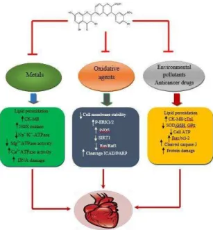
Figure 1. Hypothesized mechanisms of protective effect of silymarin against chemical-induced cardiac toxicity
In a study by Psotova et al (2002), silymarin, and its three constituents including silybin, silydianin and silychristin, protected rat heart microsomes and mitochondria against iron-dependent doxorubicin induced lipid peroxidation. The antilipoperoxidation activity of silymarin could be due to the presence of taxifolin with chelating and antioxidant activities and the ability of chemically unidentified polyphenols, which consisting 30% of silymarin, to bind transition
metals and quench ROS (59).
In vitro study on rat cardiomyocytes also showed
that silymarin and its constituents silibinin,
dehydrosilibinin, silychristin and silydianin increased the cell ATP level and displayed a dose-dependent cytoprotection against doxorubicin. The elevated ATP level in cardiomyocytes could be used for regeneration of antioxidation systems (GSH) in the course of oxidative stress induced by chemotherapeutics (57).
Cisplatin
Cisplatin is an effective chemotherapy drug used to treat several types of cancer. Different tumors, including testicular, ovarian, cervical, bladder and lung cancers as well as solid tumors resistant to other treatment regimens can be treated by cisplatin. Studies have reported that cisplatin therapy is usually associated with cardiotoxicity including arrhythmia, cardiomyopathy and congestive heart failure. It has been established that oxidative stress and apoptosis have important roles in cardiotoxicity induced by cisplatin which limits the clinical use of this drug. Silymarin (100 mg/kg, orally, for ten days) protected heart against cisplatin-induced myocardial
injury by reducing the activity of serum biochemical
markers including lactate dehydro genase (LDH),
creatine kinase isoenzyme MB (CK-MB) and cardiac
troponin I (cTnI) through its anti-lipid peroxidation activity. Therefore, silymarin induced stabilization of cardiac membranes and prevented the leakage of cardiac enzymes. In addition, silymarin inhibited the lipid peroxidation due to the presence of free hydroxyl groups at C5 and C7 which react with peroxy radicals. Moreover, silymarin increased the activities of endogenous antioxidant enzymes such as SOD. This effect led to a significant increase in the cellular antioxidant defense system. Silymarin also diminished oxidative mitochondrial DNA damage due to the free radical scavenging property (60).
As a result, silymarin may have a great potential as a novel therapeutic agent to prevent cardiac toxic effects induced by anti-cancer drugs.
Conclusion
Natural plant compounds have recently been the focus of researchers in order to protect the development of oxidative stress-induced cardiac dysfunction. The availability, lower price and less toxic effects of natural products compared with synthetic medications, make them as simple and excellent choice in the treatment of cardiac diseases. Silymarin
extracted from S. marianum has attracted great
attention because of its antioxidative, antitumor and anti-inflammatory properties. In this review article, efforts have been made to introduce some animal and
in vitro studies in scientific databases on the topic of the
protective effect of silymarin against cardiotoxicity induced by chemicals. Although there are an increasing number of studies indicating the beneficial actions of silymarin in various diseases, the number of studies relating to the potential cardiac protective effects of silymarin against chemical- induced toxicity is rather limited. Based on the results of some important investigations, silymarin acts as a potent protective agent in different types of intoxication induced by metals, environmental pollutants, oxidative agents and drugs such as doxorubicin. Some mechanisms including anti-inflammatory, free radical scavenging,
improvement of antioxidant defense systems,
membrane stabilizing, iron chelating activity and inhibition of apoptosis are involved in silymarin cardiac protective effects (Figure 1).
In conclusion, based on the current review, silymarin has a broad spectrum of cardiac protective activities against toxicities induced by chemicals. Because human reports are rare, further studies are required to establish the utility of silymarin in protection against cardiac toxicity.
Acknowledgment
References
1. Negi A, Kumar J, Luqman S, Shanker K, Gupta M, Khanuja S. Recent advances in plant hepatoprotectives: A chemical and biological profile of some important leads. Med Res Rev 2008; 28: 746-772.
2. Luper S. A review of plants used in the treatment of liver disease: Part 1. Altern Med Rev 1998; 3: 410-421. 3. Pepping J. Milk thistle: Silybum marianum, . Am J Health System Pharm 1999; 56: 1195-1197.
4. Karimi G, Vahabzadeh M, Lari P, Rashedinia M, Moshiri M. Silymarin, a promising pharmacological agent for treatment of diseases. Iran J Basic Med Sci 2011; 14: 308-317.
5. Dixit N, Baboota S, Kohli K, Ahmad S, Ali J. Silymarin: A review of pharmacological aspects and bioavailability enhancement approaches. Indian J Pharmacol 2007; 39: 172-179.
6. Karimi G, Hassanzadeh M, Mehri S. Protective effects of Silymarin against free radical-induced erythrocyte lysis, J Altern Complementary Med. 2006; 3:8.
7. Nazemian F. Effect of silymarin administration on TNF- serum concentration in peritoneal dialysis patients. Phytother Res 2010; 24:1654–1657. 8. Ramakrishnan G, Lo Muzio L,Elinos-Báez CM,Jagan S, inhibited proliferation and induced apoptosis in hepatic cancer cells. Cell Prolif 2009; 42: 229-240. 9. Taghiabadi T, Imenshahidi M, Abnous K, Mosafa F, Sankian M, Karimi G. Protective Effect of Silymarin against acrolein-induced cardiotoxicity in mice. Evid Based Complementary Altern Med 2012; 2012: Article ID 352091.
10. Muthumani M, Milton Prabu S. Silibinin potentially attenuates arsenic-induced oxidative stress mediated cardiotoxicity and dyslipidemia in rats. Cardiovasc Toxicol 2014; 14: 83-97.
11. Karimi G, Saradeghi Keisari M. Evaluation of antidepressant effect of ethanolic and aqueous extracts of Silybum marianum L. seed in mice. J Med Plants 2007; 6: 38-43.
12. Morishima C, Shuhart M, Wang CC, Paschal D, Apodaca M, Liu Y. Silymarin inhibits in vitro cell proliferation and cytokine production in hepatitis c virus infection. Gastroenterology 2010; 138: 671-681.
13. Karimi G, Ramezani M, Tahoonian Z. Cisplatin nephrotoxicity and protection by milk thistle extract in rats. Evid Based Complementary Altern Med 2005a; 2: 383-386.
14. Karimi G, Fallah Huseini H, Ramezani M, Tahoonian Z. Protective effect of silybum marianum (l.) gaertn. seeds extract and silymarin against cisplatin-induced acute nephrotoxicity in rats. J Med Plants 2005b; 4: 42-45.
15. Chu S, Chiou H, Chen P, Yang S, Hsieh Y. Silybinin inhibits the invasion of human lung cancer cells via decreased productions of urokinase-plasminogen activator and matrix metalloproteinase-2. Mol Carcinog 2004; 40: 143-149.
16. Kanter M, Meral I, Yener Z. Partial regeneration/ proliferation of the beta-cells in the islets of Langerhans by Nigella sativa L. in streptozotocin-induced diabetic rats. J Exp Med 2003; 201: 213-219. 17. Toklu H, Tunali-Akbay T, Erkanli G, Yuksel M, Ercan F, Sener G. Silymarin, the antioxidant
component of Silybum marianum, protects against burn-induced oxidative skin injury. Burns 2007; 33: 908-916.
18. Saller R, Melzer J, Reichling J, Brignoli R, Meier R. An updated systematic review of the pharmacology of silymarin. Forsch Komp Klas Nat 2007; 14: 70-80. 19. Fleming T. PDR for Herbal Medicines. New Jersy Medical Economics Company, 2000; 516-518 20. Kren V, Walterova D. Silybin and silymarin, new effects and applications. Biomed Papers 2005; 149: 29-41.
21. Rao P, Viswanath R. Cardioprotective activity of silymarin in ischemia-reperfusion-induced
myo-cardial infarction in albino rats. Exp Clin Cardiol 2007; 12: 179-187.
22. Talha J, Priyanka M A A. Hypertension and herbal plants. Int Res J Pharm 2011; 2: 26-30.
23. Sinha M, Manna P, SIL P. Terminalia arjuna protects mouse hearts against sodium fluoride-induced oxidative stress. J Med Food 2008; 11:733-740.
24. Nabavi S, Moghaddam A, Setzer W, Mirzaei M. Effect of silymarin on sodium fluoride-induced toxicity and oxidative stress in rat cardiac tissues. Anais da Academia Brasileira de Ciências 2012; 84: 1121-1126.
25. Murphy C. G. Oudit. Iron-overload cardiomyo-pathy: Pathophysiology, diagnosis, and treatment. J Card Fail 2010; 16: 888-900.
26. Wang Y, Wu M, Al-Rousan R, Liu H, Fannin J, Paturi S. Iron-induced cardiac damage: Role of apoptosis and deferasirox intervention. J Pharmacol Exp Ther 2011; 336: 56-63.
27. Borsari M, Gabbi C, Ghelfi F, Grandi R, Saladini M, Severi S. Silybin, a new iron-chelating agent. J Inorg Biochem 2001; 85: 123-129.
28. Gharagozloo M, Khoshdel Z, Amirghofran Z. The effect of an iron (III) chelator, silybin, on the proliferation and cell cycle of Jurkat cells: A comparison with desferrioxamine. Eur J Pharmacol 2008; 589: 1-7.
29. Najafzadeh H, Jalali M, Morovvati H, Taravati F. Comparison of the Prophylactic Effect of Silymarin and Deferoxamine on Iron Overload-Induced Hepatotoxicity in Rat. J Med Toxicol 2010; 6: 22-26. 30. Navidi-Shishaone M, Mohhebi S, Nematbakhsh M, Roozbehani S, Talebi A, Pezeshki Z, Eshraghi-Jazi F, Mazaheri S, Shirdavani S, Gharagozloo M, Alsaadat Moaeidi B. Co-administration of silymarin and deferoxamine against kidney, liver and heart iron deposition in male iron overload rat model. Int J Prev Med 2014; 5: 110-116.
31. Moon K, Guallar E, Navas–Acien A. Arsenic Exposure and Cardiovascular Disease: An Updated Systematic Review. Curr Atheroscler Rep 2012; 14: 542-555.
32. Manna P, Sinha MPS. Arsenic induced oxidative myocardial injury: Protective role of arjunolic acid. Arch Toxicol 2008; 82 :137-149.
33. Alamolhodaei NS, Shirani K, Karimi G. Arsenic cardiotoxicity: An overview. Env Toxicol Pharmacol 2015; 40: 1005-1014.
35. Dutta M, Ghosh A, Rangari V, Jain G, Khobragade S, Chattopadhyay A, Bhowmick D, Das T, Bandyopadhyay D. Silymarin protects against copper-ascorbate induced injury to goat cardiac mitochondria in vitro: involvement of antioxidant machnism. Int J Pharm Pharm Sci 2014; 6: 422-429.
36. Miltonprabu S, Thangapandiyan S. Epigallocatechin gallate potentially attenuates Fluoride induced oxidative stress mediated cardiotoxicity and dyslipidemia in rats. J Trace Elem Med Biol 2015; 29: 321-335.
37. Shashi A, Thapar S. Histopathology of myocardial
damage in experimental fluorosis in rabbits. Fluoride
2001; 34: 43-50.
38. Donmez N, Cinar A. Effects of chronic fluorosis on electrocardiogram in sheep. Biol Trace Elem Res 2003; 2 :115-122.
39. Nabavi S, Nabavi SF, Eslami S, Moghaddam A. In vivo protective effects of quercetin against sodium fluoride-induced oxidative stress in the hepatic tissue. Food ChemToxicol 2012; 132: 931-935. 40.Ma Y, Niu R, Sun Z, Wang J, Luo G, Zhang J, Wang J. Inflammatory responses induced by fluoride and arsenic at toxic concentration in rabbit aorta. Arch Toxicol 2012; 86: 849-856.
41. Diwan A, Dorn G. Decompensation of cardiac hypertrophy: cellular mechanisms and novel therapeutic targets. Physiology 2007; 22: 56-64. 42. Molkentin J, Dorn G. Cytoplasmic signaling pathways that regulate cardiac hypertrophy. Annu Rev Physiol 2001; 63: 391-426.
43. Bernardo B. Molecular distinction between physiological and pathological cardiac hypertrophy: experimental findings and therapeutic strategies. Pharmacol Ther 2010; 128: 191-227.
44. Takimoto E, Kass D. Role of oxidative stress in cardiac hypertrophy and remodeling. Hypertension 2007; 49: 241-248.
45. Anestopoulos I, Kavoa A, Tentesc I, Kortsarisc A, Panayiotidisd M, Lazoub A, Pappaa A. Silibinin protects
H9c2 cardiac cells from oxidative stress and inhibits phenylephrine-induced hypertrophy: potential
mechanisms. J Nut Biochem 2013; 24: 586-594. 46. Cieslak D, Lazou A. Regulation of BAD protein by PKA, PKCdelta and phosphatases in adult rat cardiac myocytes subjected to oxidative stress. . Mol Cells 2007; 24: 224-231.
47. Communal C. Norepinephrine stimulates apoptosis in adult rat ventricular myocytes by activation of the
-adrenergic pathway. Circulation 1998; 98: 1329-1334. 48. Zhou B, Wu L, Li L, Tashiro S, Onodera S, Uchiumi F, Ikejima T. Silibinin protects against isoproterenol-induced rat cardiac myocyte injury through
mitochondrial pathway after up-regulation of SIRT1. J Pharmacol Sci 2006a; 102: 387-395.
49. Zhou B, Wu L, Tashiro S, Onodera S, Uchiumi F, Ikejima T. Silibinin Protects Rat Cardiac Myocyte from Isoproterenol-Induced DNA Damage Independent on Regulation of Cell Cycle. Biol Pharm Bull 2006b; 29: 1900-1905.
50. Zhou B, Wu L, Tashiro S, Onodera S, Uchiumi F, Ikejima T. Activation of extracellular signal-regulated kinase during silibinin protected, isoproterenol-induced apoptosis in rat cardiac myocytes is tyrosine kinase pathway-mediated and protein kinase C-dependent. Acta Pharmacol Sin 2007; 28: 803-810. 51. Faroon O, Roney N, Taylor J, Ashizawa A, Lumpkin M, Plewak D. Acrolein health effects. Toxicol Ind Health 2008; 24: 447-490.
52. Stevens J, Maier C. Acrolein: sources, metabolism, and biomolecular interactions relevant to human health and disease. Mol Nut Food Res 2008; 52: 7-25. 53. Raschi E, Vasina V, Ursino M. Anticancer drugs and cardiotoxicity: insights and perspectives in the era of targeted therapy. Pharmacol Ther 2010; 125: 196-218.
54. Ado R, de Keulenaer G, Leite-Moreira A, Brs-Silva C. Cardiotoxicity associated with cancer therapy: Pathophysiology and prevention. Rev Port Cardiol. 2013;32:395-409.
55. Blum R, Carter S. Adriamycin. A new anticancer drug with significant clinical activity. Ann Intern Med 1974; 80: 249-259.
56. El-Shitany N, El-Haggar S, El-desoky K. Silymarin prevents adriamycin-induced cardiotoxicity and nephrotoxicity in rats. Food ChemToxicol 2008; 46: 2422–2428.
57. Chlopcikova S, Psotová P, Miketová P, Šimánek V. Chemoprotective Effect of plant phenolics against anthracycline-induced toxicity on rat cardiomyocytes. part I. Silymarin and its flavonolignans. Phytother Res 2004; 18: 107-110.
58. Cecen E, Dost T, Culhaci N, Karul A, Ergur B, Birincioglu M. Protective effects of silymarin against doxorubicin-induced toxicity. Asian Pacific J Cancer Prev 2011; 12: 2697-2704.
59. Psotová J, Chlopčíková S, Grambal F, Šimánek V, Ulrichová J. Influence of silymarin and its flavonolignans on doxorubicin-iron induced lipid peroxidation in rat heart microsomes and mitochondria in comparison with quercetin. Phytother Res 2002; 16: S63-S67.