Results of surgical treatment of
Results of surgical treatment of
Results of surgical treatment of
Results of surgical treatment of
Results of surgical treatment of massive localized lymphedema
massive localized lymphedema
massive localized lymphedema
massive localized lymphedema in
massive localized lymphedema
in
in
in
in
severely obese patients
severely obese patients
severely obese patients
severely obese patients
severely obese patients
Resultado do tratamento cirúrgico do linfedema maciço localizado em pacientes
Resultado do tratamento cirúrgico do linfedema maciço localizado em pacientes
Resultado do tratamento cirúrgico do linfedema maciço localizado em pacientes
Resultado do tratamento cirúrgico do linfedema maciço localizado em pacientes
Resultado do tratamento cirúrgico do linfedema maciço localizado em pacientes
obesos graves
obesos graves
obesos graves
obesos graves
obesos graves
WILSON CINTRA JÚNIOR,TCBC-SP1; MIGUEL LUIZ ANTONIO MODOLIN, ECBC-SP1; RODRIGO ITOCAZO ROCHA1; THADEU RANGEL FERNANDES1;
ARIEL BARRETO NOGUEIRA2; ROLF GEMPERLI,TCBC-SP3
A B S T R A C T A B S T R A C T A B S T R A C T A B S T R A C T A B S T R A C T
Objective Objective Objective Objective
Objective: to evaluate the importance of treatment of deformities caused by massive localized lymphedema (MLL) in the severely obese. MethodsMethodsMethodsMethodsMethods: in a period of seven years, nine patients with morbid obesity and a mean age of 33 years underwent surgical resection of massive localized lymphedema with primary synthesis. This is a retrospective study on the surgical technique, complication rates and improved quality of life. ResultsResultsResultsResultsResults: all patients reported significant improvement after surgery, with greater range of motion, ambulation with ease and more effective hygiene. Histological analysis demonstrated the existence of a chronic inflammatory process marked by lymphomonocitary infiltrate and severe tissue edema. We observed foci of necrosis, formation of microabscesses, points of suppuration and local fibrosis organization, and pachydermia. The lymphatic vessels and some blood capillaries were increased, depicting a framework of linfangiectasias. ConclusionConclusionConclusionConclusionConclusion: surgical treatment of MLL proved to be important for improving patients’ quality of life, functionally rehabilitating them and optimizing multidisciplinary follow-up of morbid obesity, with satisfactory surgical results and acceptable complication rates, demonstrating the importance of treatment and awareness about the disease.
Key words: Key words: Key words: Key words:
Key words: Lymphedema. Lymphedema/etiology. Lymphedema/pathology. Lymphedema/surgery. Obesity, morbid/ complications.
1. Department of Plastic Surgery, Clinics Hospital, Faculty of Medicine, University of São Paulo; 2. Division of Pathology, Clinics Hospital, Faculty of Medicine, University of São Paulo; 3. Department of Plastic Surgery, Faculty of Medicine, University of São Paulo.
INTRODUCTION
INTRODUCTION
INTRODUCTION
INTRODUCTION
INTRODUCTION
O
bese patients frequently have edema or even lymphedema of the lower extremities1, yet thelocalized massive lymphedema (MLL) is poorly observed and results from impaired lymphatic drainage, forming large tumor masses, especially in the lower limbs and hypogastrium2-4. MLL was preliminarily
described by Goshtarby et al.4 and Farshid et al.5, who
established the distinction with differentiated liposarcoma.
Beyond the deforming character, MLL deter-mines mobility limitation, the main reason for seeking treatment. Beside this, the huge tumor mass hinders lo-cal hygiene, thus allowing accumulation of debris and recurrent infectious episodes of cellulitis, worsening symptoms2,6,7.
This paper assesses the importance of the surgical treatment of massive localized lymphedema in severely obese patients.
METHODS
METHODS
METHODS
METHODS
METHODS
Between 2005 and 2012, nine patients were treated, six of them male. The mean age was 33 years, ranging between 19 and 57. The body mass index ranged from 44.1kg/m2 and 86.0kg/m2, with an average of 62.4.
Six patients had a deformity in the anteromedial thigh (Fi-gure 1) and three in the hypogastrium (Fi(Fi-gure 2). All reported that the onset and development of the tumor occurred after repeated episodes of skin infection, which were treated with local care and eventual antibiotic therapy, which did not prevent increase tumor.
hemostasis, one patient required volume and blood replacement for correction of instituted anemia.
All patients received antibiotic prophylaxis initiated 30 minutes prior to the procedure, maintained for up to 48 hours, and prophylaxis for deep venous thrombosis with enoxaparin beginning 12 hours after the operation and continued for seven days.
The dressings were prepared daily with surgical gauze, tapes and bandages, after cleaning of the surgical wounds.
All specimens removed were sent to histopathology.
RESULTS
RESULTS
RESULTS
RESULTS
RESULTS
All patients reported marked improvement thanks to the comfort of the withdrawal of the tumor mass, greater range of motion and greater ease of ambulation, and the possibility of more effective hygiene. The weights and vo-lumes of the removed parts are shown in Table 1. The average weight was 7.8kg and the average volume of 17.655cm3.
In all patients, we observed a marbling appearance of the subcutaneous tissue and output of
Figure 2 Figure 2 Figure 2 Figure 2
-Figure 2 - Example of MLL in the suprapubic region. Figure 1
Figure 1 Figure 1 Figure 1
-Figure 1 - Example of MLL on the right thigh.
large amount of hyaline liquid during tumor resection (Figure 3).
The skin of the tumor region had, in all cases, pachydermia features and presence of warty, scattered lesions, varying from 1 to 5 cm in size.
In the postoperative period there was drainage of large amounts of fluid, initially sero-hematic, and after three or four days, hyaline, with daily volume of about 600ml, 1270ml being the largest and 20 ml the smallest. Such drainage persisted for two to three weeks and gradually regressed spontaneously.
The length of stay ranged from one to 21 days. Two patients had local complications: one had wound dehiscence, requiring a new suture; another had wound infection, with phlegmonous cellulitis, requiring hospitalization with parenteral antibiotics. Both had good response to treatment, without further complications. These two patients had higher values of BMI, 71 and 86, respectively. There were no other complications demanding greater care, except for small dehiscences along the wound, which occurred in all cases, and healed by secondary intention.
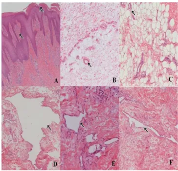
The histological results showed chronic inflammatory process, marked by lymphomonocitary infiltrate, accompanied by severe tissue edema. In some areas foci of necrosis were noted, with formation of microabscesses and points of suppuration. In other areas the organization of lo-cal fibrosis could be seen. Lymphatic vessels, as well as some blood capillaries, were increased, defining histopathological features of lymphangiectasia. The epidermis was acanthotic, featuring pachydermia (Figure 4).
DISCUSSION
DISCUSSION
DISCUSSION
DISCUSSION
DISCUSSION
The modern concepts of MLL are assigned to Farshid and Weiss5, which, in 1998, managed to gather 14
cases with lower limb injuries. These pathologists highlighted
Figure 4 -Figure 4 -Figure 4
-Figure 4 -Figure 4 - A: Skin with irregular acanthosis (two arrows) and fibrosis of skin healing pattern (arrow) - (100x magnification), B, C: Adipose tissue with vascular proliferation (arrows) and interstitial edema (200x magnification), D, F (200x magnification) and E (400x magnification): irregular dilated lymphatic vessels (arrow) and edema characteristic of lymphedema. Staining: hematoxylin-eosin.
Figure 3 -Figure 3 -Figure 3 Figure 3
-Figure 3 - A,B: marble feature of the MLL subcutaneous tissue. C,D: closing of the wound without difficulty, at the expense of a neighborhood flap.
Tabela 1 Tabela 1 Tabela 1 Tabela 1
Tabela 1 - Mensuração das peças cirúrgicas relacionadas com os valores do IMC.
P a c i e n t e P a c i e n t eP a c i e n t e P a c i e n t e
P a c i e n t e IMC (kg/mIMC (kg/mIMC (kg/mIMC (kg/mIMC (kg/m22222))))) Peso da peça (g)Peso da peça (g)Peso da peça (g)Peso da peça (g)Peso da peça (g) Medidas da peça (cm)Medidas da peça (cm)Medidas da peça (cm)Medidas da peça (cm)Medidas da peça (cm) Volume da peça (cmVolume da peça (cmVolume da peça (cmVolume da peça (cmVolume da peça (cm33333)))))
1 – NS 44,1 715 25x17x3 1275
2 – AP 53,8 5490 28x24x3 2076
3 – CT 73,1 18000 50x43x12 25800
4 – LC 54,4 5250 38x23x11 9614
5 – RB 60,5 22000 65x60x17 66300
6 – KC 86,0 13870 44x42x11 20328
7 – DT 50,0 9000 40x25x14 14000
8 – CM 69,4 6210 34x30x10 10200
9 – EP 71,0 5900 46x32x7 10304
MÉDIA 62,4 7800 - 17655
the salient features of this pathology, rendering less likely the confusion with well-differentiated liposarcoma. Still, other names such as lynphodystrophic tissue, pseudosarcomas, fibrolipoangiolipomatous hamartoma and elephantiasis nostra, can be found in the literature4,7-16.
The lower limbs are typical locations of MLL, but in some cases they also appear in the inguinal and suprapubic areas. Penile and scrotal masses were also identified8,9, but were not included in this series. MLL is
acquired in adulthood, growing insidiously and silently over the years, interfering with ambulation and hygiene, which, once committed, is a constant point of dermatitis and ulceration.
A B
The histopathological diagnosis is evidenced by widespread interstitial edema, associated with reactive fibroblast proliferation, acanthosis and dermal sclerosis, vascular and lymphatic ectasia, with inflammation. Macroscopically, the hardened tumor mass and the skin with the “orange peel” appearance feature the frequent pachydermia that accompany MLL4,7-10.
Any lymphatic obstruction may precipitate regi-onal lymphedema. In this sense, the differential diagnosis should be made with either congenital malformations and acquired conditions, including lipomas, sarcomas and other tumors, sequelae of surgery, radiation therapy, trauma and filariasis. Lipodystrophy and massive abdominal panicles may also be involved8-11.
The precise mechanism of the formation of MLL remains intact. Descriptions in the literature suggest that the formation of lymphedema is due to obstruction of the initial lymphatic system and its consequent congestion. The extravasation of protein-rich fluid to the interstitial space stimulates localized inflammatory response, hampering lymph drainage, which causes chronic lymphedema12. The
normal lymph transport depends on intrinsic contractility of the lymphatic system, with directional flow provided by the presence of valves. Extrinsic factors, such as exercise, daily activities and external compression, also play an important role to prevent lymphatic congestion. In patients with a high BMI, given the higher level of interstitial fluid resulting from a greater volume of adipose tissue, there is increased pressure, thereby compromising lymph draining. Consequently, the body condition of these patients causes a significant obstacle to mechanical lymphatic drainage10,11,
which worsens with inactivity.
All cases of MLL in obese are described in hanging areas of the abdomen and lower limbs. Therefore, it can be inferred that the lesion is a result of lymphatic stasis exacerbated by increasing of fat in hanging areas, as well as the motion reduction of the obese patient13.
Undoubtedly, in the morbidly obese local hygiene is poor due to the difficulty of access between fat-skin folds. When associated with flaking skin and natural secretions, this constitutes a fertile ground for installation of infections, generically called intertrigo. The recurrence of these infections causes lymphangitis, which increases in the lower
segments of the trunk, ie, hypogastrium and lower limbs due to the consequent stasis. Lymphangitis stasis determi-nes leakage of lymphatic fluid, which stimulates cell proliferation, both on the epidermis, which is thickened and grows warts, and in deeper layers, where the inflammatory process develops. This lymphedema framework, associated with the local inflammatory reaction, distends the tissues and increases tissue ischemia, which causes severe fibrosis with interspersed fatty tissue, generating septa and blocks. The evolution of is progressive and results in the appearance of localized tumor masses2,5,10.
Within this context, in all our patients the predisposing factor was obesity and the described etiopathogenic mechanism was triggering by infection, due to favorable local conditions. However, one cannot rule out that the same lymphatic drainage disorder is a result of obesity alone, running parallel to the two theories previously mentioned7,10.
The possibility of recurrence cannot be overlooked4,7,8,10. The presence of the pedicle at the base
of the tumor and no involvement of the deep layers below the muscle-aponeurotic fascia generally facilitate surgical resection. In our series there was a patient who demanded two reoperations, since committed tissue remained in the previous procedures due to difficulty in finding a plane of dissection, which favored recurrence. Nevertheless, the results were cosmetic and functionally appropriate, as they favored ambulation and allowed effective hygiene.
This experience reinforces the profile of MLL as rare, but it can become a serious chronic problem, rendering the patient incompetent. Unlike ordinary mild edema or lymphedema, treatment with bariatric surgery and weight loss does not excuse the complete and curative surgical removal. Obesity, and its comorbidities, does not limit the obligation of the resection of the tumor mass, despite the inherent risks. Surgical treatment requires a prolonged postoperative period, but all patients were discharged in good condition. Functional rehabilitation was achieved.
Surgical treatment of MLL was important to improve the quality of life, functionally rehabilitating and optimizing the multidisciplinary follow-up of morbid obesity, with satisfactory surgical results and acceptable complication rates, demonstrating the importance of its application in the treatment of massive localized lymphedema.
R E S U M O R E S U M O R E S U M O R E S U M O R E S U M O
Objetivo: Objetivo: Objetivo: Objetivo:
Objetivo: avaliar a importância do tratamento das deformidades ocasionadas pelo linfedema maciço localizado (LML) em obesos. Métodos:
Métodos: Métodos: Métodos:
do LML mostrou-se importante para a melhora na qualidade de vida desses pacientes, reabilitando-os funcionalmente e otimizando o seguimento multidisciplinar da obesidade mórbida, com resultados cirúrgicos satisfatórios e taxas aceitáveis de complicações, demonstrando a importância do tratamento e da consciência sobre a doença.
Descritores: Descritores: Descritores: Descritores:
Descritores: Linfedema. Linfedema/etiologia. Linfedema/patologia. Linfedema/cirurgia. Obesidade Mórbida/complicações.
REFERENCES
REFERENCES
REFERENCES
REFERENCES
REFERENCES
1. Yosipovitch G, DeVore A, Dawn A. Obesity and the skin: skin physiology and skin manifestations of obesity. J Am Acad Dermatol. 2007;56(6):901-16; quiz 917-20.
2. Warren AG, Brorson H, Borud LJ, Slavin SA. Lymphedema a comprehensive review Ann Plast Surg. 2007;59(4):464-72. 3. Scheinfeld NS. Obesity and dermatology. Clin Dermatol.
2004;22(4):303-9.
4. Goshtarby P, Dawson J, Agarwal N. Pseudosarcoma: massive localized lymphedema of the morbidly obese. Obes Surg. 2006;16(1):88-93.
5. Farshid G, Weiss SW. Massive localized lymphedema in the morbidly obese: a histologically distinct reactive lesion simulating liposarcoma. Am J Surg Pathol. 1998;22(10):1277-83.
6. Modolin ML, Cintra W Jr, Paggiaro AO, Faintuch J, Gemperli R, Ferreira MC. Massive localized limphedema (MLL) in bariatric candidates. Obes Surg. 2006;16(9):1126-30.
7. Vána J, Adamicová K, Haluska P, Celec J. Massive localized lymphedema in an extremely obese patient. Chirurg. 2002;73(4):383-6.
8. Decker P, Görtz M, Sigmund G, Kriegsmann J, Decker D. Massive localized lymphedema – a rare entity. Zentralbl Chir. 2003;128(11):985-7.
9. Oswald TM, Lineaweaver W. Limited segmental resection of symptomatic lower-extremity lymphodystrophic tissue in high-risk patients. South Med J. 2003;96(7):689-91.
10. Modolin M, Mitre AI, da Silva JC, Cintra W, Quagliano AP, Arap S, et al. Surgical treatment of lynphedema of the penis and scrotum. Clinics. 2006;61(4):289-94.
11. Asch S, James WD, Castelo-Soccio L. Massive localized lymphedema: an emerging dermatologic complication of obesity. J Am Acad Dermatol. 2008;59(5 Suppl):S109-10.
12. Barr J. Massive localized lymphedema of suprapubic origin. Plast Reconstr Surg. 2000;106(7):1663-4.
13. Hanna D, Cloutier R, Lapointe R, Desgagné Al. Abdominal elephantiasis: a case report. J Cutan Med Surg. 2004;8(4):229-32. 14. Wu D, Gibbs J, Corral D, Intengan M, Brooks JJ. Massive localized lymphedema: additional locations and association with hypothyroidism. Hum Pathol. 2000;31(9):1162-8.
15. Burri H, Loutan L, Kumaraswami V, Vijayasekaran V. Skin changes in chronic lymphatic filariasis. Trans R Soc Trop Med Hyg. 1996;90(6):671-4.
16. Bannerjee D, Williams EV, Ilott J, Monypenny IJ, Webster DJ. Obesity predisposes to increased drainage following axillary node clearance: a prospective audit. Ann R Coll Surg Engl. 2001;83(4):268-71.
Received on 15/10/2012
Accepted for publication 15/12/2012 Conflict of interest: none.
Source of funding: none.
How to cite this article: How to cite this article:How to cite this article: How to cite this article:How to cite this article:
Cintra Júnior W, Modolin MLA, Rocha RI, Fernandes TR, Nogueira AB, Gemperli R, Ferreira MC. Results of surgical treatment of massive localized lymphedema in severely obese patients. Rev Col Bras Cir. [periódico na Internet] 2014;41(1). Disponível em URL: http:// www.scielo.br/rcbc
Address for correspondence: Address for correspondence:Address for correspondence: Address for correspondence:Address for correspondence: Cintra Wilson Junior