678
Splenosis. A Diagnosis to be Considered Case Report
International Braz J Urol Vol. 32 (6): 678-680, November - December, 2006
Splenosis. A Diagnosis to be Considered
Jorge C. Ribeiro, Carlos M. Silva, Americo R. Santos
Section of Urology, São Marcos Hospital, Braga, Portugal
ABSTRACT
The term splenosis applies to the autotransplanted splenic tissue resulting from seeding in the context of past splenic trauma or surgery. We report a 42-year-old man with a history of splenectomy observed for an incidentally found retrovesical mass thought to be an ectopic testicle. The abdominal laparotomy revealed multiple focuses of pelvic splenosis. As splenosis can be diagnosed through specific imaging studies one should always consider it in differential diagnosis of a mass discovered years after splenic surgery or trauma.
Key words: spleen; wounds and injuries; splenosis; bladder Int Braz J Urol. 2006; 32: 678-80
INTRODUCTION
Ectopic splenic tissue is a cause of inciden-tally found mass leading to diagnostic confusion. It can present as a congenital accessory spleen typically localized medially to the orthotropic spleen or as a mass detected several years after a splenic surgery or trauma. These implants, resulting from seeding, can mimic a neoplasm. However, specific diagnostic tests can confirm diagnosis and avoid unnecessary surgery. In this paper we discuss a case of an inciden-tal retrovesical mass in a patient with an absent left testicle that lead us to think of an ectopic testicle.
CASE REPORT
A 42 year-old male patient with a history of Behçet disease and a motor vehicle accident 20 years before resulting in emergent splenectomy, was re-ferred due to an incidental retrovesical mass. A rou-tine abdominal ultrasound detected a solid retrovesical
neoformation independent from seminal vesicles. The CT scan confirmed a 3.7 x 2.5 cm mass (Figure-1).
No mass was identifiable on abdominal pal-pation or rectal exam. The left testicle was absent. IVU, transrectal ultrasound, and urethrocistoscopy were normal. The hypothesis of an ectopic left tes-ticle tumor emerged. Testis tumor markers (DHL, ß-HCG and α-FP) were within normal range.
679
Splenosis. A Diagnosis to be Considered
COMMENTS
Splenosis affects one to two thirds of patients submitted to splenectomy for trauma (1). Implanta-tion from seeding is most frequently in serosal sur-faces of small and large intestine, greater omentum, parietal peritoneum, mesentery, diaphragm
Figure 1 – CT scan. A 3.7 x 2.5 cm hyperdense retrovesical mass.
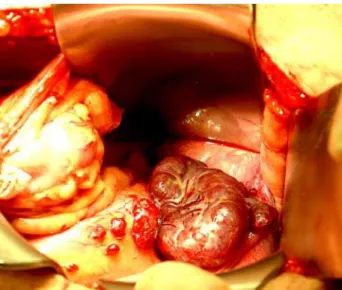
Figure 2 – Intraoperative picture revealing a large bluish-red mass and multiple small implants involving the peritoneum sur-face of pelvis suggesting ectopic splenic tissue.
undersurface and more rarely, in cases of severe trauma, intrahepatic or even intrathoracic (2,3).
Although splenosis can seldom present as a vague abdominal or testicular pain, intestinal obstruc-tion from adhesions, GI bleeding and spontaneous rupture, it usually is an incidental finding during sur-gery, either laparoscopy or imaging (2).
When present as an incidental imaging mass it has been reported on to mimic renal, adrenal or abdominal tumors, metastases, lymphoma, en-dometriosis and ectopic testicles (1-4). Although usual imaging modalities (US, CT, MRI) are helpful to lo-calize and determine the size, structure and relations with adjacent organs they are not specific.
If we had considered splenosis, signs of re-sidual splenic tissue as the absence of Howell-Jolly bodies, siderocytes, Heinz bodies and pitted red cells on peripheral blood smear a could have been of help, but their presence is still possible due to less func-tioning splenosis tissue (2,3)
More specific and diagnostic studies using agents that are sequestered by reticulendothelial tis-sue, like 99mtechnetium sufur colloid, 99mtechnetium
labeled heat-denatured autologous red blood cells or
111In-labeled platelets scans (1,2) and recently
ferumoxide-enhanced MRI (4) have been used. In conclusion, all patients with a history of spleen surgery or trauma should consider the hypoth-esis of splenosis in differential diagnosis of a newly found mass.
CONFLICT OF INTEREST
None declared.
REFERENCES
1. Pumberger W, Wiesbauer P, Leitha T: Splenosis mim-icking tumor recurrence in renal cell carcinoma: de-tection on selective spleen scintigraphy. J Pediatr Surg. 2001; 36: 1089-91.
680
Splenosis. A Diagnosis to be Considered
3. Weinstein RP, Genega EM, Dalbagni G: Splenosis mimicking transitional cell carcinoma. J Urol. 1999; 161: 1281.
4. Berman AJ, Zahalsky MP, Okon SA, Wagner JR: Dis-tinguishing splenosis from renal masses using ferumoxide-enhanced magnetic resonance imaging. Urology. 2003; 62: 748.
Accepted after revision: March 25, 2006
Correspondence address: Dr. Jorge Cabral Ribeiro
Hospital de São Marcos, Section of Urology Apartado 2242
Braga, 4701-965, Portugal Fax: + 35 125-3613334