Carcinoma of the Renal Pelvis and Ureter
Fernando Korkes, Thiago S. Silveira, Marilia G. Castro, Gustavo Cuck, Roni C. Fernandes,
Marjo D. Perez
Division of Urology and Department of Pathology, School of Medical Sciences, Santa Casa, Sao
Paulo, SP, Brazil
ABSTRACT
Objective: To assess the occurrence of upper urinary tract urothelial tumors (UUTT) in Brazil.
Materials and Methods: We performed a clinical and histopathologic study of 33 patients who were diagnosed with a malignant neoplasm in the renal pelvis or ureter in the period of 1994 to 2004, in a single institution.
Results: Among the patients with upper urinary tract carcinoma, 70% were males and 30% females, with mean age of 65 ± 16 years (ranging from 31 to 91 years). Nineteen patients presented renal pelvis tumor (58%), 9 ureteral tumor (27%) and 5 synchronic pelvic and ureteral tumors (15%). Renal pelvis tumors represented 2.8% of all the urothelial neoplasms, and 11.4% of all renal neoplasms treated in the same period. Ureteral tumors represented 1.6% of all the urothelial malignan-cies surgically managed in these 11 years. Tobacco smoking was the most common risk factor, and analgesic abuse was not reported by those patients. Most carcinomas were high-grade and muscle-invasive. Mean time to diagnosis was 7 months, being hematuria the most common symptom.
Conclusions: A high association was also found between UUTT and bladder urothelial carcinoma. UUTT were mostly seen in men in their seventies and related to a high overall and cancer-related mortality rate.
The overall disease-specific survival was 40%, much lower than found in most of the reported series.
Key words:kidney; ureter; neoplasms; transitional cell; epidemiology; Brazil
Int Braz J Urol. 2006; 32: 648-55
INTRODUCTION
Upper urinary tract tumors involving the re-nal pelvis and ureter are relatively uncommon. The great majority of these are epithelial, 80% are malig-nant and 90% are urothelial carcinomas. Renal pel-vis tumors account for approximately 7 to 10% of all renal tumors and about 5% of all urothelial tumors (1-9). Ureteral tumors are even more uncommon, occurring in one fourth of the incidence of renal pel-vis tumors (1,10).
Many factors contribute to the development of upper urinary tract urothelial tumors (UUTT), some
of them similar to bladder cancer associated factors, and the most common of these are tobacco smoking and analgesics abuse, particularly phenacetin (2,3,5,11-13). Other risk factors include papillary necrosis, chronic urinary infections, renal calculi, occupational exposure, Balkan nephropathy, thorium containing radiologic contrast medium and family associated cancer syndromes (2,3,5,12-14). The be-havior of the UUTT is also similar to the bladder urothelial carcinoma, presenting high recurrence rates and usually is multicentric (2,3,9).
cases, organ-sparring procedures are the treatment option. For UUTT, nephroureterectomy with bladder cuff removal has been the conventional treatment. More recently, with the introduction of endourological techniques, approaches that are more conservative have been advocated in selecting patients in an effort to salvage kidneys. However, different from bladder carcinoma, radical surgery is more often used in UUTT cases, as the diagnosis is commonly made at advanced stages and management by conservative measures is problematic (3).
Among the North-American population, there has been an increase in women affected, and it tends to occur at an older age (3,5,15). Large demographic stud-ies have been undertaken in several countrstud-ies (2,4,6-9,15-19) but to our knowledge, there is no study ana-lyzing data about UUTT in the Brazilian population.
The aim of the present study is to assess the occurrence of UUTT in 33 patients who underwent surgery from 1994 to 2004 in a single institution.
MATERIALS AND METHODS
Surgical pathology files of all patients who underwent surgery for primary UUTT at the Divi-sion of Urology of the authors’ institution from 1994 to 2004 have been retrospectively reviewed (33 sub-jects).
Macroscopic data were obtained from the pathological reports. The hematoxylin and eosin stained slides were reviewed by one pathologist with special expertise in the field of uropathology (MGC). Slides on each case were collected and reclassified using the criteria of the 2004 WHO grading system (5). All tumors were restaged based on TNM (tumor node metastasis) staging system, 2002 (20). All urothelial and renal tumors surgically treated during the same period in the referred institution were also revised in order to calculate disease-related preva-lence.
Further data were obtained from the hospital database and patients notes, including patients’ de-mographics, anatomical location of the tumor, sur-gery outcomes, disease recurrence, specific survival and overall survival. Contact was established with
the patient and/or family whenever possible, and the district death registry was consulted for the remain-ing cases.
Disease-specific survival was assessed by the Kaplan-Meier method and compared by the Log-Rang test.
The institutional medical ethics committee approved the present study.
RESULTS
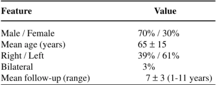
Among patients with UUTT, 70% were males and 30% females, with a mean age of 65 ± 16 years (ranging from 31 to 91 years). Ninety-one per cent of the patients were white and 9% black. The left side was affected in 61% of the cases and the right side in 39% (Table-1).
Nineteen patients had only renal pelvis tu-mor (58%), 9 had ureteral tutu-mor (27%) and 5 had both pelvic and ureteral tumors (15%). Renal pelvis urothelial carcinomas represented 2.8% of all the urothelial neoplasms surgically treated in our institu-tion during this period, and 11.4% of all renal tumors. Ureteral tumors represented 1.6% of all the urothelial malignancies surgically managed. In 50% of the pa-tients the distal ureter was affected, middle and proxi-mal ureter were respectively affected in 29% and 21% of the patients.
Association with bladder cancer was present in 30% of the patients. In 17% of them, there had been a previously treated bladder cancer (mean of 4 years previously) and in 23%, there had been a syn-chronous bladder neoplasm. In 15% of the cases, there had been synchronous ureteral and pelvic neoplasm
Table 1 – Clinical features of upper urinary tract urothelial tumors.
Feature Value
Male / Female 70% / 30%
Mean age (years) 65 ± 15
Right / Left 39% / 61%
Bilateral 03%
and in 3% of the patients, bilateral disease had been found.
Hematuria was the most common symptom, seen in 45% of the patients. Other presenting mani-festations included anemia (43%), flank pain (30%), weight loss (27%), fever (17%) pyelonephritis (17%) and palpable mass (10%). Diagnosis due to inciden-tal finding during follow-up of previous urothelial carcinoma occurred in 10% of the patients. In the cases that weight loss, the mean loss observed was 5 Kg.
Mean duration of symptoms prior to diagno-sis was 6.9 ± 4.3 months. Initial diagnosis was UUTT in 85% of the cases. In 6% of the cases, renal cancer was suspected and in 9% of the cases, the surgery was undertaken with the diagnosis of pyonephrosis, and the presence of cancer was confirmed during pathologic exam.
Regarding risk factors, tobacco smoking was referred by 66% of the patients and 33% had recur-rent urinary tract infections or calculi. In one patient there was an hereditary nonpolyposis colorectal can-cer syndrome associated (3%). Analgesic abuse was not referred to as risk factor in any patient.
Surgical treatment consisted of radical nephroureterectomy and bladder cuff removal in 65% of the patients; in 6% radical cistectomy was also performed; in 16% only distal ureterectomy and re-implant was performed, and in 13% only nephrec-tomy. Radiation therapy and chemotherapy were re-spectively combined in 6% and 12% of the cases.
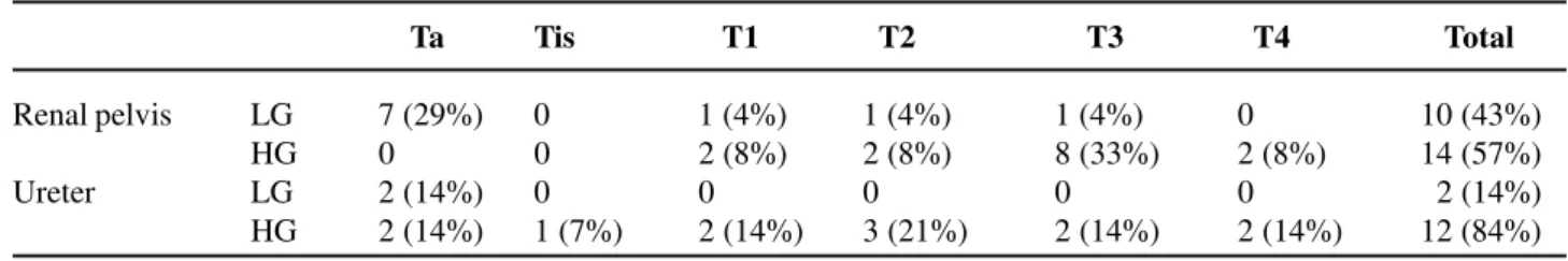
Pathological exam demonstrated high-grade malignancies in 58% of the renal pelvis neoplasms and in 86% of the ureteral neoplasms. Regarding re-nal pelvis neoplasms, pT3 was the most common
stage, observed in 37% of the patients; pTa was ob-served in 29%, pT1 in 12%, pT2 in 12% and pT4 in 8% (Table-2). In ureteral neoplasms, pTa was the most common stage, observed in 28% of the cases. Stages pTis, pT1, pT2, pT3 and pT4 occurred respectively in 7%, 14%, 21%, 14% and 14% of the cases (Table-2). In 93.9% of the patients there was an urothelial tumor, and in 6.1% a squamous cell carcinoma. Squa-mous cell differentiation was observed in 9.7% of the urothelial carcinomas (3 cases). In all the cases with squamous cell differentiation, pathological stage was pT3, and for pure squamous cell carcinomas, one had a pT3 stage and the other a pT4. All the patients with squamous cell differentiation or squamous cell carci-noma had renal calculi and/or infection associated. In terms of lymph node status, 85% of the tumors were at NX, 6% at N0 and 9% at N1-3.
During follow-up (mean 7 ±3 years, ranging from 1-11 years), 10% were alive, 30% died due to other causes, 5% died due to surgical complications and 55% died due to the malignancy. Three patients presented bladder cancer (treated endoscopically) and one patient that had a previous renal pelvis neoplasm underwent a contra-lateral distal ureterectomy 2 years later due to UUTT. Adequate follow-up was possible in 61% of the patients.
Disease-specific survival was not related to tumor grade (p = 0.31) neither to pathologic stage (p = 0.51) in the present series.
COMMENTS
In the present series, UUTT prevalence re-lated to renal and bladder cancer was similar to that
Table 2 – Distribution of renal pelvis and ureteral tumors according to histologic grade and stage.
Ta Tis T1 T2 T3 T4 Total
Renal pelvis LG 7 (29%) 0 1 (4%) 1 (4%) 1 (4%) 0 10 (43%)
HG 0 0 2 (8%) 2 (8%) 8 (33%) 2 (8%) 14 (57%)
Ureter LG 2 (14%) 0 0 0 0 0 02 (14%)
HG 2 (14%) 1 (7%) 2 (14%) 3 (21%) 2 (14%) 2 (14%) 12 (84%)
previously reported in other studies (1,2,21). The ana-tomical location of UUTT in the present study con-forms to that previously described, with almost twice as many pelvicalyceal as ureteric tumors (22). Ure-teric tumors were also more common in the distal third, followed by the middle and proximal portions of the ureter, as reported in other series (22). The in-cidence of bilateral synchronous tumors was similar to other series (1,2).
UUTT has also been found to be primarily a disease of white individuals (91% of the cases), and mostly affecting elderly men (15). The demographic characteristics of our patients showed a peak inci-dence in the seventh decade of life and male-to-fe-male ratio of 2.3:1. This is different from the lower tract disease in which the male-to-female ratio is 3 to 4:1 (3).
In the American population, Munoz et al. observed that patients with UUTT are being diagnosed at an older age, and a higher proportion of female and nonwhite individuals have been diagnosed. In our patients, such variation has not been noticed. Even though the number of patients is limited, age, ethnics and gender characteristics of the patients with UUTT remained the same during the last decade (15).
According to the WHO 2004 grading system, tumors are grading as papillary urothelial neoplasm of low malignant potential (PUNLMP), low-grade and high-grade carcinomas. There were few studies that used this system (2-4,18,19). We did not identify any PUNLMP, similarly to Olgac et al. It seems that dif-ferently from what occurs in the bladder, PUNLMP occur less frequently in upper urinary tract (19). In the present study, 86% of ureteral carcinoma and 58% of renal pelvis carcinoma were high-grade. These find-ings are similar to others investigators’ data (3,6,7,9,17-19,23), confirming that most of the pa-tients treated for UUTT present a high-grade disease. Squamous cell carcinoma has accounted for 6.1% of the UUTT, close to previously reported in other series (4,24,25). Also, as reported by Blacher et al. in our patients all the squamous cell carcinomas and the urothelial carcinoma with squamous cell dif-ferentiation occurred within the renal pelvis, and all of them were high grade and high stage diseases with extensive invasion of the renal parenchyma. All were
in pathological stage pT3 or pT4 and had an unfavor-able prognosis (25). As previously reported, all the cases were associated to calculi, chronic infection and squamous metaplasia of the neighboring epithelium (4).
In 64% of the cases, muscle-invasive disease was found, confirming the fact that unlike urothelial carcinomas of the bladder, UUTT should therefore be regarded as an aggressive, high-grade cancer, un-less proven otherwise (3,26). In the reported cases however, muscle-invasive disease was more frequent than observed in other large series, with pT2 stage or higher occurring in 42-49% of the UUTT (3,8,18,19). In our study, 13 patients (39%) had multifo-cal disease at presentation. Of these, one had bilat-eral uretbilat-eral tumor, 5 had both pelvic and uretbilat-eral tumor, 3 had pelvic and bladder tumor, 3 had ureteral and bladder tumor and 1 had carcinoma in the pelvis, ureter and bladder. The multicentric characteristic of urothelial carcinomas may be explained by several theories, but the better accepted is the so-called field effect, suggesting that the entire urothelial surface has undergone a neoplastic change (2).
Mean time from the beginning of symptoms to diagnosis was 7 months, being hematuria the most common symptom. Even though good screening pro-grams are not available for such tumors, adequate widespread information could lower the stage at di-agnosis. In the present study, a close association was found between UUTT and bladder urothelial cell car-cinoma. Diagnosis and follow up of these bladder tumors allowed an earlier diagnosis of 10% of upper tract carcinomas.
Long-term phenacetin abuse is a commonly reported risk factor for UUTT (12). However, in the present study it was not found to be a significant risk factor. Maybe the greater popularity of dipirone in-stead of phenacetin among Brazilians may explain this finding (27). Concerning risk factors, tobacco smoking, renal calculi and chronic infection were mostly observed.
fol-lowed (28). However, in patients studied, such crite-ria have not been fulfilled in any of the cases. In most of them, there were bulky high stage and high-grade lesions.
Overall 5-year disease-specific survival was 40%, much lower than most of the reported series, which varies from 67% to 75% (15,29,30). As stated previously, a high prevalence of muscle-invasive dis-ease and a high stage at diagnosis was observed in these patients. As pathological stage is one of the most important prognosis predictor, a poorer sur-vival would in fact be expected for the studied popu-lation (9,18).
When analyzing disease-specific survival, neither tumor grade nor stage was significant risk fac-tors in the present series. However, the poor follow-up of these patients, associated to the limited number of patients could explain this observation.
In conclusion, the studied population showed prevalence of UUTT related to other urothelial and renal neoplasms similar to the ones observed in other studies. UUTT was mostly diagnosed in men in the seventh decade of life, and tobacco consumption has been the major risk factor for UUTT in the present population. UUTT was associated to a high overall and cancer-related mortality rate.
CONFLICT OF INTEREST
None declared.
REFERENCES
1. Messing EM: Urothelial tumors of the urinary tract. In: Walsh PC, Retik AB, Vaughan (ed.), Campbell’s Urology. Philadelphia, Saunders. 2002; pp. 2732-73. 2. Holmang S, Johansson SL: Synchronous bilateral ure-teral and renal pelvic carcinomas: incidence, etiology, treatment and outcome. Cancer. 2004; 101: 741-7. 3. Olgac S, Mazumdar M, Dalbagni G, Reuter VE:
Urothelial carcinoma of the renal pelvis: a clinicopatho-logic study of 130 cases. Am J Surg Pathol. 2004; 28: 1545-52.
4. Perez-Montiel D, Wakely PE, Hes O, Michal M, Suster S: High-grade urothelial carcinoma of the renal
pel-vis: clinicopathologic study of 108 cases with empha-sis on unusual morphologic variants. Mod Pathol. 2006; 19: 494-503.
5. Eble JN, Sauter G, Epstein JE, Sesterhenn IA: World Health Organization Classification of Tumours. Pathol-ogy and Genetics of Tumours of the Urinary System and Male Genital Organs. Lyon. 2004.
6. Park S, Hong B, Kim CS, Ahn H: The impact of tu-mor location on prognosis of transitional cell carci-noma of the upper urinary tract. J Urol. 2004; 171: 621-5.
7. Chen WJ, Kuo JY, Chen KK, Lin AT, Chang YH, Chang LS: Primary urothelial carcinoma of the ureter: 11-year experience in Taipei Veterans General Hospital. J Chin Med Assoc. 2005; 68: 522-30.
8. Guinan P, Vogelzang NJ, Randazzo R, Sener S, Chmiel J, Fremgen A, et al.: Renal pelvic cancer: a review of 611 patients treated in Illinois 1975-1985. Cancer In-cidence and End Results Committee. Urology. 1992; 40: 393-9.
9. Hall MC, Womack S, Sagalowsky AI, Carmody T, Erickstad MD, Roehrborn CG: Prognostic factors, recurrence, and survival in transitional cell carci-noma of the upper urinary tract: a 30-year experi-ence in 252 patients Urology. Urology. 1998; 52: 594-601.
10. Huben RP, Mounzer AM, Murphy GP: Tumor grade and stage as prognostic variables in upper tract urothelial tumors. Cancer. 1988; 62: 2016-20. 11. Kirkali Z, Tuzel E: Transitional cell carcinoma of the
ureter and renal pelvis. Crit Rev Oncol Hematol. 2003; 47: 155-69.
12. Linet MS, Chow WH, McLaughlin JK, Wacholder S, Yu MC, Schoenberg JB, et al.: Analgesics and cancers of the renal pelvis and ureter. Int J Cancer. 1995; 62: 15-8.
13. Stewart JH, Hobbs JB, McCredie MR: Morphologic evidence that analgesic-induced kidney pathology con-tributes to the progression of tumors of the renal pel-vis. Cancer. 1999; 86: 1576-82.
14. Blaszyk H, Wang L, Dietmaier W, Hofstadter F, Burgart LJ, Cheville JC, et al.: Upper tract urothelial carci-noma: a clinicopathologic study including microsatellite instability analysis. Mod Pathol. 2002; 15: 790-7.
15. Munoz JJ, Ellison LM: Upper tract urothelial neo-plasms: incidence and survival during the last 2 de-cades. J Urol. 2000; 164: 1523-5.
urinary tract urothelial carcinoma in Taiwan. Urology. 2002; 59: 681-7.
17. Ozsahin M, Zouhair A, Villa S, Storme G, Chauvet B, Taussky D, et al.: Prognostic factors in urothelial re-nal pelvis and ureter tumours: a multicentre Rare Can-cer Network study. Eur J CanCan-cer. 1999; 35: 738-43. 18. Langner C, Hutterer G, Chromecki T, Winkelmayer I,
Rehak P, Zigeuner R: pT classification, grade, and vas-cular invasion as prognostic indicators in urothelial car-cinoma of the upper urinary tract. Mod Pathol. 2006; 19: 272-9.
19. Holmang S, Johansson SL: Urothelial carcinoma of the upper urinary tract: comparison between the WHO/ ISUP 1998 consensus classification and WHO 1999 classification system. Urology. 2005; 66: 274-8. 20. Sobin LH, Wittekind CH: TNM Classification of
Ma-lignant Tumours. New York, Wiley. 2002.
21. Fraley EE: Cancer of the renal pelvis. In Skinner DG, deKernion JB (ed.), Genitourinary Cancer. WB Saunders, Philadelphia,. 1978; pp. 134.
22. Mazeman E: Tumors of the upper excretory urinary tract. Rev Prat. 1984; 34: 2223-30.
23. Gomez J, Tamboli P, Stalon J: Urothelial Carcinomas (UC) of the upper urinary tract: a clinico pathologic
study of 70 cases from 1985 to 2000. Mod Pathol. 2001; 14: 109A (Abstract).
24. Babaian RJ, Johnson DE: Primary carcinoma of the ureter. J Urol. 1980; 123: 357-9.
25. Blacher EJ, Johnson DE, Abdul-Karim FW, Ayala AG: Squamous cell carcinoma of renal pelvis. Urology. 1985; 25: 124-6.
26. Stewart GD, Tolley DA: What are the oncological risks of minimal access surgery for the treatment of urinary tract cancer? Eur Urol. 2004; 46: 415-20.
27. Hamerschlak N, Cavalcanti AB: Neutropenia, agranu-locytosis and dipyrone. Sao Paulo Med J. 2005; 123: 247-9.
28. Gettman MT, Segura JW: Endourological management of upper tract transitional cell carcinoma. BJU Int. 2003; 92: 881-5.
29. Charbit L, Gendreau MC, Mee S, Cukier J: Tumors of the upper urinary tract: 10 years of experience. J Urol. 1991; 146: 1243-6.
30. Resseguie LJ, Nobrega FT, Farrow GM, Timmons JW, Worobec TG: Epidemiology of renal and ureteral can-cer in Rochester, Minnesota, 1950-1974, with special reference to clinical and pathologic features. Mayo Clin Proc. 1978; 53: 503-10.
Accepted after revision: July 17, 2006
Correspondence address:
Dr. Fernando Korkes Rua Pirapora, 167
São Paulo, SP, 04008-060, Brazil E-mail: [email protected]
EDITORIAL COMMENT
The authors report a study of the initial pre-sentation of upper urinary tract urothelial tumors. The findings of the present study are coincident with the ones found in literature where those tumors are
(phen-acetin, nephropathy by Chinese herbs, occupational factors, nephropathy of the Balkans) were not ob-served here due to population differences.
It is also mentioned that the presence of a squamous component confers more aggressiveness and worsens the patient’s prognostic.
It is important to highlight the high incidence of bladder neoplasia associated to upper tract tumors, requiring close watch to it during the patient’s fol-low-up period.
Dr. Luciano J Nesrallah
Division of Urology Federal University of São Paulo E-mail: [email protected]EDITORIAL COMMENT
Korkes and associates describe their retrospective, single institution study on upper tract urothelial tumors (UUTT) in 33 contemporary patients. The authors report that in long-term follow-up the disease-specific was only 40%, which highlights the aggressive nature of UUTT. Not
EDITORIAL COMMENT
The authors report basic data and outcome for 33 patients with upper urinary tract tumors treated in a hospital in Brazil. Most patients had high-grade tumors and 21 out of 33 had invasive tumors. The disease-specific survival rate was 40%. The data are consistent with other reports from Europe and North America. Patients with organ-confined tumors (stages Ta/T1/T2) have a good prognosis, patients with non-organ-confined disease (stage T3/T4) have a very poor prognosis, and this has not changed much during the last 30 years.
So what is new in the treatment of upper tract tumors? Laparoscopic nephroureterectomy is a tech-nically difficult procedure and may result in a faster
recovery but will not influence the long-term prog-nosis. Endoscopic surgery (ureteroscopy or percuta-neous surgery) may of course be excellent for patients who have small-sized low-grade tumors. Such tumors are, however, rare and have a disease-specific sur-vival close to 100% when treated with open surgery. The low number of patients with renal pel-vic and ureteral carcinoma treated at each center is one obstacle to improvement of the prognosis but with cooperation, prospective randomized studies are still possible. It would be of interest to evaluate whether preoperative chemotherapy can improve the poor prognosis among patients with stage pT3 renal pel-vic carcinoma.
Dr. Sten Holmang
Associate Professor, Department of Urology Sahlgrenska University Hospital Goteborg, S-413 45, Sweden E-mail: [email protected]unsurprisingly, 66% of the patients in the study were tobacco users.
have also demonstrated a delay in diagnosing bladder cancer (1,2). While this study was conducted in Brazil, other studies have demonstrated that Americans’ overall cancer awareness is low (3). We have also recently demonstrated that basic knowledge and public education regarding bladder cancer is low (4). So how are we to make an impact in the overall survival of patients with UUTT and bladder cancer? The authors appropriately stress that “widespread information could lower the stage at diagnosis.” The risk factors for UUTT and bladder cancer need to be publicized; patients and primary care physicians need to be educated regarding the timely evaluation of hematuria; and ultimately, screening programs for those at high-risk need to be implemented.
REFERENCES
1. Mansson A, Anderson H, Colleen S: Time lag to diagnosis of bladder cancer—influence of psychosocial parameters and level of health-care provision. Scand J Urol Nephrol. 1993; 27: 363-9.
2. Mommsen S, Aagaard J, Sell A: Presenting symptoms, treatment delay and survival in bladder cancer. Scand J Urol Nephrol 1983; 17: 163-7.
3. Breslow RA, Sorkin JD, Frey CM, Kessler LG: Americans’ knowledge of cancer risk and survival. Prev Med. 1997; 26: 170-7.
4. Nieder AM, John S, Messina CR, Granek IA, Adler HL: Are patients aware of the association between smoking and bladder cancer? J Urol. 2006; 176: 2405-8.