STEREOLOGICAL AND MORPHOMETRIC ANALYSIS OF COLLAGEN
AND SEMINIFEROUS TUBULES IN TESTES OF PATIENTS WITH
CRYPTORCHIDISM SUBMITTED OR NOT TO TREATMENT WITH
HUMAN CHORIONIC GONADOTROPHIN
LUCIANO A. FAVORITO, ARCHIMEDES HIDALGO JR, HELENA M. F. PAZOS, WALDEMAR
S. COSTA, FRANCISCO J. B. SAMPAIO
Urogenital Research Unit, State University of Rio de Janeiro, Rio de Janeiro, Brazil
ABSTRACT
Objectives: Quantify the distribution of collagen and analyze the seminiferous tubules diam-eter in the testis of patients with cryptorchidism, to verify if the previous use of human chorionic gonadotrophin (hCG) affects these structures.
Material and Methods: Samples of parenchymal tissue of cryptorchid testis obtained during peroperative biopsies were collected from 26 patients. Sixteen samples were embedded in paraffin and stained with picrosirius red to evidence fibers of collagen system. The quantification of these fibers was determined by stereological methods, using a test system M-42. To obtain seminiferous tubules diameter we used 10 of the 26 samples. These samples were embedded in Epon and the analyses were carried out in semi-thin sections, stained with toluidin blue. The selected results of each group were statistically analyzed and compared by the student’s t and Tukey-Kramer’s tests.
Results: The testicular interstitium and lamina propria of patients treated with hCG showed statistically significant less collagen system fibers, when compared to the testes of patients nontreated (0.30% versus 0.39%, p = 0.0079). The seminiferous tubules diameters were not statistically signifi-cant different between the testes of patients treated and nontreated with hCG (67.5 versus 59.35 µm, p = 0.0609).
Conclusions: hCG use in the cryptorchidism could delay, at least temporarily, a progressive growth of fibers of collagen system. We did not find statistically significant difference in the seminif-erous tubular diameters between treated and nontreated patients.
Key words: testis; cryptorchidism; human chorionic gonadotrophin; collagen; seminiferous tubules; infertility
Int Braz J Urol. 2005; 31: 562-8
INTRODUCTION
Cryptorchidism is the most frequent anomaly caused by a flaw in testicular migration. Failure to treat cryptorchidism would result in testicular atro-phy and infertility.
There are studies demonstrating histologi-cal alterations in cryptorchid testis in the first year of life, as Leydig and germinative cells decrease. Later on, the seminiferous tubules would develop atrophy in various degrees, the Sertoli cells would degenerate and hyalinosis and peritubular fibrosis may develop. Temperature increase in the cryp-torchid testis would be an important factor in the genesis of histological alterations in advanced ages (3).
Besides surgery, hormonal therapy is the only medical therapy known for non migrated testes. Cur-rently, there are 2 types of hormonal therapy: human chorionic gonadotrophin (hCG) and gonadotrophin releasing hormone (GnRH). hCG is used based on the premise that stimulation of Leydig cells will re-sult in an increase of plasmatic testosterone, causing testicular migration. GnRH would stimulate endogen secretion of LH and FSH (4).
Currently, hormonal treatment has been in-dicated with better results and safety to patients with the following characteristics: non migrated palpable testes (unilaterally or bilaterally), ages ranging from 6 months to 2 years and without associated hernia. Nevertheless, some authors support its use in early ages, even if not matching the above listed charac-teristics, with or without orchiopexy. Orchiopexy would be combined to hormonal treatment as a way to prevent the deleterious effects of androgen insuf-ficiency, common in cryptorchidism (5,6). Others use it as a way to facilitate surgery, since its use would increase testicular volume and improve its vascularization (7).
The alterations caused by cryptorchidism in germinative cells are widely known and described with details in the literature; conversely, little infor-mation is known about the alterations in interstitium and in seminiferous tubules and their respective con-sequences.
The objectives of this work is to character-ize, through stereology, if hCG use in cryptorchidism affects collagen distribution in the testes, and charac-terize possible morphologic alterations in cryptorchid testes, comparing seminiferous tubules diameter in patients submitted or not to hCG treatment previously to surgery.
MATERIALS AND METHODS
We analyzed 26 testes, 11 of patients who were treated with hCG before orchiopexy and 15 from patients nontreated with hCG.
The hCG was given by intramuscular injec-tion twice a week for 5 weeks, according to follow-ing protocol: patients until 2 years old received in-jections of 250 UI (total - 2500 UI), patients between 2 and 6 years old received injections of 500 UI (total - 5000 UI) and patients over 6 years old received in-jections of 1000 UI (total - 10000 UI) (8).
Free and informed consent was granted for all cases in this study. The research protocol was as-sessed and approved by the Bioethics Committee of the hospital where the orchiopexies were performed. Testicular samples were obtained through a 5 mm long incision in the tunica albuginea, near the superior pole of the testis. The material was fixed in 10% formalin buffered and embedded in paraffin or fixed in 2.5% glutaraldehyde and embedded in Epon. Both were maintained in the fixative solution at 4oC for 24 hours.
Seminiferous tubules diameter assessment was performed in 10 patients. Four patients had uni-lateral cryptorchidism and 6 patients had biuni-lateral cryptorchidism. Five patients had been treated with hCG previously to orchiopexy and 5 had not. Semi-thin sections were obtained from the material and embedded in Epon. From each sample, 10 sections were obtained with a 30 µm interval and were stained with toluidin blue. The diameters were determined using the Image Pro, Media Cybernetics computer-assisted morphometry software.
monitor. From stereological principles in isotropic tissue, the area distribution of a given structure, as determined on a two-dimensional section of the struc-ture, is proportional to the volume distribution of the structure. The volume density of the histological com-ponents was calculated as Vv = Pp/Pt, where Vv is the volume density, p is the tissue component under consideration, Pp is the number of test points associ-ated with p, and Pt is the number of points of the test system (9).
The results obtained were analyzed and com-pared in the Graphpad prism software (Graphpad) through Kolmogorov-Sminorv test to verify if each group was under a normal distribution. The non-paired student’s t test was used to determinate if the differ-ences between the groups were significant.
RESULTS
Table-1 indicates the location of the testes included in the study.
The results of seminiferous tubules diameters in patients treated and nontreated with hCG did not show statistically significant difference (67.5 versus 59.35 µm, p = 0.0609), Figures 1 and 2, Table-2.
The testicular interstitium and lamina propria of patients treated with hCG showed statistically sig-nificant less collagen system fibers, Figures 3 and 4, when compared to the testes of patients nontreated (0.30% versus 0.39%, p = 0.0079).
Table 1 – Location of the testes studied and treated (hCG +) or nontreated (hCG -) patients with human chori-onic gonadotrophin previously to surgery.
Inguinal External Ring Abdominal Canal (supra scrotal)
hCG + 6 3 2
hCG - 6 5 4
Table 2 – The seminiferous tubules diameters in 5 pa-tients treated and 5 papa-tients nontreated with hCG.
Age (years) Diameter (µµµµm)µ
Treated Nontreated
4 75.76 60.29
5 62.19 59.39
5 60.81
7 75.79 56.64
7 55.65
8 63.06 64.79
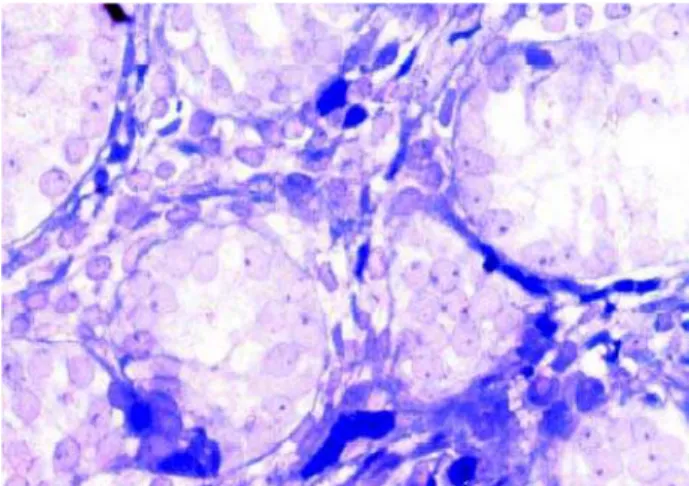
Figure 1 – Photomicrography of testicular parenchyma of a nontreated patient (Toluidin blue, X40).
COMMENTS
The efficiency of hCG as therapy for cryp-torchidism has been broadly discussed in its clinical and histological aspects. The testicular migration pro-cess have been studied by some authors, most of them focusing on the consequences of the changes that occur in the germinative epithelium (5,10,11). Most authors believe that on this aspect, the hCG use would be beneficial to patients.
Most studies on this subject relate a substan-tial collagen increase in the testes of elderly patients, accompanying testicular atrophy expected in this age, independently from cryptorchidism being or not a pre-vious condition (12). Nevertheless, some studies chal-lenge this report as a general rule, showing some cases in which 80 year old men presented histologic testicu-lar parameters simitesticu-lar to those of young men (13).
The literature describes an interaction be-tween extracellular matrix, tubular wall and germi-native cells, being very important to their normal de-velopment. (12,14).
In cryptorchid testes, some studies have been demonstrated an important increase in collagen quan-tity, resulting in interstitial fibrosis and in a thick-ened lamina propria of seminiferous tubules (3,15). Treatment with hCG would cause temporary histo-logic changes in testis, nevertheless, without deter-mining permanent damage (16).
Analyses of the groups with and without treat-ment with hCG showed a significant difference be-tween then, compatible to a possible prevention of collagen increase in patients submitted to hCG, re-ducing interstitial fibrosis and thickening of the lamina propria of the seminiferous tubules and, consequently, avoiding alterations in germinative cells (3,5).
The question is if the collagen alteration con-tributes in any way to prevent germinative cell alter-ations and, consequently, any worsening in fertility levels in cryptorchid patients without treatment. An-other possibility to prove the hCG efficiency in treat-ment of cryptorchid testes is the determination of se-miniferous tubules diameter (13,17). We compared our results of the seminiferous tubules diameter in cryp-torchid patients treated and nontreated with hCG to the normal male patients studied by Aya et al. (18). We observed that the mean diameter in treated and nontreated patients of our study was smaller than that of the normal children studied by Aya et al. (18). In this study, normal 7 year old children present a mean diameter of 88.7 µm, while in our study, the mean di-ameter in patients with the same age was 75.79 µm for treated patients and 56.14 µm for nontreated patients. In conclusion, the results of the present study suggest that hCG use as treatment for cryptorchid-ism, when employed in an early stage in life, could temporarily delay a progressive collagen increase. Relatively to tubular diameter, our results show that
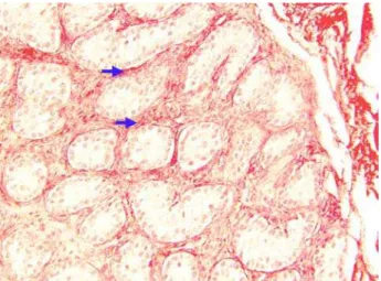
Figure 3 – Photomicrography of testicular parenchyma of a nontreated patient (picrosirius red, X400).
there is no statistically significant change between treated and nontreated patients.
ACKNOWLEDGEMENTS
This work was supported by Rio de Janeiro Foundation for Research Support - FAPERJ, and National Council for Scientific and Technological De-velopment - CNPq, Brazil.
CONFLICT OF INTEREST
None declared.
REFERENCES
1. Gracia J, Gonzales N, Gomez ME, Plaza L, Sanchez J, Alba J: Clinical and anatomopathological study of 2000 cryptorchid testes. Br J Urol. 1995; 75: 697-701. 2. Nistal M, Riestra ML, Paniagua R: Correlation between testicular biopsies (prepubertal and postpubertal) and spermiogram in cryptorchid men. Hum Pathol. 2000; 31: 1022-30.
3. Lala R, Matarazzo P, Chiabotto P, Gennari F, Cortese MG, Canavese F, et al.: Early hormonal and surgical treatment of cryptorchidism. J Urol. 1997; 157: 1898-901.
4. Gill B, Kogan S: Cryptorchidism. Current concepts. Pediatr Clin North Am. 1997; 44: 1211-27.
5. Hadziselimovic F, Herzog B: Treatment with a lutein-izing hormone-releasing hormone analogue after suc-cessful orchiopexy markedly improves the chance of fertility later in life. J Urol. 1997; 158: 1193-5. 6. Noh PH, Cooper CS, Snyder HM 3rd, Zderic SA,
Can-ning DA, Huff DS: Testicular volume does not predict germ cell count in patients with cryptorchidism. J Urol. 2000; 163: 593-6.
7. Polascik TJ, Chan-Tack KM, Jeffs RD, Gearhart JP: Reappraisal of the role of human chorionic gonadot-rophin in the diagnosis and treatment of the nonpalpable testis: a 10-year experience. J Urol. 1996; 156: 804-06.
8. Favorito LA, Toledo J: Study of testicular migration after treatment with human chorionic gonadotropin in patients with cryptorchidism. Braz J Urol. 2001; 27: 270-4.
9. Mandarim-de-Lacerda CA: Stereology in the normal and pathological morphologic research. Biomed Res. 1998; 9: 153-63.
10. Dunkel L, Taskinen S, Hovatta O, Tilly JL, Wikstrom S: Germ cell apoptosis after treatment of cryptorchid-ism with human chorionic gonadotrophin is associated with impaired reproductive function in the adult. J Clin Invest. 1997; 100: 2341-6.
11. Kennedy WA II, Huff D, Snyder HM 3rd: The value of testis biopsies in cryptorchidism. Contemp Urol. 1998; 4: 46-57.
12. Richardson LL, Kleinman HK, Dym M: Altered base-ment membrane synthesis in the testis after tissue in-jury. J Androl. 1998; 19: 145-55.
13. Paniagua R, Nistal M: Morphological and histometric study of human spermatogonia from birth to the onset of puberty. J Anat. 1984; 139: 535-52.
14. Yazama F, Esaki M, Sawada H: Immunocytochemis-try of extracellular matrix components in the rat semi-niferous tubule: electron microscopic localization with improved methodology. Anat Rec. 1997; 248: 51-62. 15. Santamaria L, Martinez-Onsorbe P, Paniagua R, Nistal M: Laminin, type IV collagen and fibronectin in nor-mal and cryptorchid human testes. An immunohis-tochemical study. Int J Androl. 1990; 13: 135-46. 16. Lackgren G, Ploen L: The influence of human
chori-onic gonadotrophin (hCG) on the morphology of the prepubertal human undescended testis. Int J Androl. 1984; 7: 39-52.
17. Schindler AM, Diaz P, Cuendet A, Sizonenko PC: Cryptorchidism: a morphological study of 670 biop-sies. Helv Paediatr Acta. 1987; 42: 145-58.
18. Aya M, Ogata T, Sakaguchi A, Sato S, Matsuo N: Tes-ticular histopathology in congenital lipoid adrenal hy-perplasia: a light and electron microscopic study. Horm Res. 1997; 47: 121-5.
Received: June 28, 2005 Accepted after revision: September 8, 2005
Correspondence address: Dr. Luciano Alves Favorito
Av. 28 de Setembro 87, fundos, prédio FCM, térreo Rio de Janeiro, RJ, 20551-030, Brasil
EDITORIAL COMMENT
The most important finding of the present study has been the amelioration at collagen contents in testes subjected to hCG administration. On the other hand, the effects of hCG were previously assessed through parameters such as the number of sper-matogonia, or Ad spermatogonia per tubular cross section (1,2). Therefore, the types of collagens sub-jected to alteration, and the means and mechanisms of this alteration require further clarification.
One of the mechanisms involved in the regu-lation of collagen production is G-protein linked sig-nal transduction. Adenylyl cyclase over-expression through cAMP elevating agents promotes an antifibrotic phenotype in pulmonary fibroblasts (3). The lutropin/choriogonadotropin receptor belongs to the family of G protein-coupled receptors. Binding of hCG to the receptor stimulates adenylyl cyclase activity (4). Stimulation of adenylyl cyclase activity might have involved in the decrease of collagen con-tent in testes subjected to hCG.
According to one of the current theories, un-descended testis is associated with a decrease in sym-pathetic, but an increase in parasympathetic tonus. Since the neurotransmitters involved in autonomic nervous system also act through receptors coupled to G- proteins, and undescended testis is associated with less stimuli to activate adenylyl cyclase, the differ-ences in signaling has been suggested to account for the alterations in an undescended testis (5,6).
The place of hormones in the process of de-scent and in the treatment of undescended testis that has been remained controversial, appears to remain controversial unless the definite understanding of the mechanism. The differences in collagen expressions may not only be important for the evaluation of the effects of hormones, but also may form another path-way that helps to unravel the mechanism of descent.
REFERENCES
1. Cortes D, Thorup J, Visfeldt J: Hormonal treatment may harm the germ cells in 1 to 3 year-old boys with cryptorchidism. J Urol. 2000; 163: 1290-2.
2. Hadziselimovic F, Zivkovic D, Bica DT, Emmons LR: The importance of mini-puberty for fertility in cryp-torchidism. J Urol. 2005; 174: 1536-9.
3. Liu X, Ostrom RS, Insel PA: c AMP-elevating agents and adenylyl cyclase overexpression promote an antifibrotic phenotype in pulmonary fibroblasts. Am J Physiol Cell Physiol. 2004; 286: C1089-99.
4. Jaquette J, Segaloff DL: Temperature sensitivity of some mutants of the Lutropin/choriogonadotropin re-ceptor. Endocrinology. 1997; 138: 85-91.
5. Tanyel FC: The descent of testis and reason for failed descent. Turk J Pediatr. 2004; 46 (Suppl): 7-17. 6. Tanyel FC: Obliteration of processus vaginalis:
aber-rations in the regulatory mechanism result in an in-guinal hernia, hydrocele or undescended testis. Turk J Pediatr. 2004; 46 (Suppl): 18- 27.
Dr. Feridun Cahit Tanyel
EDITORIAL COMMENT
Human chorionic gonadotropin (hCG) has been used in cryptorchidism for decades. The main goal of the hormonal treatment is to avoid operation or to make operation easier. In addition, it has been presumed that hCG might have beneficial effects on testicular function. Previously it has been observed that hCG enhances the maturation of gonocytes and even increases the amount of them. The article pre-sented in this issue shows that hCG treatment also prevents interstitial fibrosis at least in short term.
Surprisingly few studies, all with low num-ber of patients, have been performed dealing with the long term effects of the hCG treatment. In some stud-ies, it has been proposed that the beneficial effects of the hormone treatment on testicular function may last to the adulthood (1). However, there is also a certain controversy about the safety of the hormone treat-ment. For example in our own follow-up study (2) it turned out that the testes with preoperative hCG treat-ment were smaller in early adulthood than those with-out preoperative hormonal therapy. Histological analysis (3) suggested that hCG therapy initially in-creases the amount of germ cells, but after the test-osterone level decreases along with the withdrawal of the therapy, part of the germ cells meet apoptotic cell death. Similar effect can be seen in hormone with-drawal therapy of hormone dependent cancers. Thus it seems to be important in histological analyses to look, what is the time interval between hCG treat-ment and sample collection. Is the initially positive response later turning to opposite? Also the patient
age at treatment may be an important factor. In a study (4) hCG treatment had harmful long term conse-quences especially if it had been given to patients younger than three years of age. This is especially worrying, because patients should be treated at a young age to get optimal result in respect of fertility. It seems that studies with bigger amounts of patients treated at a young age and with a long term follow-up are needed before we can decide for sure if hormonal therapy has beneficial or harmful effects on testicular function in adulthood. Meanwhile at least I am favoring surgical therapy without hormonal pre-treatment.
REFERENCES
1. Hadziselimovic F, Herzog B: Treatment with a lutein-izing hormone-releasing hormone analogue after suc-cessful orchiopexy markedly improves the chance of fertility later in life. J Urol. 1997; 158: 1193-5. 2. Taskinen S, Wikstrom S: Effect of age at operation,
location of testis and preoperative hormonal treatment on testicular growth after cryptorchidism. J Urol. 1997; 158: 471-3.
3. Dunkel L, Taskinen S, Hovatta O, Tilly JL, Wikström S: Germ cell apoptosis after treatment of cryptorchid-ism with human chorionic gonadotropin is associated with impaired reproductive function in the adult. J Clin Invest. 1997; 100: 2341-6.
4. Cortes D, Thorup J, Visfeldt J: Hormonal treatment may harm the germ cells in 1 to 3-year-old boys with cryptorchidism. J Urol. 2000; 163: 1290-2.