555
CHRONIC HEMATOCELE OF THE VAGINALIS SAC Case Report
International Braz J Urol
Official Journal of the Brazilian Society of Urology
Vol. 31 (6): 555-557, November - December, 2005
IDIOPATHIC CHRONIC HEMATOCELE OF THE VAGINAL SAC
CARLOS ALVAREZ-ALVAREZ, LUIS A. FARINA-PEREZ, CELSO R. BARROS
Departments of Pathology (CAA), Urology (LAFP) and Radiology (CRB), Hospital POVISA, Vigo, Spain
ABSTRACT
We report a 39-year-old male who presented non-traumatic testicular swelling and pain. Physical examination and sonography presented a suspicion of testicular tumor and both surgical exploration and inguinal orchiectomy were performed. Hematocele may both clinically and sonographically resemble a testicular tumor. The diagnostic study of choice is magnetic resonance, establishing the diagnosis and differentiating it from neoplasms.
Key words: testis; hematocele; magnetic resonance imaging Int Braz J Urol. 2005; 31: 555-7
INTRODUCTION
Differential diagnosis of a scrotal mass in-cludes inflammatory conditions, malignant tumors and traumatic lesions including hematomas. Acute he-matocele is commonly associated with testicular trauma, but some cases may be idiopathic. The clini-cal presentation is that of a testicular tumor, and the main clinical significance lies in the difficulty encoun-tered in excluding malignant lesions preoperatively (1). Correct management includes early recognition and treatment to preserve the testicle.
CASE REPORT
A 39-year-old man with a history of meningitis presented a 1-month history of testicular pain and scrotal swelling. The patient did not refer previous trauma in the area. Scrotal examination revealed an enlarging hard, nontender mass in the right side. Testicular tumor markers were within normal range. Ultrasonography showed an extra testicular mass with solid hyperechoic areas separated by irregular
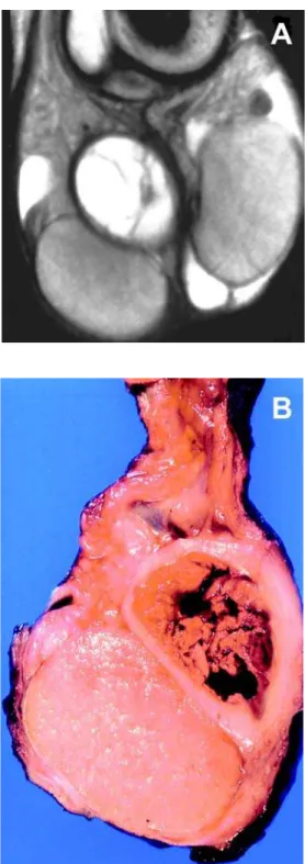
septations, suggesting an abscess or hematocele (Figure-1). Magnetic resonance imaging (MRI) revealed a well-defined encapsulated right solid mass of high signal intensity on T1-weighted images in the region of the epididymal body (Figure-2). During the surgical exploration, an encapsulated hematoma in the close vicinity of the testicle and epididymis was observed. As the lesion was severely attached to the spermatic cord, testicle could not be saved, and inguinal orchiectomy was performed. Gross examination of the resected specimen showed a cavity containing blood clots with a thick capsule that isolated the lesion from the surrounding normal structures (Figure-2). Histopathological examination showed abundant macrophages, hemosiderin, and dystrophic calcification, consistent with an organized hematoma. The diagnosis was idiopathic chronic hematocele.
COMMENTS
previ-556
CHRONIC HEMATOCELE OF THE VAGINALIS SAC
ous history of testicular trauma, and are admitted to the hospital due to acute testicular pain (2).
According to an etiological basis, hematoceles can be classified as idiopathic and secondary, although most of the idiopathic are thought to be secondary to asymptomatic trauma or infection as well. Idio-pathic or spontaneous bleeding seems to be more common in elderly patients (1), and hematoma is the result of blood leakage into the vaginalis sac. Non-traumatic secondary hematoceles can be pro-duced by hematological alterations or vasculitis (3). The etiology of the hematocele formation in our patient is unknown.
Hematocele is difficult to diagnose preop-eratively because its symptoms may mimic cysts or neoplasms. Testicle exploration shows a mass, and
Figure 2 – A) MRI showing a hemorrhagic encapsulated lesion in the region of the epididymal body. B) Surgical specimen. He-matoma in the paratesticular area is pushing the testis toward the periphery.
557
CHRONIC HEMATOCELE OF THE VAGINALIS SAC
CONFLICT OF INTEREST
None declared.
REFERENCES
1. Leibovitch I, Ramon J, Ben Chaim J, Nass D, Goldwasser B: Chronic hematocele complicating renal failure and hemodialysis. J Urol. 1991; 146: 162-4. 2. Altarac S: Management of 53 cases of testicular trauma.
Eur Urol. 1994; 25: 119-23.
3. Pascual Regueiro D, Garcia de Jalon Martinez A, Trivez Boned MA, Sancho Serrano C, Gracia Montoliu S, Rioja Sanz LA: Idiopathic superinfected giant he-matocele. Actas Urol Esp. 2003; 27: 645-8.
ultrasonography is very helpful for screening of he-matocele, but presents some difficulties to its final diagnosis (2). MRI has a higher sensitivity, allows clear demonstration of blood, and can be regarded as mandatory for the diagnosis. Surgical explora-tion may be needed when the diagnosis is in ques-tion.
Conservative approach usually produces un-satisfactory results (2). Correct management includes early recognition and complete evacuation of the he-matoma to avoid testicular compression (2), prevent-ing epididymo-orchitis, abscess formation and necro-sis, as unresorbed hematocele can eventually be in-fected.
Received: April 15, 2005 Accepted after revision: June 20, 2005
Correspondence address: Dr. Carlos Álvarez-Álvarez
Department of Pathology, Hospital POVISA C/Salamanca 5, Vigo, 36211, Spain
Fax: + 34 9 8642-1439