Laparoscopic hepatectomy: indications and results from
18 resectable cases
Hepatectomia laparoscópica: indicações e resultados em 18 casos ressecados
Sergio Renato Pais-Costa1, Sergio Luiz Melo Araujo1, Olímpia Alves Teixeira Lima2, Alexandre Chartuni Pereira Teixeira1
ABSTRACT
Objective: To evaluate the early and late results from laparoscopic hepatectomy procedures at a tertiary hospital in Brasília (DF), Brazil.
Methods: The authors report on a series of 18 patients (11 women) who underwent laparoscopic hepatectomy performed by a single surgical team at Santa Lúcia Hospital, in Brasília, between June 2007 and December 2010. Age ranged from 21 to 71 years (median = 43 years). There were eleven women and seven men. Nine patients had benign diseases and nine had malignant lesions. The lesion diameter ranged from 1.8 to 12 cm (mean: 4.96 cm). Results: Six major hepatectomy procedures and 12 minor hepatectomy procedures were performed. The mean duration of the operation was 205 minutes (range: 90 to 360 minutes). The mean intraoperative blood loss was 300 mL (range: 100 to 1,500 mL). Two patients received a transfusion (11%). There was one conversion to open surgery. There was no death and no patient underwent reoperation. The postoperative morbidity rate was 11% (n = 2). One patient presented with a minor complication (lobar pneumonia) while other presented with two major complications (intraoperative bleeding and incisional hernia). The median length of hospital stay was 4 days (range: 2 to 11 days). The median time to return to normal activities was 13 days (range: 7 to 40 days). Conclusion: Laparoscopic hepatectomy is a safe surgical approach for treating both benign and malignant hepatic lesions. This small series showed no mortality, low morbidity and good cosmetic results.
Keywords: Laparoscopy; Hepatectomy; Liver neoplasms/surgery; Neoplasm metastasis
RESUMO
Objetivo: Avaliar os resultados precoces e tardios das hepatectomias laparoscópicas realizadas em um hospital terciário, em Brasília (DF).
Métodos: Os autores relatam uma série de 18 pacientes (11 mulheres) submetidos à hepatectomia laparoscópica, realizada por uma única equipe cirúrgica do Hospital Santa Lúcia, em Brasília, entre Junho de 2007 e Dezembro de 2010. A idade variou de 21 a 71 anos com
mediana de 43 anos. Havia onze mulheres e sete homens. Nove casos apresentavam lesão benigna e nove, lesão maligna. O diâmetro da lesão variou de 1,8 a 12 cm (média: 4,96 cm). Resultados: Seis hepatectomias maiores e 12 hepatectomias menores foram realizadas. O tempo cirúrgico médio foi de 205 minutos (variação de 90 a 360 minutos). A média de sangramento intraoperatório foi de 300 mL (variação de 100 a 1.500 mL). Dois pacientes foram transfundidos. Houve uma conversão para cirurgia aberta. Não houve óbitos e nenhum paciente foi reoperado. A morbidade pós-operatória foi de 11% (n = 2). Um indivíduo apresentou uma complicação menor (pneumonia lobar), e outro teve duas complicações maiores (sangramento intraoperatório e hérnia incisional). A duração mediana de internação foi de 4 dias (variação de 2 a 11 dias). O tempo mediano de retorno às atividades diárias foi de 13 dias (variação de 7 a 40 dias). Conclusão: A hepatectomia laparoscópica é um método cirúrgico seguro para tratamento de lesões hepáticas benignas e malignas. Nesta pequena série, não houve óbitos, a taxa de morbidade foi baixa, e o resultado estético foi bom.
Descritores: Laparoscopia; Hepatectomia; Neoplasias hepáticas/ cirurgia; Metástase neoplásica
INTRODUCTION
Laparoscopic hepatectomy (LH) was first described in 1992 by Gagner et al.(1) and it remains an appealing
concept: major surgery with a potential for bleeding, carried out using a minimally invasive approach. Azagra et al.(2) performed the first anatomical laparoscopy,
which consisted of a successful left lateral sectionectomy (LLS) or sectorectomy (segments II-III) in a patient with hepatic adenoma (HA) of segments II and III. LH was the last bastion to fall to laparoscopic surgery (LS) because of a combination of the anatomical complexity of this surgical approach and the lack of surgeons with experience in both laparoscopy and hepatic surgery. In
Study carried out at Hospital Santa Lúcia – Brasília (DF), Brazil.
1 Hospital Santa Lúcia – Brasília (DF), Brazil.
2 Universidade de Brasília – UNB, Brasília (DF), Brazil; Hospital Santa Lúcia – Brasília (DF), Brazil.
Corresponding author: Sergio Renato Pais Costa – SEPS 710/910, conjunto D, sala 330 – CEP 70390-108 – Brasília (DF), Brazil – E-mail [email protected] The authors declare there is no conflict of interest.
general lines, LH offers several advantages over an open surgery. The main advantages are less postoperative pain, early mobilization, lower incidence of ileus, earlier resumption of oral intake, and shorter hospital stay. Initially, minor hepatectomy for superficial lesions was performed with great confidence. With advances in laparoscopic instruments and parenchymal transection devices, together with greater experience of complex laparoscopic hepatobiliary resections, the use of both right and left major laparoscopic hepatic resections has grown(1-27).
Over recent years, LH has been performed with unfortunate morbidity but with low mortality in reference centers(3-7,15-17). Recently a few authors have taken the view
that LH should be the preferred approach towards both benign and malignant hepatic lesions(3-5,16,17). Even
difficult-to-reach lesions in the right hepatic lobe may be resectable by means of a safe and satisfactory laparoscopic approach, with low conversion rates and morbidity(7,16,20,22,27).Thus, the
laparoscopic approach has begun a new era in minimally invasive hepatobiliary surgery.
On the other hand, except for a few studies, most papers have reported on limited numbers of patients. Nevertheless, it has been established that for selected patients, and when performed by a team with expertise in both hepatic and advanced laparoscopic surgery, LH is safe and produces results identical to those from open operations(4,5,16,17,23). In Brazil, although there have only
been a few anecdotal case reports(19,20,25,26) and few small
series have been reported(21-23,27), Machado et al.(23) showed
that LH was efficacious, with good results regarding colorectal metastasis (CRM).
OBJECTIVE
The aim of the present study was to describe both the short- and long-term results of LH used to treat benign and malignant liver disease, performed by a single surgical team in a private reference hospital in Brasília (DF), Brazil.
METHODS
Between June 2007 and December 2010, 18 consecutive LHs were performed at the Hospital Santa Lúcia, Brasilia. All resections were performed by a single surgical team. Nine LH procedures were performed to treat benign hepatic lesions, and nine for malignant lesions. The indications for laparoscopic resection of benign liver tumors were preoperative diagnosis of HA with 5 cm of diameter or more or cystadenoma; uncertain diagnosis on imaging or biopsy; and presence of symptoms. The laparoscopic approach was chosen because of the size and location of the lesions. Large tumors, tumors close to
major vascular structures, and tumors located in central positions were excluded from this sample.
Liver resections were defined in accordance with the International Hepato-Pancreato-Biliary Association (IHPBA) terminology, derived from the Couinaud classification. Subsequently, major hepatectomy was defined as the resection of three or more segments. Ultrasonography, computed tomography and magnetic resonance imaging were performed on all patients. For malignant lesions, PET-scans were also performed. Tumor markers CEA, AFP, and Ca 19.9 were assayed in all cases. The surgical technique for laparoscopic hepatic resection was determined case by case, in accordance with previously described technical principles(6,7,18,20-23,27).
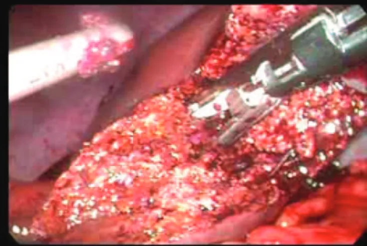
In general, the procedures were performed with carbon dioxide pressure control over the pneumoperitoneum, with a positive pressure of 12 mmHg. A 30-degree laparoscope was used with four or five port sites (Figure 1) depending on the case, and in accordance with the surgeon’s preference and site of the lesion. Liver transection (Figure 2) was always performed using Ligasure (10 mm size, Valleylab, Boulder, USA). Small vascular or biliary ducts were sealed using the Ligasure, while major structures were sealed using metal clips. Portal pedicles and hepatic veins were divided using a linear stapler (Endogia, 30 or 45 mm, vascular type), in accordance with Gumbs et al.(13).
Except for two cases in which the patients presented with an abdominal incision due to prior open surgery (one case of right subcostal and one case of median laparotomy, according to Figure 1), the surgical specimen was resected by means of a Pfannenstiel incision. The surgical specimen was placed into a plastic bag or glove. Abdominal drainage was generally not performed. When necessary, suction drains were used (in three cases).
Demographic characteristics of 18 patients and lesion etiology are shown in chart 1. There were 11 women and seven men. The mean age was 43 years (range: 21 to 71 years). Five patients (30%) were younger than 40 years. The lesion was solitary in nine patients (50%), while six patients presented two (35%) or more lesions (15%). Right-side lesions were predominant, with ten cases, while
the left lobe was involved in eight cases. The etiology of the hepatic lesions was as follows: adenoma (n = 3), metastasis (n = 7), hepatocarcinoma (HCC) (n = 2), nodular focal hyperplasia (NFH) (n = 2), hepatic abscess (n = 2), hemangioma (n = 1) and biliary cystadenoma (n = 1). The main symptoms were pain (n = 8), palpable mass (n = 6), discomfort (n = 5) and early satiety (n = 4). The lesion diameter ranged from 1.8 to 12 cm (mean: 4.96 cm).
Preoperative radiological investigations showed that 15 patients presented with a solid liver tumor (80%), and three patients had cystic lesions (20%). Eight out of 17 patients underwent tumor biopsy (percutaneously in 6 cases, laparoscopically before starting liver resection in 1 case, and laparoscopically in 1 case). In this series, tumor biopsy allowed us to obtain a certain diagnosis in four cases (50%, i.e., in four out of eight biopsies).
In ten patients, the surgical indication was malignant or premalignant disease. There were two cases of HCC, two of non-colorectal non-neuroendocrine metastases (NCNEM), five of colorectal metastases (CRM), and one of hepatic cystadenoma. In the other eight patients with benign disease, the most common indications were presence of symptoms, HA (diameter > 5 cm), uncertain preoperative diagnosis, or failure of percutaneous treatment of hepatic abscess.
Among the benign solid tumors, typical preoperative features of HA and hemangioma were found in all patients (n = 4). All presented with symptoms, such as pain and discomfort. One case presented with multiple adenomatosis with three lesions: this patient reported having made abusive use of anabolic steroids. Histological
Figure 2. Transection of hepatic parenchyma with Ligasure 10 mm – right hemi-hepatectomy
Case Gender Age Etiology Number Diameter
of the largest lesion (cm)
Site -
Hepatic segment(s) ASA
1 Male 71 Hepatic pyogenic abscess 1 10 II/III/IV 2
2 Female 68 Hepatic pyogenic abscess 3 5 II/III/VI 2
3 Female 63 Hepatic cystadenoma 1 12 VI/VII/VIII 1
4 Female 21 Hepatic adenoma 3 3 VI/VII 1
5 Female 38 Hepatic adenoma 1 5 VI 1
6 Female 23 Hepatic adenoma 1 5 VI/VII 1
7 Male 58 Focal nodular hyperplasia 1 6 II/III/III 1
8 Male 63 Hepatocellular carcinoma 1 3 VI/VII 2
9 Male 61 Colorectal metastasis 2 3.5 II/III/IV 1
10 Female 30 Focal nodular hyperplasia 1 8 V 1
11 Female 32 Non colorectal metastasis 2 3 II/III 1
12 Female 43 Non-colorectal metastasis 3 3 VI/VII 1
13 Male 63 Hepatocellular carcinoma 1 3 V 2
14 Female 43 Colorectal metastasis 3 4 V/VI/VII 1
15 Female 54 Colorectal metastasis 2 3 II/III 1
16 Male 50 Colorectal metastasis 3 3 II/III 2
17 Female 53 Colorectal metastasis 1 4 VI/VII 1
18 Male 45 Hepatic hemangioma 1 10 II/III/IV 1
Chart 1. Demographic characteristics and etiology of lesions
examination confirmed the preoperative diagnosis in all these patients. Neither of the cases of NFH was diagnosed preoperatively, since both cases presented with atypical radiological findings. The indication for liver resection in these patients was an uncertain diagnosis (differential with HCC) in one case, and right upper quadrant pain due to a bulky hanging tumor located in the V hepatic segment in the other case. Both cases of hepatic abscess had previously been unsuccessfully treated by means of percutaneous drainage. Subsequently, LH was performed because both lesions had central locations within the hepatic lobe, with partial destruction of the parenchyma.
RESULTS
The laparoscopic procedure was completed in 17 patients (94%). One patient who presented with a giant hemangioma (10 cm) underwent open conversion due to massive intraoperative bleeding. The types and details of the hepatectomy procedures are shown in chart 2 and chart 3. They included 6 major hepatectomy procedures (Figure 3) and 12 minor hepatectomy procedures (Figure 4). Two patients required postoperative blood transfusions. Three patients underwent surgical drainage of the liver bed using suction drains. The drains were taken out on the 4th or 5th postoperative days. There was
no case of gas embolism in this series.
The mean duration of the operation was 205 minutes. For the initial cases (n = 5), the mean duration of the operation was greater than in the subsequent operations (257 versus 197 minutes). However, most
of the major hepatectomy procedures (80%) were performed in the latter cases. There was no mortality in this series. Postoperative complications occurred in two patients who underwent one right hemi-hepatectomy and one left hemi-hepatectomy (11%) (Chart 3). One patient presented infectious left lobar pneumonia that was treated with antibiotics. Other patient who experienced intraoperative bleeding and underwent open conversion finally presented with an incisional hernia (90th postoperative day). There was no biliary
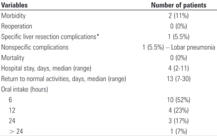
leakage or hepatic insufficiency. There was no case of reoperation in this series. Oral intake was resumed on the first postoperative day in all except one patient who underwent open conversion. The median hospital stay was 4 days (range: 2 to 11 days). All the patients except one used low doses of common analgesics, such as dipirone, during their postoperative course (1 or 2 days). One patient used narcotic analgesia during the postoperative period. The median time taken for the patients to return to their normal activities was 13 days (range: 7 to 40 days). Characteristics of the postoperative period are shown in table 1.
The mean follow-up time in this series was 14 months (median: 15 months; range: 1 to 27 months). All the symptomatic patients achieved complete symptom relief.
Type of resection Number of patients
Right hemi-hepatectomy 2
Left hemi-hepatectomy 4
Bisegmentectomy of segments II+III 4 Bisegmentectomy of segments VI +VII 5 Monosegmentectomy of segment V 2 Monosegmentectomy of segment VI 1
Total 18
Chart 2. Types of hepatectomy performed
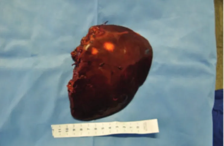
Figure 3. Surgical specimen – left hemi-hepatectomy (segments II-III-IV) from giant multiseptated abscess in left lobe
Feature Number of patients
Vascular clamping 1 (18)
Intraoperative blood loss, mL, mean (range) 300 (100-1500) Transfusions received 2 (18) Duration of surgery, min, mean (range) 240 (90-360) Mean weight of specimen, g, mean (range) 285 (57-1040)
Chart 3. Surgical features
Among the cancer patients, there was no recurrence. All of them experience good quality of life.
DISCUSSION
LH is a progression in the continuing evolution of minimally invasive surgery (MIS), in general, and laparoscopic liver surgery, in particular. Advances in expertise related to laparoscopic procedures, ongoing technological advances in laparoscopic devices, and increased patient awareness of availability of these techniques have created evolving interest in applications of these techniques in LH(16).
The surgical skills required for LH have evolved in parallel with the adaptation of laparoscopic techniques to this procedure. Hilar dissection, biliary or vascular repair, mobilization of the liver, and transection of the parenchyma are more technically demanding and potentially more dangerous than other previously
reported laparoscopic procedures. Anatomical
hemi-hepatectomy requires a clear understanding of general liver anatomy, experience in advanced hepatobiliary surgery and, the ability to dissect major vascular and biliary structures using a laparoscopic approach(10,14,16,21,23).
Despite the various obstacles and challenges, LH shows great advantages over open hepatectomy. The major advantages of LH are those of all laparoscopic surgical procedures. LH causes less tissue damage, and this has been associated with lower levels of postoperative pain, fewer peritoneal adhesions, shorter hospital stay, and an earlier return to daily activity(3,4,7,11).Additionally,
two recently published case-control studies(16,17) and one
cohort study(5) showed that LH provided lower blood
loss, reduced morbidity, fewer operative complications overall and, specifically with regard to malignant disease, no significant difference either in tumor recurrence or in long-term survival(15- 17,29,30).Furthermore, the cosmetic
advantages are excellent when LH is performed. It is particularly important when performed to treat benign disease(3,14,24).Earlier resumption of oral intake is also
a great advantage, considering that hepatectomy is a major surgical procedure. For these reasons, the laparoscopic approach should be taken into account, both for benign and for malignant liver disease management(4,5,8,12,16,17,23).
According to some authors(3,11,14,17,23), the use of the
laparoscopic route should not modify or broaden the indications for either benign or malignant liver disease. The same principles applied to open hepatic surgery must be respected. Therefore, especially for benign disease, it should be reserved for symptomatic lesions, specific complications, or even uncertain diagnoses (differential with primary or metastatic neoplasms). In particular, for HA, because of the high risk of rupture and malignant degeneration, patients should be offered more liberal surgical resections(14).
Despite the initial skepticism about the use of LH to treat malignant neoplasms, currently it is frequently performed since it is a safe and efficient procedure. Some authors(4,11,15-17) took the view that LH is as safe as
conventional open hepatectomy.
For left lesions, some authors considered LH to be the initial approach in reference centers, performed by surgeons with high levels of expertise(3-5,15,17).Campos et
al.(4) recently published a single series of left laparoscopic
resections in which the clear advantages of the laparoscopic approach were observed. More recently, in a cohort study that compared laparoscopy and open LLS, Carswell et al.(5) observed that laparoscopy was
superior because of the lower need for postoperative opiate analgesia and shorter postoperative hospital stay. In the present study, it was noted that opiates were used and, specifically regarding the LLS procedures, none of the patients presented complications on the third postoperative day. It was also observed in the present series that the results were similar, without postoperative opiate administration, for all except one patient who underwent open conversion (left hemi-hepatectomy for a giant hemangioma). Specifically regarding left resections, only one patient experienced a complication (intraoperative bleeding and incisonal hernia).
In a series of 78 patients, Zhang et al.(24) observed
totally successful laparoscopic liver resections with no conversion to open procedures, and only four patients received transfusions. In the present series, there was one perioperative complication (intraoperative bleeding), and only two patients received transfusions (in a case of major right hepatectomy due to a large cystadenoma of 12 cm in diameter and one case of left hemi-hepatectomy due to a giant hemangioma).
Variables Number of patients
Morbidity 2 (11%)
Reoperation 0 (0%)
Specific liver resection complications* 1 (5.5%) Nonspecific complications 1 (5.5%) – Lobar pneumonia
Mortality 0 (0%)
Hospital stay, days, median (range) 4 (2-11) Return to normal activities, days, median (range) 13 (7-30) Oral intake (hours)
6 10 (52%)
12 4 (23%)
24 3 (17%)
> 24 1 (7%)
Table 1. Postoperative course
Although the rate of conversion to open surgery has ranged from 0 to 15%(14,23), it depends on the type of
resection, experience of the team, and volume of the lesion. The present open conversion rate is similar to that found in literature.
Although the initial experiences of right liver resection were technically demanding, some authors(5,16,21,22,27) have
taken the view that the laparoscopic approach should be the preferred choice, even for posterior right lesions (segments VI-VII). In the present series, despite the fact that the sample was small, there were more cases of right resections, including two cases of formal right hepatectomy, five cases of posterior right sectionectomy (SVI-VII), two cases of segmentectomy of segment V, and one case of segmentectomy of segment VI. The major advantage of LH for resecting posterior right lesions is that it avoids the large open incision that is generally necessary to access posterior pedicles(7,27),
although right-side hepatic resections not only are technically more difficult, but also produce higher conversion rates than left resections(11,16,19,23).LH for
right lesions can be considered feasible and safe, as Cho et al.(7) have shown in a recent study. For the posterior
right sectionectomy (PRS) in the present series, an intra-hepatic approach was preferred, as already described in Brazil by Machado et al.(22).This technique
was previously published by the authors of the present article(27). This is safe and minimizes the intraoperative
bleeding: in the present series, none of the five patients who underwent PRS received any transfusions and the intraoperative bleeding was minimal.
In one of the largest series, with 300 minimally invasive liver resections (MILR) that were compared with open procedures (among which there were 64 cases of right hepatectomy and 8 cases of extended right hepatectomy), Koffron et al.(16) observed that
the laparoscopic approach was superior to the open technique. The advantages were the duration of the operation (99 versus 182 minutes), blood loss (102 versus
325 mL), transfusion requirement (2 out of 300 cases
versus 8 out of 100 cases), length of hospital stay (1.9
versus 5.4 days), overall operative complications (9.3
versus 22%), and local malignant recurrence (2 versus
3%). These authors concluded that the outcomes from MILR compared favorably with those of the standard open operation. In a previously published study by the present author(29) on a single series of cases of open
hepatectomy (OH) due to metastasis (n = 30 cases), in which almost all of the patients were operated by the senior author (Costa SRP), the overall mortality was 3% while the morbidity rate was 46%. The reoperation rate was 16%, and 44% of the patients received transfusions. The median blood loss was 800 mL. Although it was difficult to compare the outcomes between the
laparoscopic series and a contemporaneous open series, given the higher proportion of major hepatic resections
(66 versus 35%) and greater severity of metastatic
disease (age, nutritional state, and associated diseases) in the open series, and without any specific statistical test to compare these samples, the laparoscopic series had some advantages. Respectively, there was less mortality
(0 versus 3%), morbidity (46 versus 11%), transfusions
(44 versus 11%), blood loss (300 versus 800 mL), and
reoperation (0 versus 16%).
In Brazil, a few authors(18-23,25-27) published studies on
LH. Machado et al.(21) observed both low morbidity and
zero mortality in LH procedures performed due to CRM, and their results were similar to the present series. This has also been observed in more recent studies, in which mortality was generally zero while general morbidity ranged from 0 to 10%(3,4,16,17,21,27).However, with regard
to right resections, the morbidity is proportionally higher than in left resections, as described by Koffron et al.(16), which suggests that right lesions might be
more difficult to treat. Cho et al.(7) observed an overall
morbidity rate of 28% when considering only the right liver resections. In the present series, there was no reoperation, thus differing from what some authors have previously reported(23). However, major hepatectomy
only accounted for 30% (n = 6) of the present series, which may have contributed towards the low morbidity rate in this series(7).
To date, with regard to malignant disease, studies suggested that there is no difference between LH and OH in relation to port-site metastasis, free margins, local-systemic recurrence, or even survival rates(11,12,16,17,21,29,30).
However, there have only been a few match-controlled studies, with no ideal level of evidence. Historical series have shown no difference between LH and OH performed on malignant disease.
Nevertheless, such findings should be viewed with caution, and new studies need to be conducted in order to answer these unresolved questions.
CONCLUSION
LH is a safe surgical approach towards focal hepatic lesions consisting of either benign or malignant disease. In the small series presented here, both zero mortality and low morbidity were associated with a good cosmetic result.
REFERENCES
1. Gagner M, Rheault M, Duluc JL. Laparoscopic partial hepatectomy for liver tumour [abstract]. Surg Endosc. 1992; 6: 99.
3. Ardito F, Tayar C, Laurent A, Karoui M, Loriau J, Cherqui D. Laparoscopic liver resection for benign disease. Arch Surg. 2007;142(12):1188-93; discussion 1193.
4. Robles Campos R, Marín Hernández C, López Conesa A, Abellán B, Pastor Pérez P, Parrilla Paricio P. [Laparoscopic resection of the left segments of the liver: the “ideal technique” in experienced centres?]. Cir Esp. 2009;85(4): 214-21. Spanish.
5. Carswell KA, Sagias FG, Murgatroyd B, Rela M, Heaton N, Patel AG. Laparoscopic versus open left lateral segmentectomy. BMC Surg. 2009;9:14..
6. Cherqui D, Husson E, Hammoud R, Malassagne B, Stéphan F, Bensaid S, et al. Laparoscopic liver resections: a feasibility study in 30 patients. Ann Surg. 2000;232(6):753-62.
7. Cho JY, Han HS, Yoon YS, Shin SH. Outcomes of laparoscopic liver resection for lesions located in the right side of the liver. Arch Surg 2009;144(1):25-9.
8. Cugat E, Marco C. [Laparoscopic liver surgery. A mature option?]. Cir Esp. 2009;85(4):193-5. Spanish.
9. D´Albuquerque LAC, Herman P. Hepatectomia por videolaparoscopia. Realidade? Arq Gastroenterol. 2006;43(3):243-6.
10. Dulucq JL, Wintringer P, Stabilini C, Berticelli J, Mahajna A. Laparoscopic Liver resections: a single center experience. Surg Endosc. 2005:19(7):886-91. 11. Gagner M, Rogula T, Selzer D. Laparoscopic liver resection: benefits and
controversies. Surg Clin North Am. 2004;84(2):451-62.
12. Gigot JF, Glineur D, Santiago Azagra J, Goergen M, Ceuterick M, Morino M, Etienne J, Marescaux J, Mutter D, van Krunckelsven L, Descottes B, Valleix D, Lachachi F, Bertrand C, Mansvelt B, Hubens G, Saey JP, Schockmel R; Hepatobiliary and Pancreatic Section of the Royal Belgian Society of Surgery and the Belgian Group for Endoscopic Surgery. Laparoscopic liver resection for malignant liver tumours: preliminary results of a multicenter European study. Ann Surg. 2002;236(1):90-7.
13. Gumbs AA, Gayet B, Gagner M. Laparoscopic liver resection: When to use the laparoscopic stapler device. HPB (Oxford). 2008;10(4):296-303.
14. Herman P, Coelho FF, Lupinacci RM, Perini MV, Machado MAC, D’Albuquerque LAC, et al. Ressecções hepáticas por videolaparoscopia. ABCD Arq Bras Cir Dig. 2009;22(4):226-32.
15. Kofron AJ, Geller D, Gamblin TC, Abecassis M. Laparoscopic liver surgery: shifting the management of liver tumors. Hepatology. 2006;44(6):1694-700. 16. Kofron AJ, Auffenberg BS, Kung R, Abecassis M. Evaluation of 300 minimally
invasive liver resections at a single institution. Ann Surg. 2007;246(3): 385- 92; discussion 392-4.
17. Lee KF, Cheung YS, Chong CN, Tsang YYY, Ng WWC, Ling E, et al. Laparoscopic versus open hepatectomy for liver tumours: a case control study. Hong Kong Med J. 2007;13(6):442-8.
18. Machado MAC, Makdissi FF, Surjan RC, Herman P, Teixeira AR, Machado MCC. Laparoscopic resection of the left liver segments using the intrahepatic Glissonian approach. Surg Endosc. 2009;23(11):2615-9.
19. Kalil AN, Giovenardi R, Camargo SM. Hepatectomia regrada por videolaparoscopia. Rev Col Bras Cir. 1999;25(4):287-9.
20. Machado MAC, Makdissi FF, Surjan RCT, Teixeira ARF, Bacchella T, Machado MCC. Hepatectomia direita por videolaparoscopia. Rev Col Bras Cir. 2007;34(3):189-92.
21. Machado MAC, Makdissi FF, Almeida FAR, Luiz-Neto M, Martins ACA, Machado MCC. Hepatectomia Laparoscópica no Tratamento das Metástases Hepáticas. Arq Gastroenterol. 2008;45(4):330-2.
22. Machado MA, Makdissi FF, Galvão FH, Machado MC. Intrahepatic Glisssonian approach for laparoscopic right segmental liver resections. Am J Surg. 2008;196:e38-42.
23. Machado MAC, Makdissi FF, Herman P, Surjan RC. Intrahepatic glissonian approach for pure laparoscopic left hemihepatectomy Journ J Laparoendosc Adv Surg Tech A. 2010;20(2):141-2.
24. Zhang L, Chen YJ, Shang CZ, Zhang HW, Huang ZJ. Total laparoscopic liver resection in 78 patients. World J Gastroenterol. 2009;15(45):5727-31. 25. Wiedekher JC, Ekermman M, Kondo W, Silveira FP, Fedrizzi F, Reimann A.
Segmentectomia lateral esquerda laparoscópica em hemangioma hepático. Rev Bras Videocir. 2004;2(2):83-7.
26. Costa SRP, Araujo SM, Lima AOT, Lobo M. Hemi-hepatectomia esquerda laparoscópica para o abscesso hepático piogênico. Brasília Méd. 2010;48(2):216-25.
27. Costa SRP, Araújo SM, Teixiera AO, Pereira AC. Setorectomia posterior direita laparoscópica no tratamento dos tumores-hepáticos. ABCD Arq Bras Cir Dig. 2010;23(4):275-9.
28. Costa SRP, Horta SHC, Henriques AC, Waisberg J, Speranzini MB. Hepatectomia para o tratamento de metástases colorretais e não-colorretais: Análise Comparativa em 30 casos operados. Rev Bras Colo-Proctol. 2009; 29(2):216-25.
29. Nguyen KV, Geller DA. Is laparoscopic liver resection safe and comparable to open liver resection for hepatocellular carcinoma? Ann Surg Oncol. 2009;16(7):1765-7.