A brief contextualization on IgG4 tubulointerstitial nephritis
based on a case report in south Brazil
Uma breve contextualização sobre a nefrite tubulo-intersticial por
IgG4 com base em um relato de caso no sul do Brasil
Authors
Karla Lais Pêgas 1,2
Eduardo Cambruzzi 3,4,5 Gisele Lobato 6
1 Santa Casa de Porto
Alegre.
2 Hospital N. Sra. da
Conceição.
3 Universidade Federal do
Rio Grande do Sul.
4 Universidade Luterana do
Brasil.
5 Instituto de Cardiologia
Fundação Universitária de Cardiologia.
6 Hospital Ernesto Dornelles.
Submitted on: 08/25/2015. Approved on: 10/27/2015.
Correspondence to:
Karla Lais Pêgas. Santa Casa de Porto Alegre. Rua Sarmento Leite, nº 187, 2º Andar, Porto Alegre, RS, Brazil. CEP: 51-3214-8409 E-mail: [email protected] DOI: 10.5935/0101-2800.20160036
A Doença relacionada a IgG4 (IgG4RD) é um processo inflamatório recente de etiolo-gia supostamente autoimune, que se carac-teriza por níveis séricos elevados de IgG4, um denso infiltrado mononuclear rico em plasmócitos IgG4 positivos e fibrose es-toriforme. A nefrite túbulo-intersticial é a manifestação renal mais comum, com diferentes graus de disfunção renal e acha-dos clínicos variáveis. Aqui, os autores de-screvem um novo caso de nefrite túbulo-intersticial associada a IgG4 (NTIgG4), e discutem critérios clínicos e patológicos. Paciente masculino, 72 anos, foi admitido no serviço hospitalar com queixa clínica de astenia, perda de força, emagrecimento e anosmia. A história prévia incluía Diabetes mellitus tipo 2. Os dados laboratoriais in-cluíam anemia normocrômica, proteinúria e elevação da creatinina. A ultrassonogra-fia/tomografia computadorizada renal bi-lateral revelou um parênquima heterogê-neo, com zonas densas e difusas irregula-res, áreas de fibrose nos polos superiores e hidronefrose. A biópsia renal mostrou um infiltrado mononuclear intersticial denso, com mais de 50 plasmócitos por campo de grande aumento, áreas irregulares de fibrose fibroblástica e colagênica, tubulite focal e glomérulos normais. A imunofluo-rescência revelou deposição granular leve de IgG e C3c na membrana basal tubular. A imuno-histoquímica foi positiva para CD138, cadeias leves Kappa e lambda, e IgG4 (cerca de quarenta e cinco plasmóci-tos IgG4 positivos por campo de grande aumento). O nível sérico de IgG4 estava aumentado. O diagnóstico de NTIgG4 foi então estabelecido. O paciente recebeu cor-ticoterapia e controle rigoroso da glicemia com insulina, com melhoria significativa dos sintomas e dos níveis de creatinina.
RESUMO
Palavras-chave: diagnóstico; imunidade; patologia; rim.
IgG4-related disease (IgG4RD) is a re-cent inflammatory process of supposed autoimmune etiology, which is characte-rized by elevated serum IgG4 levels, den-se lymphoplasmacytic infiltration rich in IgG4-positive plasma cells and storiform fibrosis. Tubulointerstitial nephritis is the most common renal manifestation, with different degrees of kidney dysfunction and variable clinical findings. Herein, the authors describe a new case of IgG4 tubu-lointerstitial nephritis (IgG4TN), and dis-cuss clinic and pathologic criteria. Male patient, 72 years-old, was admitted on hospital service with clinical complaint of asthenia, loss of strength, emaciation, and anosmia. Previous history included type 2
diabetes mellitus. Laboratorial data inclu-ded normochromic anemia, proteinuria, and creatinine elevation. Bilateral kidney ultrassonography/computed tomography revealed a heterogenous parenchyma, with diffuse irregular dense zones, areas of fibrosis on upper poles, and hydrone-phrosys. Kidney biopsy showed a dense interstitial lymphoplasmacytic infiltrate, with more than 50 plasma cell per high power field, irregular areas of fibroblastic and collagenous fibrosis, focal tubulitis, and normal glomeruli. Immunofluores-cence revealed mild granular deposition of C3c and IgG in the tubular basement membrane. Immunohistochemestry was positive for CD138, lambda and Kappa light chains, and IgG4 (around forty five IgG4 positive plasma cells per high power field). IgG4 serum level was increased. The diagnosis of IgG4TN was then esta-blished. The patient received corticothe-rapy and strict control of glycemia with insulin, with marked improvement of symptoms and creatinine levels.
ABSTRACT
INTRODUCTION
IgG4-related disease (IgG4RD) is a recently described systemic inflammatory syndrome that can involve multiple organs and/or determine elevation in serum total IgG or IgG4 levels.1,2 Sarles et al.3 firstly
reported patients with sclerosing pancreatitis and hyperglobulinemia, and suggested that the process was related to an autoimmune disease. IgG4RD can compromise liver, lacrimal glands, lymph nodes, mediastinum, breast, meninges, eye, skin, urinary bladder, gastrointestinal tract, and salivary glands. IgG4 tubulointerstitial nephritis (IgG4TN) can determine kidney disfunction and show storiform fibrosis and infiltration by high numbers of IgG4-positive plasma cells.4-6 Herein, the authors describe
a new case of IgG4TN and describe clinic and morphologic findings and diagnostic criteria of this uncommon process.
CASE REPORT
Male patient, 72 years, was admitted on hospital service with clinical complaint of asthenia, loss of strength, emaciation, and anosmia. On physical examination, no significant alterations were identified. Previous history included type 2 diabetes mellitus, and was negative for hypertension, smoking, and urinary infections. Laboratorial data included normochromic anemia (hemoglobin: 10.8 d/dL), lymphocytosis (62%), glycemia of 174 mg/dL, proteinuria (2.3 g/ day), creatinine elevation (2.1 mg/dL), creatinine clerance of 48 mL/min, total proteins equal to 9.3 g/ dL, albumin of 3.73 g/dl, IgG of 1,080 mg/dL, and IgA of 327 mg/dL.
Liver function tests showed normal levels. Rheumatoid factor, antineutrophil cytoplasmic antibodies, antimicrosomal antibody, HIV, HCV, and HBsAg were negative. Radiological chest imaging identified mediastinal lymphadenopathy and focal bilateral opacities suggesting granulomatous disease. Central nervous system computed tomography revealed no significant alterations.
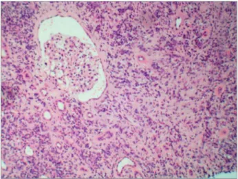
Bilateral kidney ultrassonography/computed tomography revealed a heterogenous parenchyma, with irregular diffuse dense zones, areas of fibrosis on upper poles, and hydronephrosys. No signs of obstructive urinary disease or neoplasm were identified. Kidney biopsy showed a dense interstitial lymphoplasmacytic infiltrate (Figures 1 and 2), with more than 50 plasma
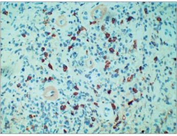
cell per high power field, irregular areas of fibroblastic and collagenous fibrosis (Figure 3), focal tubulitis, and normal glomeruli. Immunofluorescence revealed mild granular deposition of C3c and IgG in the tubular basement membrane. Immunohistochemestry was positive for CD138, lambda and Kappa light chains, and IgG4 (around forty five IgG4 positive plasma cells per high power field/figure 4).
Figure 1. IgG4 tubulointerstitial nephritis: Kidney biopsy showing a dense inflammatory infiltrate and fibrosis, hematoxylin-eosin, 40x.
Figure 2. IgG4 tubulointerstitial nephritis: Kidney parenchyma exhibiting a dense interstitial lymphoplasmacytic infiltrate and storiform fibrosis, with normal glomeruli, hematoxylin-eosin, 100x.
Figure 3. IgG4 tubulointerstitial nephritis: Numerous plasma cells in a high power field, hematoxylin-eosin, 400x.
Figure 4. IgG4 tubulointerstitial nephritis: IgG4 positive plasma cells in kidney interstitium, streptavidin-peroxidase polymer, 200x.
DISCUSSION
Tubulointerstitial nephritis is characterized by inflam-matory infiltration affecting tubules and interstitium of the kidney parenchyma, without compromising glomeruli and vessels.1,2,5,6 Tubulointerstitial nephritis
has two main forms of clinical presentation: acute, which is characterized by sudden onset and rapid de-cline of renal function, and chronic, which is charac-terized by slow decline of renal function. Most com-mon etiologic factors include drug toxicity, metabolic disorders, heavy metals, infections, and immunologic disorders.1,2,5,6
IgG4TN is a recent described interstitial nephropathy that can be associated to multiple organs involvement, and some data suggests IgG4 can be related to an autoimmune disorder.1,2,7,8 The
diagnosis of IgG4TN can be established based on clinic, radiologic, laboratorial and morphologic parameters. IgG4 production is associated with
T-helper cells type 2 activity, and IL4, IL13, IL10 and IL12 imbalance.1,2,7,8
Raissian et al.9 described that the main diagnostic
criteria for IgG4TN include: a) histological evidence of tubulointerstitial nephritis exhibiting numerous plasma cells, in which 10 IgG4-positive plasma cells are found in high power fields in the most exuberating zones, b) mandatory presence of immune complex deposits in a tubulointerstitial nephritis identified by immunofluorescence, immunohistochemestry or electron microscopy, c) elevated serum IgG4 or total IgG levels, d) imaging studies revealing small peripheral low-attenuation cortical nodules, diffuse patchy involvement of the kidneys, round/wedge-shaped lesions, or diffuse marked kidney enlargement, and e) evidences of other organ involvement, such as autoimmune pancreatitis, sclerosing cholangitis, sialadenitis, inflammatory aortic aneurysm, or inflammatory mass in any organ.
The majority of patients developing IgG4TN are males, with an average age of 65 years, with clinic findings of acute or progressive chronic kidney disorder.2,5,9,10 Clinical data includes fever, fatigue,
abdominal pain, proteinuria, and anorexia.2,5,10
Magnetic resonance can reveal iso/hypointense kidney lesions on T1-weighted images and hypointense areas on T2-weighted images.8,10,11,12 Serum levels of IgG4/
IgG are elevated around 85% of patients, which can be accompanied by elevated serum creatinine level and decreased C3, C4 or CH50 levels.8,10,11,12
On gross examination, the involved kidney reveals white, firm, homogeneous areas. The main histologic feature of IgG4TN is the presence of more than 10 IgG4-positive plasma cells per high-power field in the compromised areas, but more than 30 IgG4-positive plasma cells per high-power field can be found too.2,7,9,11 Histologic findings also include
lymphocytic infiltrate, some eosinophils, and variable degree of fibrosis. Inflammatory cells usually decrease in number with increasing fibrosis.2,7,9,11
Interstitial fibrosis is frequently zonal, determines collagenous bundles deposition encircling mononuclear inflammatory cells, with clear demarcation between compromised and non affected areas, and is more prominent at the center than peripheral zones. It is mandatory imaging data correlation in kidney biopsy due these aspects.2,5,7,9,11
changes are inespecific, except in cases related to membranous glomerulonephritis.7,6,9,11,13 No specific
vascular changes are associated with IgG4TN. IgG/ IgG4 deposits in the tubular basement membrane in a granular pattern are commonly identified in IgG4TN. Kappa and lambda light chains can be found too, with focal C1q and IgM staining, and the deposits in tubular basement membrane are restricted to the affected areas.1,2,5,8,11
The extension of fibrotic areas is accompanied with more IgG4 deposition in tubular basement membrane. Immunohistochemestry technique on paraffin sections is fundamental to quantify the infiltrating IgG4-positive plasma cells.2,8,9,11,14
Raissian et al.9 proposed three patterns to classify
the pattern of inflammation and fibrosis. The pattern A is characterized by less than 10% of interstitial fibrosis without expansive process. Pattern B exhibits expansive interstitial fibrosis and a more severe interstitial lymphoplasmacytic infiltrate. Pattern C shows a collagen-rich fibrosis with occasional inflammatory cells. Electron microscopy can be employed to evaluate the collagen deposition between fibroblasts. Table 1 shows some cases of IgG4TN found in the international literature and comparable to the reported case.
Differential diagnosis includes lupus nephritis, diabetic nephropathy, anti-neutrophil cytoplasmic
Authors Age/Gender Clinical Findings IgG4 serum
level Previous History Treatment/Outcome
Fukuhara et al.7 63/F
Incidental cystic tumor at the upper pole of the right kidney and multiple low attenuation areas in the left kidney
218 mg/dL No previous medical history
Surgery/Prednisolone 30 mg/day/
Asymptomatic
Miyata et al.6 69/M Anorexia, weight loss,
lower abdominal pain 2,750 mg/dL
Hypertension, Early-stage colon cancer
Prednisolone 55 mg/day/Proteinuria persisted for over 2 months
Nishikawa et al.15 51/F Incompatible Renal
Transplant 426 mg/dL
Bronchial asthma, Renal transplant
Methylprednisolone 16mg/day/Follow-up kidney biopsy revealed a markedly decrease in plasma cell infiltration
Wu et al.8 71/M
Diabetic nephropathy, polyarthralgia, high gammaglobulinemia, palpable lymph nodes in the neck and axillary region
532 mg/dL Diabetes mellitus
Prednisolone 30 mg/day, Cyclophosphamide 50 mg/day/Laboratory parameters returned to reference range
Stylianou et al.13 63/M
Weakness, anorexia, weight loss, cervical lymphadenopathy, sialadenitis
1,210 mg/dL
Hypertension, vitiligo, alithiasic choleystitis
Methylprednisolone 36mg/day/Rapid improvement of symptoms and renal function
Wada et al.14 59/M
Edema of lower extremities, jaundice, upper abdominal tenderness
1,920 mg/dL No previous medical history
Prednisolone 40 mg/day/Rapid improvement of symptoms
Otani et al.12 58/M Rectal cancer,
lymphadenopathy 2,990 mg/dL
Sinusitis, allergic rhinitis
Prednisolone 20 mg/ day/Improvement of symptoms
Present Report 72/M
Asthenia, loss of strength, emaciation, and anosmia
376 mg/dL Diabetes mellitus
Prednisolone 54 mg/ day/Improvement of symptoms
antibody associated glomerulonephritis, Sjögren Syndrome, chronic pyelonephritis, membranous glo-merulonephritis, drug-induced interstitial fibrosis, granulomatous lesions, and idiopatic tubulointersti-tial nephritis.5,9,11,12,13,15 The patients compromised
wi-th IgG4TN can response to steroid/immunowi-therapy around 90% of cases. Rebiopsy can shows evidences of attenuating fibrosis areas and decrease in lympho-plasmacytic infiltrate.5,8,10,13,14,15
REFERENCES
1. Kawamura E, Hisano S, Nakashima H, Takeshita M, Saito T. Immunohistological analysis for immunological response and mechanism of interstitial fibrosis in IgG4-related kidney disea-se. Mod Rheumatol 2015;25:571-8. DOI:http://dx.doi.org/10. 3109/14397595.2014.1001474
2. Kuroda N, Nao T, Fukuhara H, Karashima T, Inoue K, Tani-guchi Y, et al. IgG4-related renal disease: clinical and patholo-gical characteristics. Int J Clin Exp Pathol 2014;7:6379-85. 3. Sarles H, Sarles JC, Muratore R, Guien C. Chronic
inflamma-tory sclerosis of the pancreas-an autonomous pancreatic disea-se? Am J Dig Dis 1961;6:688-98.
4. Deshpande V, Chicano S, Finkelberg D, Selig MK, Mino--Kenudson M, Brugge WR, et al. Autoimmune pancreatitis: a systemic immune complex mediated disease. Am J Surg Pa-thol 2006;30:1537-45. DOI:http://dx.doi.org/10.1097/01. pas.0000213331.09864.2c
5. Kawano M, Saeki T. IgG4-related kidney disease-an update. Curr Opin Nephrol Hypertens 2015;24:193-201. DOI:http:// dx.doi.org/10.1097/MNH.0000000000000102
6. Miyata KN, Kihira H, Haneda M, Nishio Y. IgG4-Related Tubu-lointerstitial Nephritis Associated with Membranous Nephropathy in Two Patients: Remission after Administering a Combination of Steroid and Mizoribine. Case Rep Nephrol 2014;2014:678538. DOI: http://dx.doi.org/10.1155/2014/678538
7. Fukuhara H, Taniguchi Y, Matsumoto M, Kuroda N, Fukata S, Inoue K, et al. IgG4-related tubulointerstitial nephritis ac-companied with cystic formation. BMC Urol 2014;14:54. DOI: http://dx.doi.org/10.1186/1471-2490-14-54
8. Wu Q, Nakazawa R, Tanaka H, Endoh M, Fukagawa M. A Retrospectively Diagnosed Case of IgG4-Related Tubulointers-titial Nephritis Showing Good Renal Outcome and Patholo-gical Progress. Case Rep Nephrol 2013;2013:953214. DOI: http://dx.doi.org/10.1155/2013/953214
9. Raissian Y, Nasr SH, Larsen CP, Colvin RB, Smyrk TC, Taka-hashi N, et al. Diagnosis of IgG4-related tubulointerstitial nephritis. J Am Soc Nephrol 2011;22:1343-52. DOI: http:// dx.doi.org/10.1681/ASN.2011010062
10. Saeki T, Kawano M, Mizushima I, Yamamoto M, Wada Y, Nakashima H, et al. The clinical course of patients with IgG4--related kidney disease. Kidney Int 2013;84:826-33. PMID: 23698232 DOI: http://dx.doi.org/10.1038/ki.2013.191 11. Tang X, Zhu B, Chen R, Hu Y, Zhang Y, Zhu X, et al.
Eva-luation of diagnostic criteria for IgG4-related tubulointersti-tial nephritis. Diagn Pathol 2015;10:83. DOI: http://dx.doi. org/10.1186/s13000-015-0311-3
12. Otani M, Morinaga M, Nakajima Y, Tomioka H, Nishii M, Inoue Y, et al. IgG4-related Kidney Disease in Which the Uri-nalysis, Kidney Function and Imaging Findings Were Normal. Intern Med 2015;54:1253-7. DOI:http://dx.doi.org/10.2169/ internalmedicine.54.3259
13. Stylianou K, Maragkaki E, Tzanakakis M, Stratakis S, Gakio-poulou H, Daphnis E. Acute Interstitial Nephritis and Mem-branous Nephropathy in the Context of IgG4-Related Disea-se. Case Rep Nephrol Dial 2014;5:44-8. DOI:http://dx.doi. org/10.1159/000369924
14. Wada Y, Saeki T, Yoshita K, Ayalon R, Kamimura K, Nakano M, et al. Development of IgG4-related disease in a patient diag-nosed with idiopathic membranous nephropathy. Clin Kidney J 2013;6:486-90. DOI:http://dx.doi.org/10.1093/ckj/sft062 15. Nishikawa K, Takeda A, Masui S, Kanda H, Yamada Y, Arima