Authors
João Rodolfo Teló Timm 1
Cristina Karohl 1 Mariane dos Santos 1
Maysa Lucena de Souza 1
Rafael Zancan 1 Rafael de Almeida 2
Francisco Veríssimo Veronese 1,3
1 Universidade Federal do
Rio Grande do Sul. 2 Universidade Federal de
Pelotas.
3 Hospital de Clínicas de
Porto Alegre.
Submitted on: 08/04/2015. Approved on: 10/26/2015.
Correspondence to:
Francisco Veríssimo Veronese. Hospital de Clínicas de Porto Alegre.
Rua Ramiro Barcelos, nº 2350, Porto Alegre, RS, Brazil. CEP: 90035-003 E-mail: fveronese@hcpa. edu.br
Effect of cholecalciferol supplementation on urine
podo-cyte-associated messenger RNAs in patients with chronic
kidney disease
Efeito da suplementação de colecalciferol nos RNA mensageiros
urinários associados ao podócito em pacientes com doença renal
crônica
Introdução: A vitamina D reduz a albu-minúria em pacientes com doença renal crônica (DRC), mas o seu efeito sobre os podócitos glomerulares ainda não é claro.
Objetivos: Avaliar se a suplementação de colecalciferol reduz os RNAm urinários associados ao podócito em pacientes com DRC. Métodos: Vinte e sete pacientes com DRC estágios 2 a 4 e níveis sub-ótimos de 25-hidroxi-vitamina D [25(OH)D] sérica foram tratados com colecalciferol por seis meses. Foram medidos antes e após a in-tervenção a 25(OH)D sérica e o RNAm urinário da nefrina, podocina, podoca-lixina, receptor transitório potencial do canal de cátions, subfamília C, membro 6 (TRPC6), fator A de crescimento do en-dotélio vascular (VEGF-A) e fator de cres-cimento transformador beta (TGF-β1).
Resultados: A TFGe reduziu em média 4,71 mL/min/1,73 m2 (p = 0,010 vs.
bas-al), sendo 28 ± 16 mL/min/1,73 m2 aos seis
meses. Os RNAm dos produtos do podó-cito na urina não tiveram alteração sig-nificativa após o tratamento. Entretanto, pacientes que atingiram níveis de 25(OH) D ≥ 20 ng/mL aos 6 meses tiveram tendên-cia de redução do RNAm da nefrina e da podocina na urina; nos pacientes em que a 25(OH)D permaneceu < 20 ng/mL houve aumento significativo da podocalixina, e tendência de maior expressão do RNAm da nefrina e da podocina. Conclusão: A reposição de colecalciferol por seis meses não teve efeito sobre os RNAm associad-os ao podócito nestes pacientes com DRC avançada. O efeito protetor da vitamina D ou seus análogos sobre o podócito glo-merular deve ser investigado em estágios mais precoces da DRC e com maior tem-po de tratamento.
R
ESUMOPalavras-chave: fator A de crescimento do endotélio vascular; fator de crescimento transformador beta1; insuficiência renal crônica; podócitos; vitamina D.
Introduction: Vitamin D reduces albuminuria in patients with chronic kidney disease (CKD) but its effects on glomerular podocytes are not entirely understood. Objective: To evaluate if cholecalciferol supplementation reduces the levels of podocyte-associated urine mRNAs in patients with CKD.
Methods: A total of 27 patients with stages 2 to 4 CKD and suboptimal serum vitamin D [25(OH)D] levels were treated with cholecalciferol for 6 months. Serum 25(OH) D level, estimated glomerular filtration rate (eGFR), proteinuria, and urine mRNA of nephrin, podocin, podocalyxin, transient receptor potential cation channel 6, vascular endothelial growth factor A, and transforming growth factor beta were assessed before and after intervention. Results: eGFR declined at an average rate of -4.71 mL/ min/1.73 m2 (p = 0.010 vs. baseline), being 28 ± 16 mL/min/1.73 m2 at six months. No changes in proteinuria or mineral and bone metabolism parameters were observed after cholecalciferol supplementation. Urinary podocyte-associated mRNAs did not change significantly after treatment. However, patients who achieved 25(OH)D level > 20 ng/mL at six months showed a trend of reduction of urinary nephrin and podocin mRNA levels; in patients with 25(OH)D that remained < 20 ng/mL there was a significant increase in urinary podocalyxin, and a trend of higher expression of urinary nephrin and podocin mRNA. Conclusion: Six months of cholecalciferol supplementation had no effect on urine podocyte-associated mRNA profile of patients with advanced CKD. The protective effect of vitamin D or its analogues on the glomerular podocyte should be investigated in early stages of CKD with a longer treatment period.
A
BSTRACTKeywords: podocytes, renal insufficiency, chronic; transforming growth factor beta1; vascular endothelial growth factor A; vitamin D.
I
NTRODUCTIONSuboptimal serum vitamin D level is a common finding in patients with chronic kidney disease (CKD).1,2 Several observational studies have demonstrated an association between vitamin D deficiency and increased albuminuria, lower glomerular filtration rate (GFR), and higher mortality risk.3-5 Both experimental and clinical studies have suggested that vitamin D receptor (VDR) activation can reduce albuminuria, glomerular hyperfiltration, podocyte loss, glomerulosclerosis, interstitial fibrosis, and even all-cause and cardiovascular mortality.6-12 Therefore, vitamin D could have antiproteinuric and renoprotective effects possibly through different mechanisms, including fibrotic, anti-inflammatory, and anti-apoptotic anti-inflammatory, and anti-apoptotic pathways.13
Active vitamin D and its analogs may reduce albuminuria through podocyte protection.7,8 Experimental studies demonstrated that vitamin D 1-α-hydroxylase and VDR are expressed on podocyte suggesting that these cells are able to synthesize active vitamin D, 1.25-dihydroxyvitamin D, which can act via paracrine and/or autocrine signaling pathways.14 1.25-dihydroxyvitamin D interacts intracellularly with the VDR, and this complex is translocated to the nucleus and binds to vitamin D response elements stimulating transcription of slit diaphragm-associated proteins such as nephrin and podocin.9,15
In diabetic knockout mice for VDR more renal fibronectin and pro-fibrotic factors and less nephrin were expressed compared to diabetic wild-type mice, while vitamin D administration in culture podocytes increased nephrin expression.16 In addition, vitamin D attenuated podocyte damage, reducing the index of podocyte fusion, apoptosis and loss in urine in experimental models of diabetes15 and adriamycin-induced nephrosis.8
Podocyte detachment, as viable or apoptotic cells, leads to podocyte loss in urine and ultimately to proteinuria. Podocyturia can be quantified by measuring podocyte byproducts or its fragments in urine. Although this non-invasive diagnostic tool is not yet used in clinical practice, it has been suggested that podocyturia can be more accurate than proteinuria in the detection of glomerular filter damage, disease activity and progression.17 To date, no clinical studies have evaluated the effect of cholecalciferol supplementation as a protective
strategy to attenuate podocyte injury in patients with established CKD of different etiologies that present suboptimal serum levels of 25(OH)D. Therefore, we hypothesized that cholecalciferol supplementation may reduce urinary excretion of podocyte-associated mRNAs in this patient population.
M
ATERIAL ANDMETHODSThis single-center, open-label prospective intervention study recruited 27 adult patients with CKD from August 2013 to May 2014. Inclusion criteria were age ≥ 18 years, estimated GFR (eGFR) between 15 and 89 mL/min/1.73m2 measured according to the Chronic Kidney Disease Epidemiology Collaboration (CKD-EPI) equation,18 stable kidney function in the previous three months, protein to creatinine ratio (Pr/Cr) in a random urine sample higher than 0.5, and a serum concentration of 25(OH)D < 30 ng/ mL. Exclusion criteria were acute intercurrent illness, current treatment with immunosuppressive drugs or other vitamin D preparations including calcitriol or analogs, pregnancy, positivity for HIV, hepatitis B or hepatitis C antibodies, and kidney transplantation.
This study was approved by the Research Ethics Committee of the HCPA and performed according to the 1975 Declaration of Helsinki. All participants provided written informed consent prior to enrollment. The Research Ethics Committee is registered in the Brazilian Human Research Protection Committee of the National Ministry of Health under Institutional Review Board number 00000921.
INTERVENTION
All patients received oral cholecalciferol for 6 months at doses recommended by Kidney Disease Outcomes Quality Initiative (K-DOQI) guidelines.19 Treatment was prescribed as follows: a) 25(OH)D < 5 ng/mL: 50.000 IU a week for 12 weeks, followed by 10.000 UI a week; b) 25(OH)D between 5 and 15 ng/mL: 50.000 UI a week for 4 weeks, followed by 10.000 UI a week; c) 25(OH)D between 16 to 30 ng/mL: 10.000 UI a week.
CLINICAL CHARACTERISTICS AND LABORATORY PARAMETERS
The following clinical data were collected from each participant: age, gender, race, mean systolic (SBP) and diastolic blood pressure (DBP), body mass index (BMI) calculated by weight and height (kg/m2), etiology of CKD, and current medications. Laboratory tests were performed at baseline and at 3 and 6 month follow-up. The following variables were assessed at baseline: blood glucose, creatinine, calcium, phosphorus, total cholesterol, HDL cholesterol, and triglycerides (Spectrophotometric, Roche Diagnostics, Rotkreuz, Switzerland), LDL cholesterol (Friedewald formula), albumin, intact parathyroid hormone measured by chemiluminescence (Global Siemens Headquarters, Muenchen, Germany). Serum and urine creatinine were measured using the Jaffe reaction (Modular P Roche Diagnostic, Mannheim, Germany). eGFR was calculated using the CKD-EPI formula, and proteinuria was quantified using the Pr/Cr. Serum 25(OH)D levels were evaluated using the DiaSorin 25OH Vitamin D immunoassay on a LIAISON™ auto-analyzer (DiaSorinInc, Northwest, MN, USA). Calcium and phosphorus levels were reassessed at 3 months, and serum intact parathyroid hormone, calcium, phosphorus, 25(OH)D, and creatinine levels and urinary Pr/Cr were reassessed at 6 months.
PODOCYTE-ASSOCIATED MRNA LEVELS
Podocyte-associated mRNA expression in the urinary sediment cells of a morning urine sample (whole stream) was determined by real-time polymerase chain reaction (RT-PCR), as previously described.20 Samples were screened for the following mRNA transcripts: nephrin, podocin, podocalyxin, transient receptor potential cation channel 6 (TPC6), vascular endothelial growth factor (VEGF-A) as an endothelial proliferation marker, and transforming growth factor beta (TGF-ß1) as a marker of intra-renal fibrosis. Messenger RNA levels were measured at baseline and after six months of cholecalciferol supplementation. In brief, mRNA was extracted using the QIAmp™ RNA Mini Kit (Qiagen Inc. Chatsworth, CA, USA), according to the manufacturer’s instructions. Total RNA was quantified using a NanoDrop™ 1000 Spectrophotometer v.3.7 (Thermo Fisher Scientific, DE, USA) and RNA purity was determined by the ratio of absorbance at 260 and 280 nm ratio. RNA was reverse-transcribed using the cDNA High
Capacity Kit (Applied Biosystems, Foster City, CA, USA), according to manufacturer’s instructions, until a volume of 20 μL was achieved. RT-PCR was performed using a Taqman Universal PCR Master Mix, containing AmpliTaq Gold™ DNA polymerase, Amperase UNG, passive reference (ROX), buffer solution and dNTPs (Applied Biosystems, Foster City, CA, USA), in addition of specific primers for the amplification of the following genes: NPSH1, nephrin (ID:Hs00190446_m1); NPSH2, podocin (ID:Hs00387817_m1); podocalyxin (ID:Hs01574644_m1); TRPC6 (ID:Hs00989190_
m1); VEGF-A (ID:Hs00173626_m1) and TGF-ß1
(ID:Hs00998133_m1), according to manufacturer instructions (Applied Biosystems, Foster City, CA, USA). 18s rRNA (Taqman™ PDAR) was used as an endogenous control. RT-PCR was performed in duplicate in 96-well plates containing 2 μL cDNA.
Reactions were cycled at 50ºC for 2 minutes, 60ºC for 30 minutes followed by denaturation at 95ºC for 5 minutes, and 40 cycles at 94ºC for 20 seconds and 62ºC for 60 seconds. Amplification was performed in an ABI PRISM 7000 SDS thermocycler (Applied Biosystems, Foster City, CA, USA). The relative quantification of target gene expression was perfor-med using the 2-ΔΔct comparative method where CT (threshold cycle) value is defined as the point where a statistically significant increase in the fluorescence has occurred.
S
TATISTICALANALYSISContinuous variables were described as mean and standard deviation (SD) or median and interquartile range, while categorical variables were expressed as proportions. Normality of distribution was determined using the Kolmogorov-Smirnov and Shapiro Wilk tests. Messenger RNA values were log-transformed to conform to normality assumptions. Continuous variables were compared using independent t-test, Mann-Whitney or Kruskal Wallis tests. Pre- and post-treatment results were compared using paired
t-tests or Wilcoxon’s signed rank test. The association between urinary podocyte-associated mRNAs, serum 25(OH)D, eGFR and proteinuria was evaluated using Spearman correlation coefficients.
multiple comparisons. GEE was used to evaluate between-group differences and changes over time in addition to group-by-time interactions. Results were expressed as median and confidence intervals (95%CI). Data were analyzed using the SPSS software for Windows (version 18.0, SPSS Inc., Chicago, IL). Results were considered significant at p < 0.05.
R
ESULTSDEMOGRAPHIC AND CLINICAL CHARACTERISTICS
The demographic and clinical characteristics are summarized in Table 1. Ten (37%) patients had chronic glomerulonephritis because they were referred from the Glomerular Disease outpatient clinic. Fifteen (55.6%) patients had stage 4 CKD, while the remainder had an eGFR ≥ 30 mL/min/1.73 m2. Approximately half of the patients were using angiotensin converting enzyme (ACE) inhibitors or angiotensin II receptor antagonists (ARA-2). All patients presented with suboptimal serum 25(OH)D levels (< 30 ng/mL). After six months of oral cholecalciferol supplementation, serum 25(OH)D levels increased by 90.4% in patients with baseline 25(OH) D levels < 20 ng/mL (p < 0.001), and a smaller but significant 66% increase in serum 25(OH)D was also noted in patients with baseline 25(OH)D ≥ 20 ng/mL (p
= 0.005); those increases reflect the effectiveness of the prescribed treatment.
eGFR decreased significantly over the follow-up period (-4.71 mL/min/1.73 m2, p < 0.010 vs. baseline). Patients with an eGFR between 15-29 mL/min/1.73 m2 showed serum levels of 25(OH)D of 19.7 ± 6.0 and 28.9 ± 9.9 ng/mL at baseline and after 6 months, respectively (p < 0.001). In patients with an eGFR ≥ 30 ml/min/1.73 m2, the corresponding values were 18.2 ± 7.2 and 26.7 ± 13.4 ng/mL (p = 0.042). Urine Pr/ Cr did not change after six months of cholecalciferol supplementation, as shown in Table 2.
Bone and mineral metabolism parameters (calcium, phosphorus and intact parathyroid hormone) did not differ between baseline and six-month follow-up. However, a trend toward increasing levels was observed in both parathyroid hormone (p = 0.086) and serum calcium (p = 0.084) levels.
PODOCYTE-ASSOCIATED MRNAS EXPRESSION AFTER CHOLECALCIFEROLSUPPLEMENTATION
Urinary excretion of podocyte-associated mRNA did not change significantly from baseline to post-treatment assessment (Table 3). To evaluate the
effect of achieving higher levels of serum 25(OH)D compared to lower levels on the urinary expression of podocyte-associated mRNAs, the sample was classified according to 25(OH)D status (< 20 ng/ mL or ≥ 20 ng/mL). Also, patients were categorized according CKD stage: stage 4 CKD (eGFR 15-29 mL/min/1.73 m2) or stage 2-3 CKD (eGFR ≥ 30 mL/ min/1.73 m2) at the end of follow up to determine if better or worse renal function could account for differences in mRNA expression.
Both groups - with higher or lower levels of 25(OH) D at 6 months - showed a non-significant variation in the mRNA expression of podocyte proteins and growth factors. Similarly, urinary mRNA expression of podocyte proteins and growth factors did not change in patients with better kidney function (eGFR ≥ 30 ml/min/1.73 m2).
Although the log10 mRNA of nephrin, podocin and podocalyxin (but not of the remaining podocyte mRNAs) decreased from baseline to the 6-month follow-up, these values did not differ between groups or showed group-by-time interactions. This was the case for nephrin, whose levels decreased from 4.48 (3.03-5.93) to 2.79 (1.46-4.12); (p value = 0.08) in patients with 25(OH)D ≥ 20 ng/mL, and increased from 3.12 (2.41-3.10) to 4.61 (2.83-6.40); (p value = 0.07) in patients with 25(OH)D levels < 20 ng/ mL. Podocin mRNA also decreased from 3.43 (2.54-4.32) to 2.50 (1.21-3.15), (p value = 0.08) in patients with 25(OH)D levels ≥ 20 ng/mL, but did not show an inverse trend in patients with vitamin D < 20 ng/mL. Podocalyxin expression did not change in patients with higher 25(OH)D levels, but increased significantly at follow-up in patients with low serum 25(OH)D levels: 2.71 (2.10-3.42) vs. 3.63 (2.64-4.52), (p = 0.009).
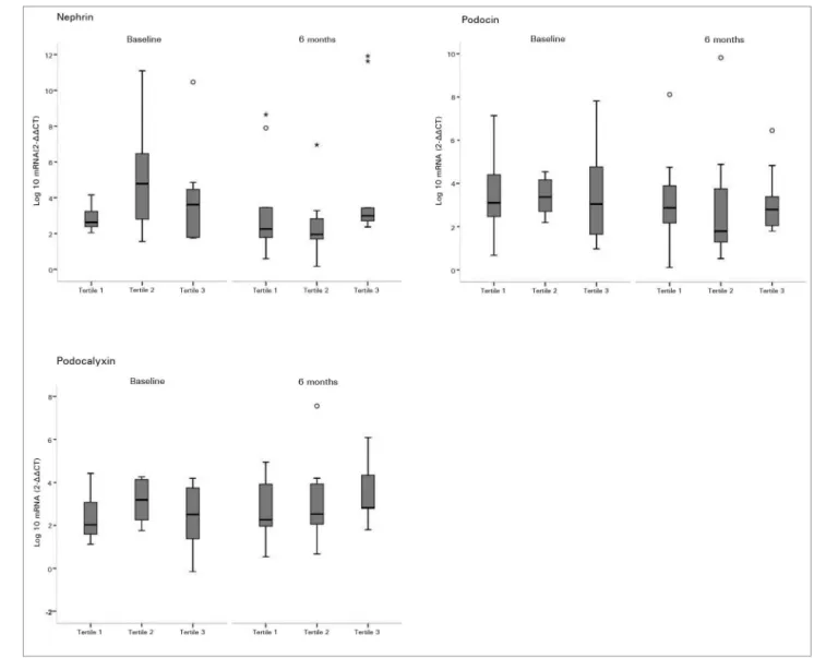
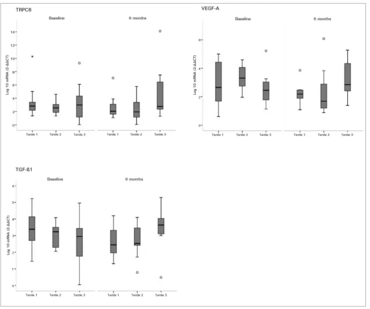
Analyzing the expression of each podocyte mRNA according to the tertile of serum 25(OH)D levels at baseline and after 6 months of cholecalciferol supplementation, correlation between higher 25(OD)D levels and lower mRNA expression of podocyte-associated mRNAs was not observed in any tertile. As shown in Figure 1 and Figure 2, the variability of each gene in both periods was small across the distribution of 25(OH)D tertiles.
CORRELATIONSBETWEENPODOCYTE-ASSOCIATEDMRNAS, 25(OH)D, PROTEINURIAANDKIDNEYFUNCTION
N = 27
Age (years) 56 ± 13
Gender (female) 14 (52)
Ethnicity (Caucasian) 17 (63)
BMI (kg/m2) 28.7 ± 5.4
Etiology of CKD
Hypertension 7 (26)
Diabetes 5 (18.5)
Glomerulonephritis 10 (37)
Other 5 (18.5)
CKD Stage
2 2 (7.4)
3 10 (37)
4 15 (55.6)
Hypertension 23 (85)
SBP (mmHg) 140 ± 21
DBP (mmHg) 86 ± 10
Use of ACE-I 15 (55.5)
Use of ARA-2 14 (51.8)
BMI: Body Mass Index; CKD: Chronic kidney disease; SBP: Systolic blood pressure; DBP: Diastolic blood pressure; ACE-I: Angiotensin-converting enzyme inhibitor; ARA-2: angiotensin II receptor antagonist. Values are expressed as mean ± DP or number of patients (%).
TABLE 1 CLINICALANDDEMOGRAPHICCHARACTERISTICSOFPATIENTSWITHCHRONICKIDNEYDISEASE
Baseline 6 months p
eGFR (mL/min/1.73m2) 32.74 ± 15.56 28.03 ± 16.30 0.010
Pr/Cr (urine) 2.53 ± 2.14 2.62 ± 2.51 0.855
Serum calcium (mg/dL) 8.83 ± 0.60 8.98 ± 0.68 0.084
Serum phosphorus (mg/dL) 3.70 ± 0.49 3.98 ± 0.74 0.204
Intact PTH (pg/mL) 222 ± 162 286 ± 221 0.086
Serum albumin (g/dl) 4.08 ± 0.30 4.10 ± 0.22 0.101
25(OH)D (ng/mL) 19 ± 7 28 ± 11 0.003
eGFR: estimated glomerular filtration rate; Pr/Cr: protein/creatinine index in random urine sample; PTH: parathyroid hormone. Data are expressed as mean ± DP.
TABLE 2 LABORATORYEVALUATIONOFKIDNEYFUNCTION, PROTEINURIA, MINERALANDBONEMETABOLISMATBASELINEAND
AFTERCHOLECALCIFEROLSUPPLEMENTATION
mRNA Baseline 6 months p
Nephrin 3.24 (2.30-4.46)† 2.71 (1.86-3.43) 0.349
Podocin 3.10 (2.25-4.53) 2.60 (1.79-3.89) 0.400
Podocalyxin 2.72 (1.75-3.74) 2.79 (1.97-4.18) 0.109
TRPC6 2.82 (1.93-3.97) 2.53 (1.52-5.46) 0.665
VEGF-A 2.85 (1.82-3.86) 2.41 (1.52-3.83) 0.239
TGF-ß1 3.16 (2.26-3.67) 3.05 (2.32-3.94) 0.923
TABLE 3 URINARYPODOCYTE-ASSOCIATEDMRNASEXPRESSIONATBASELINEANDAFTERCHOLECALCIFEROL
SUPPLEMENTATIONINCHRONICKIDNEYDISEASEPATIENTS
Figure 1. Urine mRNA of nephrin, podocin, and podocalyxin according to tertiles of serum 25(OH)D - Effects of cholecalciferol supplementation on urine mRNA of nephrin, podocin, and podocalyxin according to tertiles of serum 25(OH)D level both before and after intervention. Tertile 1 included subjects with lowest serum 25(OH)D level (< 23 ng/mL), tertile 2 included subjects with middle 25(OH)D level (23-30 ng/mL), and tertile 3 included subjects with highest 25(OH)D level (> 30 ng/mL). In the box plots, cross bars and horizontal bars represent median, minimum and maximum ranges of each Log10 urine mRNA. There was no significant correlation between 25(OH)D tertiles and mRNA expression of podocyte proteins (p value > 0.05).
at the 6-month follow-up. Similarly, no correlations were observed between mRNA expression and eGFR or proteinuria. Serum 25(OH)D was positively and significantly correlated with urinary protein excretion both at baseline (r = 0.517, p = 0.008) and at the six-month follow-up (r = 0.539, p = 0.005).
D
ISCUSSIONThe present study did not find an improvement in the mRNA profile of podocyte-associated byproducts in urine after 6 months of cholecalciferol supplementation. Proteinuria did not change as well. However, those with higher serum levels of 25(OH)D at 6 months (≥ 20 ng/ml) showed a trend toward decreased nephrin and podocin
expression, while the opposite pattern was observed in patients with low 25(OH)D levels.
Figure 2. Urine mRNA of TRPC6, VEGF-A, and TGF-ß1 according to tertiles of serum 25(OH)D - Effects of cholecalciferol supplementation on urine mRNA of TRPC6, VEGF-A, and TGF-ß1 according to tertiles of serum 25(OH)D level both before and after intervention. Tertile 1 included subjects with lowest serum 25(OH)D level (< 23 ng/mL), tertile 2 included subjects with middle 25(OH)D level (23-30 ng/mL), and tertile 3 included subjects with highest 25(OH)D level (> 30 ng/mL). In the box plots, cross bars and horizontal bars represent median, minimum and maximum ranges of each Log10 urine mRNA. There was no significant correlation between 25(OH)D tertiles and mRNA expression of expression of podocyte proteins (p value > 0.05).
expression, thus preserving podocyte structure and function.7-9,14-16,23,24
In animal models of induced nephropathy, vitamin D has been found to decrease the amount of podocytopenia, podocyturia, podocyte hypertrophy and apoptosis, increase the expression of nephrin, podocin, α3β1 integrin and dystroglycan, and suppress TGF-β1 and TRPC6 expression.6,25-28 These findings suggest that the combined effects of vitamin D or its analogues on the glomerular podocyte may be critical for renoprotection, which have significant impact in reducing albuminuria and chronic kidney damage.
TRPC6 is a slit diaphragm protein whose expression increases as a result of podocyte injury in proteinuric nephropathies. ACE inhibitors
Low serum 25(OH)D and 1,25(OH)2D3 are usually associated with higher rates of albuminuria in CKD patients.29 Liu et al.30 evaluated the effects of three months of calcitriol therapy in patients with IgA nephropathy, while de Zeeuw et al.11 evaluated the effects of paricalcitol in type 2 diabetes individuals in the VITAL study. These studies reported sustained post-treatment reductions in proteinuria and albuminuria, respectively, without an increase in adverse events such as hypercalcemia.
However, both drugs may be associated with adverse events that cannot be neglected, such as hypercalcemia, hypercalciuria, and hypoparathyroidism; in the VITAL study, for example, a higher proportion of patients using 2 μg of paricalcitol was withdrew from the study because of adverse events (p = 0.018 vs. placebo).11 Agarwal et al.31 performed a randomized placebo-controlled trial of paricalcitol in patients with stages 3 or 4 CKD and secondary hyperparathyroidism. The authors reported a significant reduction in proteinuria, even after adjusting for several covariates such as
diabetes mellitus and use of angiotensin blockers. Alborzi et al.10 identified similar effects of paricalcitol on albuminuria of patients with stages 2 or 3 CKD, regardless of its effects on parathyroid hormone suppression.
These data were confirmed in a recent systematic review, which found that calcitriol and paricalcitol reduced proteinuria by 16% in patients with CKD, while controls showed a 6% increase.32 As our patients have more advanced chronic kidney damage and higher levels of proteinuria, we could not demonstrate such an association, probably because serum 25(OH)D and albuminuria correlate at earlier stages of kidney disease.
The protective effects of vitamin D on the loss of glomerular filtration rate and the progression of kidney disease are yet to be demonstrated in clinical trials. De Boer et al.5 evaluated 1705 elderly subjects with predominantly normal kidney function, and found that a 10 ng/mL reduction in the serum concentration of 25(OH)D increased the risk of rapid GFR loss by 25%. In the present study, eGFR decreased significantly by a mean of 4 ml/min/1.73 m2 following six months of cholecalciferol treatment. These findings are in agreement with those of Liu
et al.,30 who reported an increase in serum creatinine and a decrease in eGFR in calcitriol-treated patients. However, the effects of vitamin D on the creatinine
metabolism of patients with CKD must also be considered. Vitamin D is known to increase creatinine generation leading to higher serum creatinine levels, with no real effect on GFR.33,34 Clinical trials in early stages of CKD are still required to determine the long-term renoprotective effects of vitamin D on renal function and patient morbidity and mortality.
To evaluate the distribution of each urine podocyte mRNA according to baseline and post treatment serum 25(OH)D, the tertiles of serum 25(OH)D (< 23, 23-30 and > 23-30 ng/mL) were determined. Theoretically, we would expect higher levels of podocyte byproducts in urine in the lower tertile of vitamin D at baseline. Conversely, lower levels of podocyte mRNAs in the highest tertile of 25(OH)D would be assumed after cholecalciferol supplementation, suggesting a putative effect of vitamin D on “morphologic and functional recovery” of glomerular epithelial cells, and consequently lower podocyturia. However, the erratic distribution of podocyte mRNAs throughout the tertiles did not confirm our main hypothesis.
It is possible that a higher threshold for 25(OH) D levels or a longer supplementation period may be required to restore gene transcription and the expression of podocyte proteins in the glomerulus. In addition, cholecalciferol replacement may not be effective to reduce proteinuria in the advanced CKD. Otherwise, the degree of podocyte injury in more advanced CKD, which is associated with a higher percentage of glomerular sclerosis, may result in irreversible podocyte damage and cell death through apoptotic or non-apoptotic mechanisms.
There are several limitations in this study. The small sample size and the absence of a control group might preclude any conclusions regarding the actual effects of cholecalciferol on podocyturia, and the course of urine podocyte mRNAs during the progression of CKD. Another limitation is the heterogeneity of the study group regarding different etiologies and stage of CKD, as both of which may have implications for podocyte injury and recovery. However, the method used to detect podocyte injury in the present sample, that is, quantification of podocyte-associated mRNAs in urine, offers an accurate and non-invasive, albeit indirect, measure of podocyturia, with none of the risks associated with invasive methods such as renal biopsy.
the supplementation (which may need to reach a certain cutoff before any effect is seen), a longer duration of treatment, and/or the beginning of vitamin D at earlier stages of CKD, rather than the baseline 25(OH)D levels itself. In addition, the use of active forms of vitamin D or its analogous such as calcitriol or paricalcitol, instead of the substrate cholecalciferol, maybe needed to maximize the direct activation of the VDR in podocyte cells leading to a biologic response.
We can also consider that cholecalciferol is not properly converted to active calcitriol in more advanced stages of CKD. This lower conversion of native vitamin D to calcitriol can be related to inhibition of 1-alpha-hydroxilase in the kidney due to higher levels of fibroblast growth factor 23 via activation of the ERK1/2 signaling pathway35 besides the loss of functional renal mass.
C
ONCLUSIONIn conclusion, six months of cholecalciferol supplementation had no effect on urine podocyte mRNA profile, proteinuria or kidney function in patients with CKD stages 2 to 4. However, patients with higher 25(OH)D levels showed a trend in reduction of nephrin and podocin mRNAs following treatment. Further studies are needed to evaluate the protective effects of vitamin D or its analogues on podocytes, perhaps at earlier stages of CKD and with a longer follow-up.
D
ECLARATION OF INTERESTThe authors report no conflicts of interest.
A
CKNOWLEDGEMENTSWe thank the Research Support Fund of the Hospital de Clínicas de Porto Alegre (FIPE/HCPA) for the financial support, the Brazilian Coordination for the Improvement of Higher Education Personnel (CAPES) for the Master’s scholarship awarded to João Rodolfo Teló Timm, and the Laboratory of Molecular Biology Applied to Nephrology for the technical support.
R
EFERENCES1. LaClair RE, Hellman RN, Karp SL, Kraus M, Ofner S, Li Q, et al. Prevalence of calcidiol deficiency in CKD: a cross-sectio-nal study across latitudes in the United States. Am J Kidney Dis 2005;45:1026-33. PMID: 15957131 DOI: http://dx.doi. org/10.1053/j.ajkd.2005.02.029
2. Diniz HF, Romão MF, Elias RM, Romão Júnior JE. Vitamin D deficiency and insufficiency in patients with chronic kid-ney disease. J Bras Nefrol 2012;34:58-63. DOI: http://dx.doi. org/10.1590/S0101-28002012000100009
3. Pilz S, Iodice S, Zittermann A, Grant WB, Gandini S. Vitamin D status and mortality risk in CKD: a meta-analysis of pros-pective studies. Am J Kidney Dis 2011;58:374-82. DOI: http:// dx.doi.org/10.1053/j.ajkd.2011.03.020
4. de Boer IH, Ioannou GN, Kestenbaum B, Brunzell JD, Weiss NS. 25-Hydroxyvitamin D levels and albuminuria in the Third National Health and Nutrition Examination Survey (NHA-NES III). Am J Kidney Dis 2007;50:69-77. PMID: 17591526 DOI:http://dx.doi.org/10.1053/j.ajkd.2007.04.015
5. de Boer IH, Katz R, Chonchol M, Ix JH, Sarnak MJ, Shlipak MG, et al. Serum 25-hydroxyvitamin D and change in estimated glomerular filtration rate. Clin J Am Soc Nephrol 2011;6:2141-9. DOI: http://dx.doi.org/10.2215/CJN.02640311
6. Mizobuchi M, Morrissey J, Finch JL, Martin DR, Liapis H, Akizawa T, et al. Combination therapy with an angiotensin-converting enzy-me inhibitor and a vitamin D analog suppresses the progression of renal insufficiency in uremic rats. J Am Soc Nephrol 2007;18:1796-806. DOI: http://dx.doi.org/10.1681/ASN.2006091028
7. Kuhlmann A, Haas CS, Gross ML, Reulbach U, Holzinger M, Sch-warz U, et al. 1,25-Dihydroxyvitamin D3 decreases podocyte loss and podocyte hypertrophy in the subtotally nephrectomized rat. Am J Physiol Renal Physiol 2004;286:F526-33. PMID: 14600034 DOI:http://dx.doi.org/10.1152/ajprenal.00316.2003
8. Lydia A, Asanuma K, Nonaka K, Takagi M, Jeong KH, Koda-ma F, et al. Effects of 22-oxa-calcitriol on podocyte injury in adriamycin-induced nephrosis. Am J Nephrol 2012;35:58-68. DOI: http://dx.doi.org/10.1159/000334626
9. Deb DK, Wang Y, Zhang Z, Nie H, Huang X, Yuan Z, et al. Mo-lecular mechanism underlying 1,25-dihydroxyvitamin D regulation of nephrin gene expression. J Biol Chem 2011;286:32011-7. PMID: 21803771 DOI: http://dx.doi.org/10.1074/jbc.M111.269118 10. Alborzi P, Patel NA, Peterson C, Bills JE, Bekele DM, Bunaye
Z, et al. Paricalcitol reduces albuminuria and inflammation in chronic kidney disease: a randomized double-blind pilot trial. Hypertension 2008;52:249-55. PMID: 18606901 DOI:http:// dx.doi.org/10.1161/HYPERTENSIONAHA.108.113159 11. de Zeeuw D, Agarwal R, Amdahl M, Audhya P, Coyne D,
Ga-rimella T, et al. Selective vitamin D receptor activation with paricalcitol for reduction of albuminuria in patients with type 2 diabetes (VITAL study): a randomised controlled trial. Lan-cet 2010;376:1543-51. PMID:21055801 DOI: http://dx.doi. org/10.1016/S0140-6736(10)61032-X
12. Zheng Z, Shi H, Jia J, Li D, Lin S. Vitamin D supplementation and mortality risk in chronic kidney disease: a meta-analysis of 20 observational studies. BMC Nephrol 2013;14:199. DOI: http://dx.doi.org/10.1186/1471-2369-14-199
13. Kim CS, Kim SW. Vitamin D and chronic kidney disease. Korean J Intern Med 2014;29:416-27. DOI:http://dx.doi. org/10.3904/kjim.2014.29.4.416
14. Wang Y, Zhou J, Minto AW, Hack BK, Alexander JJ, Haas M, et al. Altered vitamin D metabolism in type II diabetic mouse glo-meruli may provide protection from diabetic nephropathy. Kidney Int 2006;70:882-91. DOI:http://dx.doi.org/10.1038/sj.ki.5001624 15. Wang Y, Deb DK, Zhang Z, Sun T, Liu W, Yoon D, et al. Vita-min D receptor signaling in podocytes protects against diabetic nephropathy. J Am Soc Nephrol 2012;23:1977-86. DOI: http:// dx.doi.org/10.1681/ASN.2012040383
16. Zhang Z, Sun L, Wang Y, Ning G, Minto AW, Kong J, et al. Renoprotective role of the vitamin D receptor in diabetic ne-phropathy. Kidney Int 2008;73:163-71. DOI: http://dx.doi. org/10.1038/sj.ki.5002572
17. Yu D, Petermann A, Kunter U, Rong S, Shankland SJ, Floe-ge J. Urinary podocyte loss is a more specific marker of on-going glomerular damage than proteinuria. J Am Soc Ne-phrol 2005;16:1733-41. DOI: http://dx.doi.org/10.1681/ ASN.2005020159
19. National Kidney Foundation. K/DOQI clinical practice guideli-nes for bone metabolism and disease in chronic kidney disease. Am J Kidney Dis 2003;42:S1-201.
20. Rodrigues PG, Bringhenti RN, do Nascimento JF, Joelsons G, dos Santos M, Pereira S, et al. Expression patterns of podocyte-asso-ciated mRNAs in patients with proliferative or non-proliferative glomerulopathies. Int J Clin Exp Pathol 2014;7:2185-98.
21. Nagpal S, Na S, Rathnachalam R. Noncalcemic actions of vitamin D receptor ligands. Endocr Rev 2005;26:662-87. DOI:http://dx.doi.org/10.1210/er.2004-0002
22. Molina P, Górriz JL, Molina MD, Peris A, Beltrán S, Kanter J, et al. The effect of cholecalciferol for lowering albuminuria in chronic kidney disease: a prospective controlled study. Nephrol Dial Trans-plant 2014;29:97-109. DOI: http://dx.doi.org/10.1093/ndt/gft360 23. Li YC. Podocytes as target of vitamin D. Curr Diabetes Rev
2011;7:35-40. DOI: http://dx.doi.org/10.2174/157339911794273964 24. Wang Y, Borchert ML, Deluca HF. Identification of the
vita-min D receptor in various cells of the mouse kidney. Kidney Int 2012;81:993-1001. DOI: http://dx.doi.org/10.1038/ki.2011.463 25. Sonneveld R, Ferrè S, Hoenderop JG, Dijkman HB, Berden JH,
Bindels RJ, et al. Vitamin D down-regulates TRPC6 expression in podocyte injury and proteinuric glomerular disease. Am J Pathol 2013;182:1196-204. DOI:http://dx.doi.org/10.1016/j. ajpath.2012.12.011
26. Xiao HQ, Shi W, Liu SX, Zhang B, Xu LX, Liang XL, et al. Podocyte injury is suppressed by 1,25-dihydroxyvitamin D via modulation of transforming growth factor-beta 1/bone morphogenetic protein-7 signalling in puromycin amino-nucleoside nephropathy rats. Clin Exp Pharmacol Physiol 2009;36:682-9. DOI: http://dx.doi.org/10.1111/j.1440-1681.2008.05133.x
27. Zou MS, Yu J, Nie GM, He WS, Luo LM, Xu HT. 1, 25-dihydroxyvitamin D3 decreases adriamycin-induced po-docyte apoptosis and loss. Int J Med Sci 2010;7:290-9. DOI: http://dx.doi.org/10.7150/ijms.7.290
28. Zhang X, Song Z, Guo Y, Zhou M. The novel role of TRPC6 in vitamin D ameliorating podocyte injury in STZ-induced dia-betic rats. Mol Cell Biochem 2015;399:155-65. DOI: http:// dx.doi.org/10.1007/s11010-014-2242-9
29. Isakova T, Gutiérrez OM, Patel NM, Andress DL, Wolf M, Levin A. Vitamin D deficiency, inflammation, and albuminuria in chro-nic kidney disease: complex interactions. J Ren Nutr 2011;21:295-302. DOI: http://dx.doi.org/10.1053/j.jrn.2010.07.002
30. Liu LJ, Lv JC, Shi SF, Chen YQ, Zhang H, Wang HY. Oral calci-triol for reduction of proteinuria in patients with IgA nephropathy: a randomized controlled trial. Am J Kidney Dis 2012;59:67-74. DOI: http://dx.doi.org/10.1053/j.ajkd.2011.09.014
24. Wang Y, Borchert ML, Deluca HF. Identification of the vita-min D receptor in various cells of the mouse kidney. Kidney Int 2012;81:993-1001. PMID: 22278022 DOI: http://dx.doi. org/10.1038/ki.2011.463
25. Sonneveld R, Ferrè S, Hoenderop JG, Dijkman HB, Berden JH, Bindels RJ, et al. Vitamin D down-regulates TRPC6 expression in podocyte injury and proteinuric glomerular disease. Am J Pa-thol 2013;182:1196-204. PMID: 23385000 DOI:http://dx.doi. org/10.1016/j.ajpath.2012.12.011
26. Xiao HQ, Shi W, Liu SX, Zhang B, Xu LX, Liang XL, et al. Podo-cyte injury is suppressed by 1,25-dihydroxyvitamin D via modula-tion of transforming growth factor-beta 1/bone morphogenetic pro-tein-7 signalling in puromycin aminonucleoside nephropathy rats. Clin Exp Pharmacol Physiol 2009;36:682-9. PMID: 19594554 DOI: http://dx.doi.org/10.1111/j.1440-1681.2008.05133.x 27. Zou MS, Yu J, Nie GM, He WS, Luo LM, Xu HT. 1,
25-dihydroxyvitamin D3 decreases adriamycin-induced po-docyte apoptosis and loss. Int J Med Sci 2010;7:290-9. DOI: http://dx.doi.org/10.7150/ijms.7.290
28. Zhang X, Song Z, Guo Y, Zhou M. The novel role of TRPC6 in vitamin D ameliorating podocyte injury in STZ-induced dia-betic rats. Mol Cell Biochem 2015;399:155-65. DOI: http:// dx.doi.org/10.1007/s11010-014-2242-9
29. Isakova T, Gutiérrez OM, Patel NM, Andress DL, Wolf M, Levin A. Vitamin D deficiency, inflammation, and albuminuria in chro-nic kidney disease: complex interactions. J Ren Nutr 2011;21:295-302. DOI: http://dx.doi.org/10.1053/j.jrn.2010.07.002