Carotid body tumor (paraganglioma):
report of two cases treated surgically
Tumor de corpo carotídeo (paraganglioma):
relato de dois casos submetidos a tratamento cirúrgico
Nelson Mesquita Junior1
*
, Rogério Santos Silva1, José Henrique Agner Ribeiro1, Lislaine Cruz Batista1, Emanuelle Melania Stedille Bringhentti1, Bruno Benjamin Brunini de Souza1, Lisiane Cristine da Mota Cabral1
Abstract
A carotid body tumor is a rare neoplasm, generally benign, that predominantly afects people between their fourth and ifth decades of life. It manifests as a pulsatile and generally painless cervical mass with irm consistency, located below the angle of the jaw. It can progress to the extent that it causes localized pain, dysphagia, hiccups, hoarseness and hypersensitive carotid body syndrome. his article reports the cases of two female patients diagnosed with this tumor who were treated surgically. he irst was treated with block resection of the tumor, while the second patient, who had an early stage tumor, was treated with subadventitial resection of the lesion.
Keywords: carotid body tumor; paraganglioma; carotid glomus tumor.
Resumo
O tumor de corpo carotídeo é uma neoplasia rara, geralmente benigna, que acomete, sobretudo, indivíduos entre a quarta e a quinta décadas de vida. Manifesta-se pela presença de massa cervical consistente localizada abaixo do ângulo da mandíbula, pulsátil e comumente indolor. Pode evoluir para dor local, disfagia, soluços, rouquidão e síndrome do corpo carotídeo hipersensível. Este artigo relata os casos de duas pacientes diagnosticadas com essa neoplasia e submetidas ao tratamento cirúrgico. A primeira foi submetida a uma ressecção em bloco do tumor, enquanto a segunda, com estadiamento mais precoce, foi tratada com uma ressecção subadventicial da lesão.
Palavras-chave: tumor de corpo carotídeo; paraganglioma; tumor de glomo carotídeo.
1 Faculdade Evangélica do Paraná – FEPAR, Curitiba, PR, Brazil.
Financial support: None.
Conlicts of interest: No conlicts of interest declared concerning the publication of this article. Submitted: November 03, 2015. Accepted: February 05, 2016.
Nelson Mesquita Junior, Rogério Santos Silva et al.
INTRODUCTION
The carotid body is a structure of elliptical shape, 3 to 4 mm in size, that is located at the bifurcation of the common carotid, at the level of its adventitial layer.1-4
Its functions are chemoreception and baroreception and because of this tumors that originate from it are called chemodectomas, i.e., chemoreceptor tumors.2,5-7
Carotid body tumors are neoplasms that arise from paraganglion cells and although they are well-delimited, they are unencapsulated tumors and are highly vascularized by branches of the external carotid artery and their “vasa vasorum”.2-4,8-12
While rare, they are the most common paragangliomas that occur in the head and neck area (60-70%).5,13
In the great majority of cases these neoplasms are hypervascularized, of familial origin and slow growing, and there is no predilection for either sex. They predominantly occur in patients in their fourth
and ifth decades of life and the great majority are
benign; however, a large proportion of authors report malignancy in 5 to 6% of cases.1,6,14,15 Symptoms are
varied. They can be asymptomatic or manifest as a slow-growing tumor that is painless and pulsatile, in the side of the neck, close to the angle of the jaw, and occasionally lead to complaints of hoarseness, deglutition problems and symptoms of carotid sinus syndrome.5,6,16
DESCRIPTIONS OF CASES
The irst patient was a 28-year-old white female
from Loanda, PR, Brazil, who was seen at a clinic in the Hospital Universitário Evangélico de Curitiba with a history of bilateral tumors in the cervical region, with onset 5 years previously. She had had a surgical biopsy of the left cervical region 4 months previously and referred with a diagnosis of carotid aneurysm. She stated that she had no history of dysphagia, dysphonia, ischemic attacks or weight loss. During the physical examination, a scar approximately 3 cm long was observed at the anterior margin of the sternocleidomastoid muscle, on the left. Palpation revealed two ovoid masses, one measuring 3 x 4 cm, on the left, and the other measuring 2 x 2 cm, on the right (Figure 1). They were located within the right and left carotid triangles and had an elastic consistency, were painless and pulsatile and were mobile in the lateral direction, but immobile longitudinally. They did not produce thrills or murmurs. Examination of the oral
cavity did not ind evidence of lesions or adenopathies
in other sites. The neurological examination showed that the cranial pairs were undamaged.
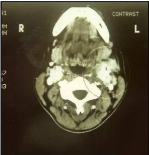
The patient was admitted to hospital and underwent examinations with color Doppler ultrasonography and computed tomography (Figure 2), which were suggestive of carotid body tumor and this diagnosis
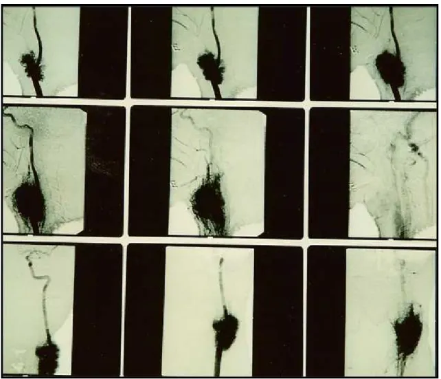
was later conirmed by arteriography (Figure 3). On the
left, it could be observed that the external carotid was occluded at the origin. The tomographic examination ruled out signs of invasion of the base of the skull.
The patient underwent surgery under general anesthesia. An incision was made at the anterior margin of the left sternocleidomastoid muscle, revealing a solid tumor adhering to the carotid bifurcation, without involving the vagus or hypoglossal nerves. A ligature of the external carotid at the origin was observed. Since
Figure 1. Presence of bilateral tumors in topographic area
corresponding to the carotid vessels.
Figure 2. Tomographic image showing evidence of the presence
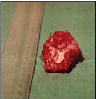
dissection was dificult, en bloc resection of the tumor
and carotid bifurcation was performed (Figure 4). A temporary Pruitt-Inahara shunt was used for cerebral protection, with interposition of the internal saphenous with proximal side-to-end anastomosis and distal end-to-end anastomosis (Figure 5). Two glands were removed for histopathological analysis. The patient recovered during the postoperative period with no
neurological deicits and was discharged on the sixth day. The histopathological analysis conirmed
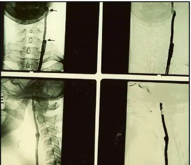
the diagnosis of carotid body tumor and found the glands to be free of involvement (Figure 6). A repeat arteriography conducted 60 days after the procedure
did not ind evidence of tumor remnants (Figure 7).
The patient refused surgery on the contralateral tumor. The second patient was a 65-year-old female from Curitiba, PR, Brazil, who complained of a tumor in the left cervical region. She reported a history of systemic
arterial hypertension and hypercholesterolemia, both under control. She also reported having a meningioma adhering to the superior sagittal sinus, which had not exhibited growth since 2009. During physical
examination, palpation identiied an ovoid, pulsatile,
and painless mass in the left carotid trigone. It was of elastic consistency, with no adherence to the deeper planes and was mobile laterally, but immobile in the longitudinal direction. There was no thrill or murmur.
In common with the irst case, examination of the
oral cavity and a neurological examination did not detect abnormalities. The patient underwent imaging exams during the preoperative period. Color Doppler ultrasonography showed an extremely vascularized tumor with dimensions of 3.9 × 3.2 cm at the level of the carotid bifurcation, with separation of the internal and external carotids (the “lyre sign”).
Figure 3. Arteriographic examination in which the rich tumoral vascularization can be seen, in addition to absence of contrast
Nelson Mesquita Junior, Rogério Santos Silva et al.
Surgical treatment was chosen. Under general anesthesia, a cervical incision similar to that described in the previous case description was made on the left. A solid mass was found adhering to the carotid bifurcation. Essential adjacent structures were isolated easily and the tumor dissected using the subadventitial technique with no intercurrent events (Figures 8 and 9). Material for histopathological analysis was removed. A Jackson-Pratt drain was
used for the irst 24 postoperative hours. The patient
recovered well during the postoperative period, with no complications, and was discharged on the second
postoperative day. Histopathological analysis conirmed
the diagnosis of carotid paraganglioma.
DISCUSSION
According to Shamblin et al.3 carotid body tumors
can be classiied into three groups depending on their
circumference and degree of adherence: Group I – small tumor with no adherence to vessels that can be resected without damaging neighboring structures; Group II – intermediate tumor with minor adherence
to vessels, which is more dificult to dissect and may
require revascularization; Group III – large tumor with
iniltration of vessels, which is almost impractical
to dissect, making resection en bloc with the carotid bifurcation and revascularization with saphenous vein or prosthetic graft necessary.6,17,18
With regard to work-up tests and examinations,
color Doppler ultrasonography is the irst choice,
because it provides information that is suggestive of the diagnosis and important for screening and differential diagnosis. Cervical computed tomography or, better still, magnetic resonance imaging, are the
Figure 4. Surgical specimen, product of resection en bloc of
tumor together with the carotid bifurcation.
Figure 5. Transoperative control arteriography, showing patency
of the saphenous vein used for reconstruction.
Figure 6. Histopathological analysis with immunohistochemical
examinations of choice for obtaining data on location, extension, correlation with adjacent structures and vascular nature of the tumor.6,12,13,19 Whole-body
scintigraphy and DNA low cytometry can be used
to investigate the possibility of metastatic lesions or multiple tumors.17,20 Biopsy is not indicated because
of the considerable chance of bleeding.5,10,12
Surgery is the treatment of choice, bearing in mind the possibility of malignant transformation, peritumoral invasion and metastasis. The most widely-used technique is subadventitial dissection
Figure 9. Appearance of surgical site after resection.
Figure 7. Control arteriography at 60 days, which does not show tumor remnants.
Figure 8. Resection of the glomus tumor using the subadventitial
Nelson Mesquita Junior, Rogério Santos Silva et al.
of the tumor (Gordon-Taylor).17,18,21 Resection en
bloc is only employed in cases in which it is not feasible to separate the tumor from the artery, and should be followed by interposition of a graft to the internal carotid.17 In 1984, Pantanowitz et al.
conducted a comparison between size of tumor and
Shamblin grades, inding that tumors up to 6 cm
in size corresponded to Shamblin grades 1 and 2 and those larger than 6 corresponded to group 3. They therefore concluded that tumors that were smaller than 6 cm and had an incomplete circumferential extension and an angle of bifurcation less than 90º were indicated for dissection and that those that should be treated with resection were larger than 6 cm and with complete circumferential extension and an angle of bifurcation greater than 90º.18,22
Tumors are considered inoperable if they surround the entire extracranial internal carotid, which makes it impossible to perform distal anastomosis with graft or prosthesis.10
During the procedure, before dissection, it is very important to identify and fully expose all nerves, avoiding excessive handling.5,23 In certain
situations, such as resection en bloc or a possible injury to the artery wall, it will be necessary to construct a shunt.17 Barbiturates and mannitol
are recommended if it is necessary to clamp the internal carotid artery.24
Overall complication rates vary from 32 to 44% and mortality rates vary from 8 to 20%.25 The greatest
cause of morbidity is injury to the cranial pairs, of which the hypoglossal, vagus and laryngeal superior are the most often affected, which can
cause paralysis that is very often deinitive.4,17,18,23
More serious complications, such as strokes and
dificult to control hemorrhages, are rarer.4
Embolization can be employed in cases of large tumors, which will reduce blood loss and the size of the tumor and improve the results of surgery, but the risk of cerebral embolism cannot be ruled out.17,18 Radiotherapy can be used for inaccessible,
partially resected and/or metastatic tumors, in cases of local recurrence and in patients in whom surgery has high rates of morbidity. However, it must be remembered that side effects can occur, such as necrosis of the jaw, brain and soft tissues.17,18
Screening for carotid body tumors is indicated for
irst-degree relatives and patients should receive
long-term follow-up, because metastatic disease can take from 10 to 20 years to appear.6
REFERENCES
1. Gaylis H, Davidge-Pitts K, Pantanowitz D. Carotid body tumors: a review of 52 cases. S Afr Med J. 1987;72(7):493-6. PMid:3660158.
2. Meyer FB, Sundt TM Jr, Pearson BW. Carotid body tumors: a subject review and suggested surgical approach. J Neurosurg. 1986;64(3):377-85. http://dx.doi.org/10.3171/jns.1986.64.3.0377. PMid:3950716.
3. Shamblin WR, Remine WH, Sheps SG, Harrison EG. Carotid body tumor (chemodectoma). Clinicopathologic analysis of ninety cases. Am J Surg. 1971;122(6):732-9. http://dx.doi.org/10.1016/0002-9610(71)90436-3. PMid:5127724.
4. Patiño FT, Acosta FG, Guzman CP, Parada JM, Almedaro SL. Tumor de cuerpo carotídeo: análisis de 96 casos. Ver Invest Clin. 1991;43:119-23.
5. Davidovic LB, Djukic VB, Vasic DM, Sindjelic RP, Duvnjak SN. Diagnosis and treatment of carotid body paraganglioma: 21 years of experience at a clinical center of Serbia. World J Surg Oncol. 2005;3(1):10. http://dx.doi.org/10.1186/1477-7819-3-10. PMid:15707500.
6. França LH, Bredt CG, Vedolin A, Back LA, Stahlke HJ Jr. Surgical treatment of the carotid body tumor: a 30 year experience. J Vasc Bras. 2003;2(3):171-6.
7. Matticari S, Credi G, Pratesi C, Bertini D. Diagnosis and surgical treatment of the carotid body tumors. J Cardiovasc Surg (Torino). 1995;36(3):233-9. PMid:7629206.
8. Franklin J, Gonçalves S, Cruz EC. Tumores do corpo carotídeo (paragangliomas). Acta Med Port. 1990;3(2):89-93. PMid:2349894.
9. Irons GB, Weiland LH, Brown WL. Paragangliomas of the neck: clinical and pathological analysis of 116 cases. Surg Clin North Am. 1977;57(3):575-83. PMid:867224.
10. Chung WB. The carotid body tumor. Can J Surg. 1979;22(4):319-22. PMid:455160.
11. Sanchez AC, Seijas EV, Matesanz JM, Trapero VL. Carotid body tumor unusual cause of transient ischemic attacks. Stroke. 1988;19(1):102-3. http://dx.doi.org/10.1161/01.STR.19.1.102. PMid:3336888.
12. Wax MK, Briant DR. Carotid body tumors: a review. J Otolaryngol. 1992;21(4):277-85. PMid:1527835.
13. Koishi HU, De La Cortina RA, Sennes LU, Tsuji DH, Frizzarini R. Paraganglioma cervical bilateral. Arch Otor Fund. 1998;2(3).
14. Gaylis H, Mieny CJ. The incidence of malignancy in carotid body tumours. Br J Surg. 1977;64(12):885-9. http://dx.doi.org/10.1002/ bjs.1800641214. PMid:588988.
15. Pantanowitz D, Davidge-Pitts K, Gaylis H, Hale MJ. Are carotid body tumours malignant? S Afr J Surg. 1990;28(3):97-9. PMid:2218758.
16. Maves MD. Vascular tumors of the head and neck. In: Bailey BJ, Johnson JT, Kohut RI, Pillsbury HC, Tardy ME, editor. Head and neck surgery-otolaryngology. Philadelphia: Lippincott; 1993. p. 1397-1409.
17. De Vasconcelos Filho JO, Oliveira RS. Tumor do corpúsculo carotídeo: revisão da literatura e apresentação de um caso. Cir Vasc Angiol. 1998;14:164-80.
18. Ferreira GP, Gaspar RJ, Pinto HC, Cintra PP, Pinto JA. Tumor de corpo carotídeo. Cir Vasc Angiol. 2000;16:171-7.
20. Sauter E, Hollier LH, Bolton JS, Ochsner JL, Sardi A. Prognostic value of DNA flow cytometry in paragangliomas of the carotid body. J Surg Oncol. 1991;46(3):151-3. http://dx.doi.org/10.1002/ jso.2930460304. PMid:2011023.
21. Gordon-Taylor G. On carotid tumours. Br J Surg. 1940;28(110):163-72. http://dx.doi.org/10.1002/bjs.18002811003.
22. Pantanowitz D, Davidge-Pitts K, Deme Triades D. The significance os the bifurcation angle in carotid body tumors. S Afr Med J. 1991;80(7):318-21. PMid:1925836.
23. Netterville JL, Reilly KM, Robertson D, Reiber ME, Armstrong WB, Childs O. Carotid body tumors: a review of 30 patients with 46 tumors. Laryngoscope. 1995;105(2):115-25. http://dx.doi. org/10.1288/00005537-199502000-00002. PMid:8544589.
24. Sousa AA, Vega MG, Carvalho GT, Scarpelli MM. Tumores do corpo carotídeo. Arq Bras Neurocirug. 1995;14:63-9.
25. Rush BF Jr. Current concepts in the treatment of carotid body tumors. Surgery. 1962;52:679-84. PMid:14495345.
*
Correspondence
Nelson Mesquita Junior Rua Deputado Heitor Alencar Furtado, 1819/1302 - Mossunguê CEP 81200-110 - Curitiba (PR), Brazil Tel.: +55 (41) 9994-5167 E-mail: [email protected]
Author information
NMJ - MSc in Principles of Surgery from Universidade Federal do Paraná (UFPR); Professor of Anatomy and Angiology and Vascular Surgery at Faculdade Evangélica do Paraná (FEPAR). RSS - MSc in Principles of Surgery from Instituto de Pesquisas Médicas (IPEM); Vascular Surgeon. JHAR - Medical student, Faculdade Evangélica do Paraná (FEPAR). LCB - Medical student, Faculdade Evangélica do Paraná (FEPAR). EMSB - Medical student, Faculdade Evangélica do Paraná (FEPAR). BBBS - Medical student, Faculdade Evangélica do Paraná (FEPAR). LCMC - Pathologist at Citolab.
Author contributions
Conception and design: NMJ Analysis and interpretation: NMJ, RSS, JHAR, LCB, EMSB, BBBS, LCMC Data collection: NMJ, RSS, JHAR, LCB, EMSB, BBBS, LCMC Writing the article: NMJ, RSS, JHAR, LCB, EMSB, BBBS Critical revision of the article: NMJ, RSS, JHAR, LCB, EMSB, BBBS Final approval of the article*: NMJ, RSS, JHAR, LCB, EMSB, BBBS Statistical analysis: N/A. Overall responsibility: NMJ