Inclusion Bodies Using Leucine Zippers
Su-Lim Choi1,2, Sang Jun Lee3,4, Soo-Jin Yeom1, Hyun Ju Kim3,4, Young Ha Rhee2, Heung-Chae Jung1, Seung-Goo Lee1,4*
1Biochemicals and Synthetic Biology Research Center, KRIBB, Yuseong-gu, Daejeon, Korea,2Department of Bioscience and Biotechnology, Chungnam National University, Yuseong-gu, Daejeon, Korea,3Infection and Immunity Biology Research Center, KRIBB, Yuseong-gu, Daejeon, Korea,4Korea University of Science and Technology, Yuseong-gu, Daejeon, Korea
Abstract
Inclusion bodies (IBs) are typically non-functional particles of aggregated proteins. However, some proteins in fusion with amyloid-like peptides, viral coat proteins, and cellulose binding domains (CBDs) generate IB particles retaining the original functions in cells. Here, we attempted to generate CBD IBs displaying functional leucine zipper proteins (LZs) as bait for localizing cytosolic proteins in E. coli. When a red fluorescent protein was tested as a target protein, microscopic observations showed that the IBs red-fluoresced strongly. When different LZ pairs with KDs of 8–1,000mM were tested as the bait and prey, the localization of the red fluorescence appeared to change following the affinities between the LZs, as observed by fluorescence imaging and flow cytometry. This result proposed that LZ-tagged CBD IBs can be applied as anin vivomatrix to entrap cytosolic proteins inE. coliwhile maintaining their original activities. In addition, easy detection of localization to IBs provides a unique platform for the engineering and analyses of protein-protein interactions inE. coli.
Citation:Choi S-L, Lee SJ, Yeom S-J, Kim HJ, Rhee YH, et al. (2014) Controlled Localization of Functionally Active Proteins to Inclusion Bodies Using Leucine Zippers. PLoS ONE 9(6): e97093. doi:10.1371/journal.pone.0097093
Editor:Anna Mitraki, University of Crete, Greece
ReceivedJanuary 1, 2014;AcceptedApril 14, 2014;PublishedJune 4, 2014
Copyright:ß2014 Choi et al. This is an open-access article distributed under the terms of the Creative Commons Attribution License, which permits unrestricted use, distribution, and reproduction in any medium, provided the original author and source are credited.
Funding:This research was supported by grants from the Intelligent Synthetic Biology Center of Global Frontier Project (2011-0031944), the Pioneer Research Program for Converging Technology (20110001745), Bio-industry Technology Development Program (No. 310006-5), the New & Renewable Energy Core Technology Program of the Korea Institute of Energy Technology Evaluation and Planning (20133030000300), and the Korea Research Institute of Bioscience and Biotechnology Research Initiative Program. The funders had no role in study design, data collection and analysis, decision to publish, or preparation of the manuscript.
Competing Interests:The authors have declared that no competing interests exist.
* E-mail: [email protected]
Introduction
Inclusion bodies (IBs) are dense, electron-refractile particles of aggregated proteins found in the cytoplasmic space of bacterial cells [1]. Hydrophobic heterologous proteins expressed at high levels in bacterial cells are likely to accumulate in IBs [2,3]. IBs vary in diameter from 0.5–1.3mm and are more dense (,1.3 mg/ mL) than many other cellular components, which make the particles easy to separate from disrupted cells by high-speed centrifugation for protein refolding [4,5].
In general, the proteins in IBs are functionally inactive. However, recent studies have shown that they are not necessarily inactive, and some exhibit substantial levels of activity inE. coli[2,3,6–8]. For example, certain enzymes fused to a viral capsid protein or an ionic self-assembling peptide generated active IBs that had high levels of catalytic activity [6–8]. Accordingly, we found that a family II cellulose binding
domain (CBD) fromCellulomonas fimi induced the formation of
active IBs when fused withb-glycoside hydrolyzing enzymes.
The enzymatic activity of these IBs was 30%–40% of that of the soluble enzymes [9]. In addition, a family IIIa CBD has also been used to form active IBs with high D-amino acid oxidase activity [10]. The family II CBD in IBs also exhibited significant binding affinity towards insoluble cellu-loses [9].
In this study, the family II CBD from C. fimi was used to
Materials and Methods
Materials
The family II CBD was cloned from the exoglucanase (cex) of
Cellulomonas fimiKCTC 9143. TheEGFPgene was obtained from the commercial plasmid pEGFP-N1 (Clontech, Mountain View, CA, USA). The pRFP plasmid, which contains the gene for monomeric red fluorescent protein 1 (mrfp) [13], was a kind gift from Dr. R. Tsien (UCSD, USA). Genes encoding two anti-parallel LZs, used for the bait and prey, were cloned from pET11a-Z-NGFP and pMRBAD-Z-CGFP [12], respectively, which were provided by Dr. L. Regan (Yale University, USA).
E. coli DH5a (Takara Bio, Ohtsu, Japan) and BL21(DE3) (Novagen, Gibbstown, NJ, USA) cells were used as the cloning host and the expression host, respectively. All restriction enzymes were purchased from Roche Applied Science (Indianapolis, IN, USA), and T4 DNA Ligase was purchased from Fermentas (Glen Burnie, MD, USA).
DNA manipulation
All primers were synthesized by Bioneer Co. (Daejeon, Korea)
(Table S1). TheEGFPgene was amplified from pEGFP-N1, and
then cloned into theNdeI andXhoI sites of pET21a (Invitrogen,
Carlsbad, CA, USA) to yield pEGFP. The EGFP-CBDgene was
prepared using overlap PCR and was inserted into theNdeI and
HindIII sites of pET21a to yield plasmid pEGFP-CBD. The bait
and prey LZs were fused to the EGFP-CBD and mRFP genes,
respectively, by overlap extension PCR (Fig. S1). The resulting
bait-EGFP-CBD and prey-mRFP genes were then inserted into pET21a to yield pCN20-CBD. pC20-CBD, a bait-less variant of
pCN20-CBD was constructed by usingEGFP-CBDinstead of
bait-EGFP-CBD. Four variants of the pCN20-CBD plasmid (pCN8-CBD, pCN31-(pCN8-CBD, pCN50-(pCN8-CBD, and pCN1000-CBD) were
constructed by introducing known mutations into the prey moiety as shown in Table 1 [12], using a QuikChange mutagenesis kit (Stratagene, La Jolla, CA, USA).
Protein expression and western blotting analysis
E. coliBL21(DE3) cells were cultivated at 37uC in LB medium containing ampicillin (50mg/mL). Protein expression was induced
with 0.5 mM IPTG when the cultures reached an OD600of 0.5,
and the cells were incubated for an additional 6 h. The cells were
harvested by centrifugation at 16,3006g for 10 min and then
disrupted by sonication on ice.
The protein expression was analyzed by SDS-PAGE and western blotting. Aliquots of cell lysates were electrophoresed on 12% SDS-polyacrylamide gels and electro-transferred to poly-vinylidene fluoride membranes (Millipore, Billerica, MA, USA). The membranes were hybridized with an anti-GFP mouse antibody (Sigma-Aldrich, St. Louis, MO, USA) and an anti-groEL antibody as the internal standard (Abcam, Cambridge, MA, USA), followed by an HRP-conjugated anti-mouse IgG goat antibody (Bio-Rad, Hercules, CA, USA) prepared in TBST buffer (20 mM Tris-HCl, 100 mM NaCl, and 0.1% Tween-20, pH 7.5) containing 5% skimmed milk. The hybridized bands were identified by colorimetric detection using an Opti-4CN substrate kit (Bio-Rad).
Imaging and fluorescence analyses
Cells were observed with an Axio Observer microscope (Carl
Zeiss, Oberkochen, Germany) at 61,000 magnification under
differential interference contrast (DIC) imaging conditions. Fluo-rescence imaging was also performed using the same microscope fitted with a GFP filter (excitation BP 470/20, beam splitter FT 493, emission BP 505–530) and a rhodamine filter (excitation BP 546/12, beam splitter FT 580, emission LP 590) for EGFP and mRFP, respectively. Image acquisition and region-of-interest analyses were performed using MetaMorph software (Molecular Devices, Sunnyvale, CA, USA). At least 5 cells per image were selected and subjected to region-of-interest analyses. All ROI data
were presented as means6standard error of the mean.
Flow cytometry
Flow cytometric analyses were performed using a FACSCalibur flow cytometer (BD Biosciences, Franklin Lakes, NJ, USA). The gate was set based on side scatter channel (SSC) and forward scatter channel (FSC) parameters, and the EGFP and mRFP signals were detected using FL1 (530/30 nm) and FL2 (585/ 42 nm) photomultiplier tubes (PMTs), respectively. The overlap of the EGFP and mRFP signals was minimized using a compensation option. A total of 104cells were counted for each sample and the data were collected using BD CellQuest Pro software (version 4.0.2; BD Biosciences). Cell sorting was performed using a FACSAria Cell Sorter (BD Biosciences) at KRIBB, Jeonbuk Branch (Jeongeup, Korea).
Electron microscopy and Zeta-potential analysis
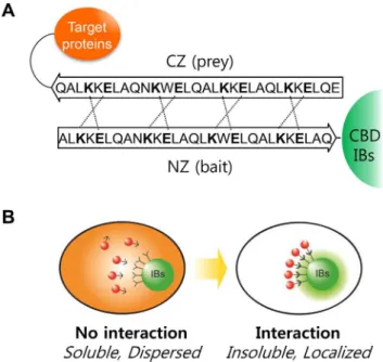
For SEM imaging, purified CBD-IBs were fixed in a mixture of 2.5% paraformaldehyde and 2.5% glutaraldehyde in a 100-mM sodium phosphate buffer (pH 7.2) for 2 h, post-fixed with 1% osmium tetroxide in the same buffer for 1 h, dehydrated in graded ethanol, substituted with isoamyl acetate, and then critical point dried in CO2. The samples were then coated with gold in a SC502 sputter coater (Quorum Technologies Ltd, East Sussex, UK) and observed under a Quanta 250 FEG scanning electron microscope (FEI, Hillsboro, OR, USA) at KRIBB (Daejeon, Korea). Figure 1. Controlled localization of functionally active proteins
to inclusion bodies (IBs) using leucine zippers (LZs). A. Interactions between anti-parallel leucine zippers (LZ). Dashed lines indicate the charge-charge interactions of glutamic acid (E) and lysine (K).B.Representation of controlled localization in bacterial cell. The red fluorescent protein, which is dispersed throughout the cytosol, is localized to IBs by a specific molecular interaction.
The size and zeta-potential of the EGFP-IBs were measured using a Malvern Zetasizer Nano ZS (Malvern Instruments, Malvern, UK) at the National Nanofab Center (Daejeon, Korea). The protein solution was diluted with 10 mM Tris-HCl (pH 8.0), and 0.75 mL of the diluted solution was added to disposable zetasizer cuvettes for the measurements. The experiments were performed in triplicate and the data were processed using Zetasizer Nano software (version 6.01; Malvern Instruments).
Results
Generation of functional IBs
The CBDs include three to four aromatic residues that are exposed to bulk liquid on the surface of the protein (http://www. pdb.org; PDB ID: 1exg) [19], which may cause rapid aggregation of the protein. As previously mentioned, C-terminal fusions of the family II CBD fromC. fimiformed active IBs retaining 30%–40% of the original activity while maintaining the ability to bind insoluble celluloses [9]. In the current study,E. colicells expressing a fusion of the CBD with EGFP exhibited one or two fluorescent IBs in microscopic images (Fig. 2A). When cells expressing either EGFP or EGFP-CBD were compared by flow cytometry, the fluorescence intensity of the EGFP-CBD cells was estimated to be 10%–20% of that in cells expressing soluble EGFP (Fig. S2), although the expression of both proteins (as detected by western blotting) was similar. When the EGFP-CBD cells were sonicated in Tris buffer (50 mM Tris-HCl, pH 8.0 and 200 mM NaCl) to break the IBs into smaller pieces, the fluorescence intensity increased up to 2 folds in proportion to the sonication time (Fig. 2B). Therefore, the IBs are estimated to contain higher amounts of properly folded/native-like protein than that observed in flow cytometry. The low detection of fluorescence in IBs is discussed further in the Discussion section.
Localization of soluble proteins to IBs
The possibility of active IBs as a matrix to recruit soluble cytosolic proteins was tested by displaying a bait LZ that can bind to prey LZs in cytosol (Figs. 1A and 1B). LZ is a super-secondary structure that generates adhesion forces between a-helices. A single LZ consists of multiple leucine residues at approximately 7-residue intervals, which forms an amphipathic alpha helix with a hydrophobic region on one side. This hydrophobic region provides an area for dimerization, allowing the motifs to combine. Therefore, fusion proteins tagged with prey LZs may form a two-stranded a-helical coiled-coil heterodimer with the bait LZ in active IBs (Fig. 1A). A monomeric red fluorescent protein 1 (mRFP1) was used as a model prey protein to take advantage of its easy detection in living cells. Thebait-EGFP-CBDandprey-mRFP1
genes were cloned into pET21a in a polycistronic manner to balance the relative expression of the bait and prey. When these
bait and prey proteins were co-expressed inE. colicells, the red fluorescence was clearly localized to the IBs (lower row in Fig. 2C), whereas the red fluorescence remained dispersed in cells without the bait LZ (upper row in Fig. 2C), showing that localization was dependent on the bait LZ.
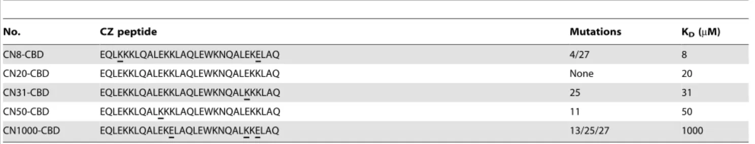
Next, the effect of LZ binding affinity was investigated using different combinations of LZs (shown in Table 1). The leucine residue is essential for duplex formation, whereas ionic interactions between oppositely charged residues affected binding affinity. We examined five different bait and prey pairs that were designed by Magliery et al. [12] with KDvalues of 8, 20, 31, 50, and 1,000mM.
Table 1.Amino acid sequences and affinity information of mutant CZs.
No. CZ peptide Mutations KD(mM)
CN8-CBD EQLKKKLQALEKKLAQLEWKNQALEKELAQ 4/27 8
CN20-CBD EQLEKKLQALEKKLAQLEWKNQALEKKLAQ None 20
CN31-CBD EQLEKKLQALEKKLAQLEWKNQALKKKLAQ 25 31
CN50-CBD EQLEKKLQALKKKLAQLEWKNQALEKKLAQ 11 50
CN1000-CBD EQLEKKLQALEKELAQLEWKNQALKKELAQ 13/25/27 1000
Mutation sites are underlined. The CZ peptide sequences and KD’s were adopted from the results of Magliery TJ et al. [12]. doi:10.1371/journal.pone.0097093.t001
Figure 2. Microscopic observation of controlled localization to CBD IBs. A.Imaging expressed EGFP and EGFP-CBD inE. coli. The left panel represents the western blot images after treatment with anti-GFP and anti-GroEL antibodies. The EGFP band is indicated by the black arrows. Scale bar = 5mm.B.Increased fluorescence following sonication of EGFP-CBD IBs.E. coli cells expressing EGFP-CBD were treated by sonication in a Tris buffer (50 mM Tris-HCl, pH 8.0, 200 mM NaCl) and the fluorescence intensity analyzed using a Cary Eclipse fluorometer. The inset represents a SEM image of the EGFP-CBD IBs.C.Microscopic images ofE. colicells with no interaction (top) and interaction (bottom) between LZs. Scale bar = 5mm.
As anticipated, more red fluorescence was observed to localize to the IBs when bait-prey pairs with smaller KDvalues were used for the co-expression experiments (Fig. 3). When region-of-interest (ROI) analysis was applied to the cellular images (Fig. 4A), red fluorescence in cytosol decreased as the prey-mRFP1 protein localized to the IBs. Consequently, the mean yield of localization to IBs, ROI2vs. ROI1, was calculated from at least five single cell images and a high yield of 0.65 was estimated for CN8 (KD= 8mM), which was nearly the same as the mean yield for
EGFP-CBD (Fig. 4B). The yield for CN1000 (KD= 1,000mM)
was approximately 0.30. Therefore, the higher the affinity of the bait for the prey, the more prey-mRFP localized to the IBs. In all the experiments, the expression levels of the bait-EGFP-CBD and prey-mRFPs were similar (as shown by SDS-PAGE analyses) (Fig. S3).
The localization of red fluorescence to IBs was also investigated by flow cytometry. When the cytometric results were drawn on FL1vs.FL2 dot plots, the mRFP intensity (FL2) decreased as the binding affinity increased (Fig. 5A), whereas the EGFP intensity (FL1) increased. For example, the mean intensity of mRFP for the CN8-CBD cell populations was about 40% of that for cells with no bait in the CBD IBs (Fig. 5B). This result was consistent with the microscopic observations in Fig. 4A.
Finally, we attempted to purify cells with IB-localized red fluorescence using a single cell sorter, the FACSAria. For this experiment, equal amounts of cells with (pCN20-CBD) and without bait (pC20-CBD) were mixed, and the specific cells within a predetermined gate (dashed areas in Fig. 5A) were recovered. The collected cells were then analyzed by western blotting using an anti-GFP antibody (Fig. 5C). Lanes 1 and 2 show the control bands for bait-EGFP-CBD and EGFP-CBD, respectively. Before sorting, both proteins were observed in the cells (lane 3), whereas after sorting, the band corresponding to bait-EGFP-CBD was enriched in the recovered cells (lane 4), indicating selective sorting of cells with red fluorescent IBs due to protein-protein interactions between the bait and prey LZs.
Figure 3. Effects of binding affinity between LZs.Microscopic images ofE. colicells containing LZ pairs with varying affinities (KD= 8, 20, 31, 50, and 1,000mM). Scale bar = 5mm.
doi:10.1371/journal.pone.0097093.g003
Figure 4. Region of interest (ROI) analysis of microscopic images. A. Cellular fluorescence decreased in proportion to the binding affinity between LZs in IBs.B. Comparison of localization yield to IBs. The fluorescence in IBs was normalized to the total cellular fluorescence, ROI2/ROI1, where ROI1is the cellular area and ROI2is the IB area of the cell. More than five cells per image were examined for the ROI analysis. Error bars show the standard deviations from 5 independent measurements of the cells.
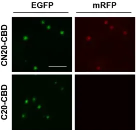
High physical stability of fluorescent IBs
The functional IB particles were extracted from the CN20-CBD cells and the C20-CBD cells by sonication and washed twice with a solution containing 0.5% Triton X-100 detergent in a Tris buffer (50 mM Tris-HCl, pH 8.0 and 200 mM NaCl). Microscopic observation showed that the CN20-CBD IBs contained both green and red fluorescent IB particles, while the bait-less C20-CBD IBs contained only green fluorescent IBs because the prey-mRFP was washed out (Fig. 6). Therefore, the interactions in the active IBs were highly specific and were maintained during sonication and washing.
The physical stability of the active IBs was investigated using a zeta potential analyzer (Zetasizer Nano). The particle size was approximately 0.45–0.5mm in diameter and the zeta potentials
were estimated at approximately 256.8 mV. Zeta potentials
larger than 640 indicate that colloidal particles are stable in solution, while particles with a zeta potential smaller than 630 tend to coagulate or flocculate easily [20]. Therefore, the IB
particles in this study remained physically stable under bothin vivo
andin vitroconditions. Discussion
Synthetic biology, an emerging field, involves the design and construction of new genetic devices for use in research and industry [15,21]. One successful device applied to metabolite production is the synthetic protein scaffold [16,17]. When a heterologous or synthetic pathway is introduced, the host cell can suffer from flux imbalance, intermediate loss, and chemical toxicity [22]. Therefore, constructing synthetic scaffolds may improve the metabolite conversion rate by increasing the local enzyme concentration and reducing intermediate loss caused by diffusion or side reactions. In this regard, CBD IBs could be useful as a synthetic matrix inE. colicells. The target proteins can be recruited to the synthetic IB matrix via bait and prey interactions between LZs (Fig. 1), which are a well-known domain consisting of Figure 5. Flow cytometric analyses of controlled localization to IBs. A.FL1vs.FL2 dot plot of cells containing LZ pairs with varying binding affinities (KD= 8, 20, 31, 50, and 1,000mM). EGFP* indicates theE. colicells that expressed soluble EGFP and soluble mRFP (the controls). The dashed line indicates the sorting gate.B.Comparison of the fluorescence intensity of cells expressing different leucine zipper pairs.C. Western blot analysis of CN20-CBD cells sorted by the FACSAria. Lane 1, CN20-CBD cells; lane 2, C20-CBD cells; lane 3, a mixture of CN20-CBD and C20-CBD cells before sorting; lane 4, a mixture of CN20-CBD and C20-CBD cells after sorting. Error bars show the standard deviations from 5 independent measurements of the cells.
only 30 amino acids. LZs such as E, K coil proteins have been used previously to immobilize active enzymes in polyhydroxybu-tyrate synthase IBs [14]. In this study, the affinities between LZs were controlled by changing the amino acid sequences. In addition, mRFP1 was used as a soluble target protein because it is easy to detect without cell disruption. Imaging and flow cytometric analyses showed that prey localization was dependent on the binding affinity between the bait and prey LZ proteins; the prey protein exhibited only marginal localization to the IBs when the KDof the LZs was 1,000mM (Fig. 3); as the KDdecreased, localization increased sharply and it reached a maximum level
when the KD was 8 or 20mM. Eventually, we established a
quantitative method to evaluate the localization of cytosolic proteins to IBsin situby using LZs with different affinities (Fig. 3), which provides useful implications for the generation of synthetic matrices with designed compositions.
Localization of EGFP to IBs resulted in a large decrease in the fluorescence signal compared to the signal for soluble EGFP (Fig. S2), which is approximately half of the activity retention observed for catalytic enzymes in a previous study. The fluorescence intensity increased 2-fold when the particles were broken into smaller pieces by sonication (Fig. 2B). Based on literature reviews and our results in Fig. S4, the reason for the decreased fluorescence in the IBs is thought to be related with the scattering of the excitation light by the highly refractile surfaces of the IB particles [23,24] and/or a shortened fluorescence lifetime in the densely packed environment [25]. In general, IBs are more dense (,1.3 mg/mL) than any other cellular component.
Investigations of protein-protein interactions (PPIs) are crucial in modern biological science research [26], and there is growing
interest in the development of high throughput technologies [18,27]. In the method developed here, proteins with different affinity of LZs localized to IBs were quantitatively analyzed in living cells using flow cytometry (Fig. 5), while the E, K coil proteins in IB fractions was detected by electrophoretic methods after cell disruption in previous study [14]. Therefore, the current method can be applied usefully for high throughput screening of PPI inhibitors, comparisons of interacting protein partners, and engineering binding affinities in bacterial cells.
Conclusions
Fluorescent proteins localized in IBs exhibited high intrinsic activity; however, their activity was somewhat suppressed when
localized to IBs formed by fusion with the CBD from C. fimi
exoglucanase. The signal intensity on microscopic images or in high throughput flow cytometry was dependent on the binding affinities of the interacting pairs. This controlled localization to IBs in living cells can be useful for the collective localization of cytosolic proteins inE. coli for sequential reactions. In addition, easy detection of protein localization to the IBs may provide a new platform for the rapid analyses of PPIs in bacterial cells.
Supporting Information
Figure S1 Construction of CN20-CBD (A) and C20-CBD (B).
(TIF)
Figure S2 Flow cytometric analyses of cells expressing EGFP and EGFP-CBD proteins. The dark green and light green signals indicate cells expressing EGFP and EGFP-CBD, respectively.
(TIF)
Figure S3 SDS-PAGE analysis of different leucine zipper proteins in E. coli cells (CN8-CBD, CN20-CBD,
CN31-CBD, CN50-CN31-CBD, and CN1000-CBD).The upper and lower arrows indicate the size of the NZ-EGFP-CBD and CZ-mRFP proteins, respectively.
(TIF)
Figure S4 Side and forward scattering analyses of E. colicells expressing EGFP (A) and EGFP-CBD (B).
(TIF)
Table S1 Primers used in this study. (TIF)
Author Contributions
Conceived and designed the experiments: SLC SJL HJK YHR SGL. Performed the experiments: SLC SJL HJK YHR SGL. Analyzed the data: SLC SJL HJK YHR SGL. Contributed reagents/materials/analysis tools: SLC SJL HJK YHR SGL. Wrote the paper: SLC SJL SJY HCJ SGL.
References
1. Palmer I, Wingfield PT (2012) Preparation and extraction of insoluble (inclusion-body) proteins fromEscherichia coli. Curr Protoc Protein Sci Chapter 6: Unit6 3. 2. Carrio MM, Corchero JL, Villaverde A (1998) Dynamics of in vivo protein aggregation: building inclusion bodies in recombinant bacteria. FEMS Microbiol Lett 169: 9–15.
3. Villaverde A, Carrio MM (2003) Protein aggregation in recombinant bacteria: biological role of inclusion bodies. Biotechnol Lett 25: 1385–1395.
4. Sabate R, de Groot NS, Ventura S (2010) Protein folding and aggregation in bacteria. Cell Mol Life Sci 67: 2695–2715.
5. Singh SM, Panda AK (2005) Solubilization and refolding of bacterial inclusion body proteins. J Biosci Bioeng 99: 303–310.
6. Ventura S, Villaverde A (2006) Protein quality in bacterial inclusion bodies. Trends Biotechnol 24: 179–185.
7. Garcia-Fruitos E, Gonzalez-Montalban N, Morell M, Vera A, Ferraz RM, et al. (2005) Aggregation as bacterial inclusion bodies does not imply inactivation of enzymes and fluorescent proteins. Microb Cell Fact 4: 27.
8. Wu W, Xing L, Zhou B, Lin Z (2011) Active protein aggregates induced by terminally attached self-assembling peptide ELK16 inEscherichia coli. Microb Cell Fact 10: 9.
9. Choi S-L, Lee SJ, Ha J-S, Song JJ, Rhee YH, et al. (2011) Generation of catalytic protein particles inEscherichia colicells using the cellulose-binding domain from
Cellulomonas fimias a fusion partner Biotechnol Bioprocess Eng 16: 1173–1179. 10. Nahalka J, Nidetzky B (2007) Fusion to a pull-down domain: a novel approach of producing Trigonopsis variabilis D-amino acid oxidase as insoluble enzyme aggregates. Biotechnol Bioeng 97: 454–461.
Figure 6. Comparison of fluorescent IBs purified from CN20-CBD and C20-CN20-CBD cells.Scale bar = 5mm.
11. Ghosh I, Hamilton AD, Regan L (2000) Antiparallel leucine zipper-directed protein reassembly: application to the green fluorescent protein. J Am Chem Soc 122: 5658–5659.
12. Magliery TJ, Wilson CG, Pan W, Mishler D, Ghosh I, et al. (2005) Detecting protein-protein interactions with a green fluorescent protein fragment reassembly trap: scope and mechanism. J Am Chem Soc 127: 146–157. 13. Campbell RE, Tour O, Palmer AE, Steinbach PA, Baird GS, et al. (2002) A
monomeric red fluorescent protein. Proc Natl Acad Sci U S A 99: 7877–7882. 14. Steinmann B, Christmann A, Heiseler T, Fritz J, Kolmar H (2010) In vivo enzyme immobilization by inclusion body display. Appl Environ Microbiol 76: 5563–5569.
15. Chen AH, Silver PA (2012) Designing biological compartmentalization. Trends Cell Biol 22: 662–670.
16. Dueber JE, Wu GC, Malmirchegini GR, Moon TS, Petzold CJ, et al. (2009) Synthetic protein scaffolds provide modular control over metabolic flux. Nat Biotechnol 27: 753–759.
17. You C, Zhang YH (2013) Self-assembly of synthetic metabolons through synthetic protein scaffolds: one-step purification, co-immobilization, and substrate channeling. ACS Synth Biol 2: 102–110.
18. Koyama-Honda I, Ritchie K, Fujiwara T, Iino R, Murakoshi H, et al. (2005) Fluorescence imaging for monitoring the colocalization of two single molecules in living cells. Biophys J 88: 2126–2136.
19. McLean BW, Bray MR, Boraston AB, Gilkes NR, Haynes CA, et al. (2000) Analysis of binding of the family 2a carbohydrate-binding module from
Cellulomonas fimi xylanase 10A to cellulose: specificity and identification of functionally important amino acid residues. Protein Eng 13: 801–809. 20. Hanaor D, Michelazzi M, Leonelli C, Sorrell CC (2012) The effects of
carboxylic acids on the aqueous dispersion and electrophoretic deposition of ZrO2. J Eur Ceram Soc 32: 235–244.
21. Khalil AS, Collins JJ (2010) Synthetic biology: applications come of age. Nat Rev Genet 11: 367–379.
22. Bentley WE, Mirjalili N, Andersen DC, Davis RH, Kompala DS (1990) Plasmid-encoded protein: the principal factor in the ‘‘metabolic burden’’ associated with recombinant bacteria. Biotechnol Bioeng 35: 668–681. 23. Young PA, Clendenon SG, Byars JM, Dunn KW (2011) The effects of refractive
index heterogeneity within kidney tissue on multiphoton fluorescence excitation microscopy. J Microsc 242: 148–156.
24. Theer P, Denk W (2006) On the fundamental imaging-depth limit in two-photon microscopy. J Opt Soc Am A Opt Image Sci Vis 23: 3139–3149. 25. Suhling K, Siegel J, Phillips D, French PM, Leveque-Fort S, et al. (2002)
Imaging the environment of green fluorescent protein. Biophys J 83: 3589–3595. 26. Phizicky EM, Fields S (1995) Protein-protein interactions: methods for detection
and analysis. Microbiol Rev 59: 94–123.