In vivo
antitumoural activity and composition of an oil extract of Brazilian propolis
Adriana Andrade Carvalho
a, Daiane Finger
b, Christiane Schinieder Machado
b, Eduardo Morgado Schmidt
b,
Patrícia Marçal da Costa
a, Ana Paula Negreiros Nunes Alves
c, Thamires Maria Fontenele Morais
d,
Maria Goretti Rodrigues de Queiroz
d, Sueli Pércio Quináia
b, Marcos Roberto da Rosa
b,
Julio Murilo Trevas dos Santos
b, Cláudia Pessoa
a, Manoel Odorico de Moraes
a, Letícia Veras Costa-Lotufo
a,
Alexandra Christine Helena Frankland Sawaya
e, Marcos Nogueira Eberlin
f, Yohandra Reyes Torres
b,⇑aDepartamento de Fisiologia, Universidade Federal do Ceará, Fortaleza, CE, Brazil bDepartamento de Química, Universidade Estadual do Centro-Oeste, Guarapuava, PR, Brazil cDepartamento de Clínica Odontológica, Universidade Federal do Ceará, Fortaleza, CE, Brazil dFaculdade de Farmácia, Universidade Federal do Ceará, Fortaleza, CE, Brazil
eDepartamento de Fisiologia Vegetal, Instituto de Biologia, Universidade Estadual de Campinas, Campinas, SP, Brazil
fLaboratório ThoMSon de Espectrometria de Massas, Instituto de Química, Universidade Estadual de Campinas, Campinas, SP, Brazil
a r t i c l e
i n f o
Article history: Received 2 June 2010
Received in revised form 16 November 2010 Accepted 6 December 2010
Available online 10 December 2010
Keywords: Propolis Edible oil extract Antitumour activity Phenolic compounds
a b s t r a c t
The present study aimed to evaluatein vivoandin vitrothe antitumoural activity of a propolis extract obtained with edible vegetable oil and its fractions and also to investigate its chemical composition by LC–MS and LC–MS/MS. To evaluate the toxicological aspects related to the propolis extract treatment, hematological, biochemical, histopathological and morphological analyses of treated animals were per-formed. All propolis extracts showed anin vivoantitumour activity in the experimental model with a moderate toxicity effect at experimental exposure levels. The oil extract was as effective as the ethanolic extract at inhibiting tumour growth.In vitroassays showed that the whole oil extract produced better inhibition of tumour cells than its fractions. LC–MS and LC–MS/MS identified four phenolic acids and three flavonoids. The anticancer potential of the oil extract of propolis has been demonstrated and the edible vegetable oil was shown as an attractive alternative solvent to extract bioactive natural propolis components.
Ó2010 Elsevier Ltd. All rights reserved.
1. Introduction
The use of propolis is ancient in traditional medicine dating back at least to 300 BC (Ghisalberti, 1979). Today, this resinous bee product continues to be used worldwide, and a broad spectrum of biological activities for propolis has been reported, including anticancer, antioxidant, inflammatory, antibiotic, and anti-fungal activities (Burdok, 1998; Marcucci, 1995). The biological effect of propolis is attributed to its natural bioactive chemicals, such as polyphenols, flavonoid aglycones, phenolic acid and their esters, caffeic acid and their esters and phenolic aldehydes and ke-tones (Orsolic & Basic, 2003).
Recently, attention is being focused on the anti-cancer activity of propolis. The studies, however, have been performed using etha-nolic or hydroalcoholic extracts (Orsolic & Basic, 2003). Ethanolic formulations of propolis prevent its consumption by people who
can not consume alcohol for medical reasons, such as diabetic patients. Several patents have dealt therefore with new methods or solvents besides ethanol to extract propolis (Kasuma & Kenichi, 2001a, 2001b; Namiki et al., 2005). These patents have reported the use of edible vegetable oils, triglycerides and fatty acids as extraction solvents for propolis. Data on the biological activity and chemical composition of oil extracts of propolis are, however, scarce. Tosi, Donini, Romagnoli, and Bruni (1996) evaluated the antimicrobial activity of commercial extracts of propolis prepared with different solvents including oils. They reported a wide range of antimicrobial activity for the oil extract and concluded that the solvent employed for the extraction of propolis influences the po-tency of its antimicrobial activity. We have compared antiprolifer-ative activity against the HL-60, MDAMB-435 and SF-295 cells lines of oil and ethanolic propolis extracts (Buriol et al., 2009) and found out that oil extracts were active against the tumour cell line tested showing higher anticancer potential against the SF-295 cell line.
The aim of this study was to investigate the effects of the oil and ethanolic extracts of propolis in experimental models. Hematolog-ical, biochemHematolog-ical, histopathological and morphological analyses of the tumour and the organs, including liver, spleen and kidney,
0308-8146/$ - see front matterÓ2010 Elsevier Ltd. All rights reserved. doi:10.1016/j.foodchem.2010.12.035
⇑ Corresponding author. Address: UNICENTRO CEDETEG: Rua Simeão Camargo Varela de Sá, 03, Vila Carli – Guarapuava – PR, CEP 85040 080, Brazil. Tel.: +55 042 3629 8324/8120.
E-mail address:ryohandra@yahoo.com(Y.R. Torres).
Contents lists available atScienceDirect
Food Chemistry
were performed to evaluate the toxicological aspects of the treat-ment. The results of this study should therefore advance the knowledge of the antitumour benefits of edible oil extracts of prop-olis and provide a better understanding of their application in the prevention/treatment of malignant tumours. Besides biological assays, the oil extract of propolis was also fractioned by chroma-tography and its fractions analysed using mass spectrometry to evaluate their chemical composition.
2. Materials and methods
2.1. Propolis and extracts
2.1.1. Samples
Propolis samples were collected in 2006 and supplied by Camp-olin and Schmidt Company from Prudentópolis city (Paraná State, Brazil). Propolis was stored at 18°C until extraction.
2.1.2. Extracts
Fifty grams of propolis were extracted in a shaker with 500 ml of canola oil or 70% ethanol, during 24 h, at room temperature. After that period, the extractive solutions were filtered. The solvent was removed from the hydro-alcoholic solution yielding EEP70. The oil extract of propolis was partitioned into 80% v/v metha-nol/water and the aqueous methanolic phase was dried in a rota-tory evaporator yielding ODEP.
2.2. Chromatography fractionation
Hundred and eighty milligrams of ODEP in methanol were ap-plied on a glass column (380 cm) containing Sephadex LH-20.
The elution was performed with methanol. A total of 80 fractions of 8 ml each were obtained. These fractions were joined in six new fractions, labeled as OLSx 1–6, through thin layer chromatog-raphy analyses on silica gel (mobile phase chloroform/acetone/ace-tic acid 18:4:1 v/v/v and visualisation of spots at UV of 254 nm with the use of FeCl3as the colour reagent).
2.3. Mass spectrometry analyses
2.3.1. ESI( )–MS data
Samples of OLSx 1–6 were analysed using a Q-Trap mass spec-trometer (Applied Biosystems) with the direct infusion of the sam-ple solutions into the electrospray ionisation source operating in the negative ion mode. Capillary and cone voltages were set to 4500 V and 50 V, respectively, with a de-solvation temperature of 100°C.
2.3.2. Liquid chromatographic mass spectrometry analyses (LC–MS) OLSx 3–6 were introduced into an HPLC (Agillent) with a
l
Bondapak C18 analytical column (Waters, 3,9300 mm,10
l
m) and detected in a Q-Trap mass analyser. ESI( )–MS was carried out with capillary and cone voltages set to 4500 and 50 V, respectively, and a de-solvation temperature of 300°C. Abinary mobile phase of acetonitrile and 1% of formic acid was em-ployed. A linear gradient was performed starting from 30% of ace-tonitrile to 100% aceace-tonitrile, in 30 min, and an elution flow rate of 1 ml/min.
2.3.3. ESI( )–MS/MS
Tandemmass spectra were acquired using a hybrid high-reso-lution and high-accuracy (5 ppm) Micromass Q-TOF mass spec-trometer (Waters) and via collision-induced dissociation at ca. 15 V. Capillary and cone voltages were set to ±3000 and ±40 V, respectively, for the negative or positive mode of ionisation. The
de-solvation temperature was 100°C; nitrogen and argon were
used as de-solvation or collision gas, respectively.
2.4. Biological assays
2.4.1. Cytotoxicity assays
The cytotoxicity of propolis extracts and fractions from ODEP was evaluated against four human tumour cell lines: HL-60 (leuke-mia), HCT-8 (colon), MDA/MB-435 (breast) and SF-295 (brain) obtained from the National Cancer Institute (Bethesda, MD, USA). The general viability of cultured cells was determined by the reduction of the yellow dye 3-(4,5-dimethyl-2-thiazolyl)-2,5-di-phenyl-2H-tetrazolium bromide (MTT) to a blue formazan product, as previously described byMosmann (1983).
The tumour cells were maintained in RPMI 1640 medium, sup-plemented with 10% fetal bovine serum, 1% penicillin and strepto-mycin at 37°C with 5% CO2. For all experiments cells were seeded
at 0.3106cells/ml (HL-60, MDA/MB-435 e SF-295) and
0.7105cells/ml (HCT-8), and incubated during 72 h with
propo-lis extracts (0.001–50
l
g/ml ODEP and EEP70) and fractions (0.001–25l
g/ml), under the conditions described above. After cen-trifugation and solution removing, MTT solution was added and the plates were incubated, centrifuged, and the solids dissolved in pure and sterile DMSO. The absorbance was measured in a plate spectrophotometer DTX-800 (Beckman Coulter) at 595 nm. Doxo-rubicin (Sigma) was used as a positive control.2.4.2. In vivo antitumoural assays
2.4.2.1. Animals.A total of 80 Swiss mice (male, 25–30 g), obtained from the central animal house of Federal University of Ceará, Brazil, were used. The animals were housed in cages with free access to food and water (conforming to a well-defined rodent diet). All ani-mals were kept under a 12:12 h light–dark cycle (lights on at 6:00 a.m.). The animals were treated according to the ethic princi-ples of animal experimentation of COBEA (Colégio Brasileiro de Experimentação Animal), Brazil. The Animal Studies Committee of the Federal University of Ceará approved the experimental protocol.
Sarcoma 180 tumour cells were maintained in the peritoneal cavities of the Swiss mice obtained from the central animal house of the Federal University of Ceará.
2.4.2.2. Determination of the effect of propolis extract on tumour growth in vivo.Ten-day-old sarcoma 180 ascites tumour cells (2 ± 106cell/500
l
l) were implanted subcutaneously into the left hind groin of the experimental mice. One day after inoculation, the propolis extracts (50 and 80 mg/kg to ODEP and EEP70) or 5-FU (25 mg/kg) were dissolved in 4% DMSO and administered intraperitoneally for 7 days. The negative control was injected with 4% DMSO. On day 8th, the mice were killed and the tumours were excised, weighed and fixed in 10% formaldehyde. The inhibition ra-tio (%) was calculated by the following formula: inhibira-tion rara-tio (%) = [(A B)/A]100, whereAis the average tumour weight ofthe negative control, andBis the tumour weight of the treated group.
2.4.3. Toxicological analysis
2.4.3.2. Determination of the effect of propolis extract on biochemical parameters.After fasting for 6–8 h, the animals were submitted to blood collection from the orbital plexus for biochemical analysis (urea and creatinine to investigate any renal function alterations; AST and ALT as liver parameter). The analysis was carried out in a semi-automatic equipment (LabQuestÒ
), using enzymatic col-ourimetric kits, while the hematological cells were quantified in a SysmexÒ
KX-21 N. The methodology of the LabQuest and Sysmex equipment are based, respectively, on the principle of absorption and impedance.
2.4.3.3. Determination of the effect of propolis extracts on hematolog-ical parameters. After fasting for 6–8 h, the animals were submitted to blood collection from the orbital plexus for hematological anal-yses. The hematological analyses were performed by an optical microscope OlympusÒ
BX 41. Hematological parameters, including the hemoglobin content, platelet count, total count of leukocytes as well as a differential count of leukocytes, such as eosinophil (%), lymphocyte (%), neutrophil (%) and monocyte (%) were measured.
2.4.3.4. Histopathology and morphological observations.After being fixed with formaldehyde, tumours, livers, spleens and kidneys were grossly examined for size or colour changes and hemorrhage. Subsequently, portions of the tumour, liver, spleen and kidney were cut into small pieces, followed by staining with hematoxylin and eosin of the histological sections. Histological analyses were performed by light microscopy. The occurrence and the extent of liver or kidney lesions attributed to drugs were recorded.
3. Results
3.1. Analysis of ODEP chemical composition by ESI( )–MS, LC–MS and LC–MS/MS
ODEP fractionation gave fractions OLSx 1–6, which were first analysed by direct infusion ESI( )–MS. Since propolis is a rich nat-ural bee product containing mainly phenolic compounds, such as cinnamic acid derivatives and flavonoids, ions were mostly pro-duced by electrospray ionisation in the negative ion mode. ESI( ) is a soft ionisation technique and mass spectra mainly detected components as their intact deprotonated molecules. ESI( )–MS provides therefore fingerprinting characterisation of propolis ex-tracts via characteristic profiles of their chemical composition in terms of the most polar and acidic or basic components (Sawaya et al., 2004a, 2007).
Fig. 1shows the ESI( )–MS fingerprints of ODEP fractions and indicates great differences in the chemical composition between the fractions. OLSx1 and OLSx2 were complex fractions with sev-eral high to medium abundant ions, such asm/z 387, 311, 341 and 275 (OLSx1). For OLSx2, the highest abundant ions were those ofm/z315, 339, with medium abundant ions ofm/z265, 325, 293 and 377. Fractions OLSx 3–6 were simpler and showed mostly a single major ion. OLSx3 showed mostly the ions ofm/z247, 231, 301 and 393 whereas OLSx4 showed those ofm/z301, 245, 393 and 287 as most abundant. Fractions OLSx5 and OLSx6 showed a major common ion ofm/z299 and other less abundant ions ofm/ z329 and 387 (OLSx5) andm/z249, 285, 311 and 387 (OLSx6).
To characterise the fractions constituents, LC–MS and LC–MS/ MS were performed. Via comparisons with the fragmentation pat-tern of previously identified compounds (Marcucci, Sawaya, Custo-dio, Paulino, & Eberlin, 2008), several components have been identified: 3,4-dihydroxi-5-prenyl-cinnamic acid (m/z247, OLSx3); dihydrokaempferide (m/z 301, OLSx3 and OLSx4); 3-prenyl-4-hydroxicinnamic acid (m/z231, OLSx3); (E)-3-{-4-hydroxy-3-[(E)-4-(2,3-dihydrocinnamoyl
oxy)-3-methyl-2-butenyl]-5-prenylphe-nyl}-2-propenoic acid (m/z447, OLSx2). Isosakuranetin (m/z285, OLSx4 and OLSx6) was identified by comparison of its MS/MS frag-mentation with a standard kindly provided by Vassya S. Bankova, (Bulgarian Academy of Science). We have identified some of those compounds previously in the oil extracts of propolis (Buriol et al., 2009).
In addition, LC–MS identified two compounds with the samem/z 299 but different retention time. Compound in OLSx2 with [M–H] ofm/z299 andtR24 min showed in its ESI( )–MS/MS a fragmenta-tion to the ion ofm/z255 via initial loss of 45 Da and ofm/z244, 200 and 145. It was identified as 3,5-diprenyl-4-hydroxicinnamic acid also known by artepellin C. ESI( )–MS/MS of the other compound with [M–H] ofm/z299 buttR14 min in OLSx5 and OLSx6 under-went fragmentation to the ion ofm/z284 via initial loss of a methyl radical and was identified as kaempferide. Other fragment ions ofm/ z164, 151, and 107 are typical of the cleavage of the flavonoids cen-tral C-ring (Cuyckens & Claeys, 2004) (Table 1).
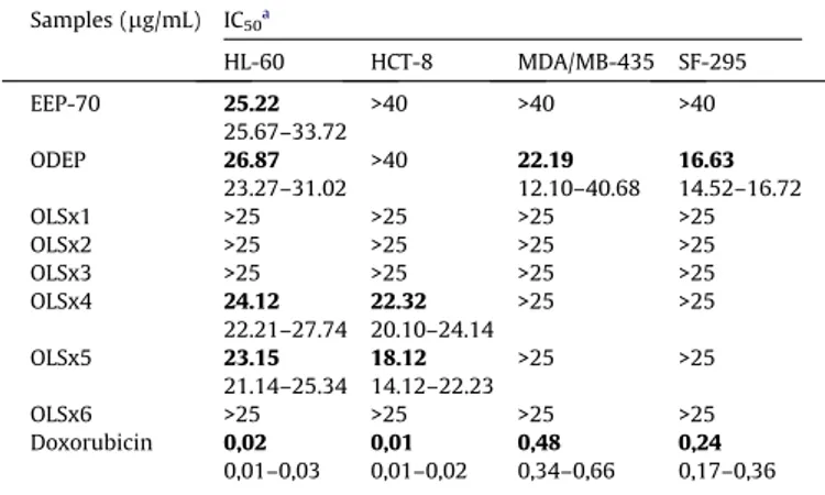
3.2. In vitro cytotoxicity assays
Table 2shows the results from thein vitrocytotoxic assays for the propolis extracts and the fractions obtained by the chromato-graphic separation of ODEP on Sephadex LH-20. The extract of propolis obtained with canola oil and dried (ODEP) displayed mod-erate cytotoxicity against leukemia (HL-60), melanoma (MDA-MB-435) and glioblastoma (SF-295) cancer cells, a better result than the ethanolic extract (EEP70). When analysing cytotoxicity from ODEP fractions, it was evident that the fractions were less active than the propolis extract (ODEP) in the cell lines evaluated, whereas OLSx4 and OLSx5 showed moderate cytotoxicity against leukemia (HL-60) and colon (HCT-8).
3.3. Effect of propolis extracts on tumour growth in vivo and on organ and body weight
To check if the cytotoxic effects observedin vitroalso occured in vivo, we used the Sarcoma 180 (S-180) model which is a mouse-originated tumour frequently used inin vivoantitumour re-lated research (Gonzaga et al., 2009).Fig. 2shows the effects of the propolis extracts on mice transplanted with S-180 tumour. There was a significant reduction of the tumour weight in all extracts tested. The differences between experimental groups were com-pared by ANOVA followed by Student Newman Keuls or Bonferroni tests (p< 0.05). On the 8th day, the average tumour weight of the control mice inoculated with sarcoma 180 was 2.05 ± 0.22 g. In the presence of EEP70, the sarcoma 180 weight was reduced to 0.94 ± 0.35 and 0.90 ± 0.22 g at doses of 50 and 80 mg/kg, respec-tively. These reductions are equivalent to inhibition ratios of 53.94% and 56.29%. In the presence of ODEP, the sarcoma 180 weight was reduced to 0.92 ± 0.14 and 0.96 ± 0.24 g at doses of 50 and 80 mg/kg, respectively. These reductions are equivalent to inhibition ratios of 54.94% and 53.35%. At 25 mg/kg, 5-FU reduced tumour weight by 51.56% within the same period. These results showed that the inhibition ratio of the ethanolic extract was the same as that of oil extract and no differences were observed when the extracts were tested at doses of 50 and 80 mg/kg. It was dem-onstrated that both extracts of propolis inhibited the Sarcoma 180 tumour growth in mice.
Fig. 1.ESI( )–MS fingerprints from ODEP fractions.
Table 1
Identified compounds in fractions from the oil extract of propolis and ESI( )–MS/MS fragments.
Compound Fraction tR m/z[M–H] Main fragments:m/z(relative abundance %)
4-Hydroxy-3-prenylcinnamic acid OLSx3 6,4 231 187 (50), 132 (100)
3,4-Dihydroxy-5-prenylcinnamic acid OLSx3 10,6 247 203 (100), 185 (10), 148 (55)
Isosakuranetin OLSx4
OLSx6
15 285 270 (10), 243 (20), 164 (100), 151 (20), 136 (20)
Kaempferide OLSx5
OLSx6
14 299 284 (100), 164 (10), 151 (20)
Artepillin C OLSx2 24 299 255 (30), 200 (100), 185 (10), 145 (10)
Dihydrokaempferide OLSx4
OLSx3
10,8 301 283 (50), 227 (30), 152 (100), 151 (30), 125 (40), 107 (20)
(E)-3-{-4-hydroxy-3-[(E)-4-(2,3-dihydrocinnamoyl oxy)-3-methyl-2-butenyl]-5-prenylphenyl}-2-propenoic acid
3.4. Effect of the propolis extracts on biochemical parameters
No significant changes in the renal (urea and creatinine levels) or liver [enzymatic activity of transaminases aspartate aminotransfer-ase (AST) and alanine aminotransferaminotransfer-ase (ALT)] parameters were seen in the animals treated with propolis extracts (data compared by AN-OVA followed by Student Newman Keuls or Bonferroni testsp< 0.05) in mice transplanted with Sarcoma 180 tumour (Fig. 3). The animals treated with 5-FU have alteration on renal and liver parameters.
3.5. Histopathology, morphological, and morphometric changes
Histopathological analyses of the tumours extirpated from the control mice showed a malignant neoplasm composed of pleomor-phic polygonal cells with little cytoplasm, some with vesicular nu-clei, and acidophilic macronucleoli. Muscular invasion, areas of coagulation necrosis and typical and atypical mitotic figures were also observed. In the tumours extirpated from treated animals, extensive areas of coagulative necrosis were observed.
The histopathological analyses of livers, removed from all groups, showed foci of microvesicular steatosis. Mild swelling of hepatocytes and focal microvesicular steatosis were observed in the negative control group. In 5-FU-treated animals intense cell swelling of hepatocytes, microvesicular steatosis, hyperplasia of Kupffer cells and hemosiderin were observed. In ODEP-treated ani-mals, moderate swelling cell hepatocyte, microvesicular steatosis, hyperplasia of Kupffer cells, inflammatory foci and bilirubin were observed. In EEP70-treated animals we found intense swelling of hepatocytes and large areas of microvesicular steatosis.
Analyses of the kidneys showed cilindrohialin, which indicates a difficulty of the renal filtration system of proteins. Severe swell-ing of the tubular epithelium was also found in all groups, includ-ing the 5-FU. All groups showed lymphoid follicles in the spleen, sometimes with large, irregular, ill-defined borders, probably re-lated to the actual tumour (sarcoma 180) that leads to this histo-logical finding. All groups showed areas of hemorrhage.
3.6. Effect of the propolis extracts on hematological parameters
The animals transplanted with Sarcoma 180 tumours treated with 5-FU showed a strong reduction on the total leukocytes (p< 0.05). Treatment with the propolis extracts demonstrated no alteration (Table 3).
4. Discussions
ESI( )–MS, LC–MS and LC–MS/MS identified several prenylated phenolic acids and flavonoids in ODEP, demonstrating that vegeta-ble oil was avegeta-ble to extract important bioactive natural phenolic compound from crude propolis. Compounds identified in the pres-ent work have been found in alcoholic and hydro-alcoholic extracts of propolis from Brazil and other countries and are related to many biological activities (Banskota, Tezuka, & Kadota, 2001; Banskota et al., 1998; Lustosa, Galindo, Nunes, Randau, & Neto, 2008; Sa-waya, Souza, Marcucci, Cunha, & Shimizu, 2004; Sawaya et al., 2002b). This is so for artepillin C, for instance, a major compound in green Brazilian propolis, for which biological activities, such as antimicrobial, antioxidant and anti-tumour, as well as increases in the immune response against leukemia have been reported (Shi-mizu, Ashida, Matsuura, & Kanazawa, 2004). Because of these bio-Table 2
In vitrocytotoxic activity of propolis extracts and fractions against tumour cell lines.
Samples (lg/mL) IC50a
HL-60 HCT-8 MDA/MB-435 SF-295
EEP-70 25.22
25.67–33.72
>40 >40 >40
ODEP 26.87
23.27–31.02
>40 22.19
12.10–40.68 16.63 14.52–16.72
OLSx1 >25 >25 >25 >25
OLSx2 >25 >25 >25 >25
OLSx3 >25 >25 >25 >25
OLSx4 24.12
22.21–27.74 22.32 20.10–24.14
>25 >25
OLSx5 23.15
21.14–25.34 18.12 14.12–22.23
>25 >25
OLSx6 >25 >25 >25 >25
Doxorubicin 0,02 0,01–0,03
0,01 0,01–0,02
0,48 0,34–0,66
0,24 0,17–0,36
a Data are presented as IC
50values and 95% confidence interval obtained by
non-linear regression for leukemia (HL-60), colon (HCT-8), breast (MDA-MB-435) and brain (SF-295) cancer cells from two independent experiments performed four times (n= 8).
logical properties, propolis, which contains artepillin C is consid-ered as a high quality propolis and the content of artepillin C is al-ready used in quality control by some companies (Funari & Ferro, 2006). By visual inspections of the chromatograms (data not shown), from both LC–MS and LC–UV (PDA), it was evident that artepillin C is commonly present in propolis from Prudentópolis, and that the oil extracts of propolis do contain significant levels of prenylated phenolic acids, including artepillin C. Several other ions in the oily extract of propolis remained unknown indicating the need to carry on further studies of the chemical composition of this promising ODEP extract.
All treatment regimens for the majority of cancers produce side effects, which makes this treatment extremely unpleasant for patients. Scientists have spent therefore efforts to develop new therapies for the treatment of cancer. Propolis has been a subject of intense research, especially in the areas of anticancer research (Banskota et al., 2000; El-khawaga Om-Ali, Tarek, & Mohammed, 2003; Padmavathi, Senthilnathan, Chodon, & Sakthisekaran, 2006). The majority of those studies evaluated the ethanol, aque-ous or methanol extracts of propolis, hence in this work we as-sessed the antitumour effect of an oil extract of propolis. We have previously reported data comparing antiproliferative activity against HL-60, MDAMB-435 and SF-295 cells lines of oil and etha-nolic propolis extracts (Buriol et al., 2009). Because a different sample of propolis (but from the same region) and different condi-tions extraccondi-tions were used, we can not directly compare the IC50 value reported earlier with the present values, but in both investi-gations the oil extracts of propolis were active against the tested tumour cell lines indicating more anticancer potential against the
SF-295 cell line than the ethanolic extracts. The present results show that the oil extract of propolis has substances with cytotoxic effects similar to the more common ethanolic extracts, making this extract attractive for applications where ethanol must be avoided. The oil extract of propolis also produced better inhibition of tumour cells than its fractions, indicating that propolis cytotoxic activity is probably a ‘‘shotgun’’ synergic effect of its many bioac-tive components. The biological activity of propolis should there-fore be highly depended on the extraction process.
In the Sarcoma 180 model assay it was demonstrated that all propolis extracts inhibited tumour growth in mice with the same inhibition ratio as the positive control 5-FU but with less side effects.
The histopathological analyses of organs removed from treated animals showed that the treatment with 5-FU, ODEP and EEP70 led to Kupffer cell hyperplasia and necrosis, which signals the presence of a toxic agent. Nevertheless, these alterations could be consid-ered reversible (Kummar, Abbas, & Fausto, 2004; Scheuer & Lefko-witch, 2000).
Renal parameters were also evaluated. The histopathological analyses of the kidneys showed focal areas of necrosis in the tubu-lar epithelium in all groups of treatment, suggesting that the treat-ment with 5-FU, ODEP and EEP70 caused kidney damage. Necrosis of the renal tubule epithelium may occur on a large scale as a con-sequence of the administration of different chemical classes (Olsen & Solez, 1994). Although modifications of kidney tissues were observed, the analysis of biochemical parameters didn’t show changes in levels of urea and creatinine. Nevertheless, blood urea was increased only a long time after renal alteration, which may Fig. 3.Effect of propolis extracts on biochemical parameters in peripheral blood from mice transplanted with sarcoma 180 tumour.
Table 3
Effect of propolis extracts on the hematological changes on peripheral blood.
Drug Dose mg/kg Hemoglobin (g/dl) Platelets (106cel/ll) Leukocytes (106cel/ll) Differential count (%)
Eosinophils Neutrophils Lymphocytes Monocytes
Salina – 12.26 ± 0.31 1240 ± 119 4.1 ± 0.31 0 44 36.00 8
5-FU 25 10.30 ± 0.20 934 ± 54a 1.2 ± 0.21a 0 52 37.00 7
EEP70 50 13.01 ± 0.40 1243 ± 55 3.96 ± 0.40 0.2 47 35.00 9
80 14.02 ± 0.50 1244 ± 45 3.87 ± 0.30 0.1 45 34.00 8
ODEP 50 12.08 ± 0.49 1343 ± 105 3.12 ± 0.34 0 39 36.60 8
80 11.76 ± 0.52 1454 ± 109 4.15 ± 1.22 0 40 35.40 7
Data are presented as mean ± SEM.
explain why the treatment in this study failed to induce any alter-ation in the blood urea levels.
Separately from hepatotoxic and nephrotoxic effects, anticancer drugs also produce delayed hematopoietic depression, as observed in treatment with methotrexate and 5-FU (Bezerra et al., 2008; Katzung, 2003). In fact, most chemotherapeutic drugs, including 5-FU, are immunosuppressive because they kill many normal cells as well as tumour cells (Bezerra et al., 2008; Takiguchi et al., 2001) and have negative side effects. One of the risks of radiation and chemotherapy in the treatment of cancer patients is the develop-ment of leukopenia, which substantially increases the risk of infec-tions. We observed herein leukopenia in the 5-FU treatment, but not in the EEP70 and ODEP treatment.
The weight of the spleens in animals treated with 5-FU was also significantly lower than in the control group, which also indicated an immunosuppressive side effect of 5-FU, but propolis treatment caused no alteration in the weight of the spleen. When comparing the histopathological analyses, we observed that all groups treated with propolis showed congestion on red pulp, which indicates a possible effect on the immodulatory system. It is well reported that the mechanism of antitumour effects elicited by propolis extracts has been attributed to its effect on the immodulatory system.
5. Conclusions
The findings in the present study indicate the potential of oil extract of propolis for the treatment of cancer. The ethanol-free vegetable oil extract of propolis displayed importantin vitroand in vivoantitumour effects due to a synergic effect of its many bio-active constituents with moderate signs of toxicity.
Acknowledgements
We wish to thank Vassya S. Bankova, (Bulgarian Academy of Science) for the isosakuranetin standard. Financial support was provided by Finep and Fundação Araucária through 10908/PPP/ 2006 and 7102/PPI phase I-2004 and phase II-2006. DF and EMS wish to thank for a scholarship from CAPES and CSM thanks for a scholarship from Fundação Araucária. MNE and AFWS wish to thank FAPESP and CNPq for the financial support. The authors also thank Silvana França dos Santos and Erivanda França for their tech-nical assistance. ACHFS thanks CAPES for a postdoctoral fellowship.
References
Banskota, A. H., Tezuka, Y., Prasain, J. K., Matsushige, K., Saiki, I., & Kadota, S. (1998). Chemical constituents of Brazilian propolis and their cytotoxic activities.Journal of Natural Products, 61, 896–900.
Banskota, A. H., Tezuka, Y., Adnyana, I. K., Midorikawa, K., Matsushige, K., Message, M., et al. (2000). Biological evaluation of propolis from Brazil, Peru, the Netherlands and China.Journal of Ethnopharmacology, 72, 239–246.
Banskota, A. H., Tezuka, Y., & Kadota, S. (2001). Recent progress in pharmacological research of propolis.Phytotherapy Research, 15, 561–571.
Bezerra, D. P., de Castro, F. O., Alves, A. P. N. N., Pessoa, C., de Moraes, M. O., Silveira, E. R., et al. (2008).In vitroandin vivoantitumor effect of 5-FU combined with piplartine and piperine.Journal of Applied Toxicology, 28, 156–163.
Burdok, G. A. (1998). Review of the biological properties and toxicity of bee propolis (Propolis).Food and Chemical Toxicology, 36, 347–363.
Buriol, L., Finger, D., Schmidt, E. M., Santos, J. M. T., Rosa, M. R., Quináia, S. P., et al. (2009). Composição química e atividade biológica de extrato oleoso de própolis: uma alternativa ao extrato etanólico.Química Nova, 32, 296–302.
Cuyckens, F., & Claeys, M. (2004). Mass spectrometry in the structural analysis of flavonoids.Journal of Mass spectrometry, 39, 1–15.
El-khawaga Om-Ali, Y., Tarek, A. S., & Mohammed, F. E. (2003). Protective role of Egyptian propolis against tumor in mice.Clinica Chimica Acta, 338, 11–16. Funari, C. S., & Ferro, V. O. (2006). Análise de própolis.Ciência e Tecnologia de
Alimentos, 26, 171–178.
Ghisalberti, E. L. (1979). Propolis a review.Bee World, 60, 59–84.
Gonzaga, M. L. C., Bezerra, D. P., Alves, A. P. N. N., Alencar, N. M. N., Mesquita, R. O., Lima, M. W., et al. (2009).In vivogrowth-inhibition of Sarcoma 180 by ana -(1,4)-glucan–b-(1,6)-glucan-protein complex polysaccharide obtained from Agaricus blazeiMurill.Journal of Natural Medicine, 63, 32–40.
Kasuma, Y., & Kenichi, I. (2001a). Propolis composition and its production. JP Pat. 20010374227 (A).
Kasuma, Y., & Kenichi, I. (2001b). Extraction of propolis extract. JP Pat. 2001017096 (A).
Katzung, G. B. (2003).Basic and Clinical Pharmacology(9th ed.). USA: McGraw-Hill Medical.
Kummar, V., Abbas, A., & Fausto, N. (2004).Robbins and Cotran pathologic basis of disease. China: W.B. Saunders.
Lustosa, S. R., Galindo, A. B., Nunes, L. C. C., Randau, K. P., & Neto, P. J. R. (2008). Própolis: Atualizações sobre a química e farmacologia. Brazilian Journal of Pharmacognosy, 18, 447–454.
Marcucci, M. C. (1995). Propolis: Chemical composition, biological properties and therapeutic activity.Apidologie, 26, 83–99.
Marcucci, M. C., Sawaya, A. C. H. F., Custodio, A. R., Paulino, N., & Eberlin, M. N. (2008). HPLC and ESI–MS typification: New approaches for natural therapy with Brazilian propolis. In N. Orsolic & I. Basic (Eds.),Scientific evidence of the use of propolis in ethnomedicine(pp. 33–54). Kerala: Research Network.
Mosmann, T. (1983). Rapid colourimetric assay for cellular growth and survival: Application to proliferation and cytotoxicity assays.Journal of Immunological Methods, 65, 55–63.
Namiki, H., Matsuka, M., Nakamura, J., Adachi, S., Watanabe, G., Okazaki, M. Propolis extract and method for extracting the same. WO Pat. 2005/094853 (A1), 2005. Olsen, S., & Solez, K. (1994). Acute tubular necrosis and toxic renal injury. In C. C. Tisher & B. M. Brenner (Eds.), Renal pathology: with clinical and functional correlations(pp. 769–809). Philadelphia: JB. Lippincott Company.
Orsolic, N., & Basic, I. (2003). Immunomodulation by water-soluble derivative of propolis: a factor of antitumor reactivity.Journal of Ethnopharmacology, 84, 265–273.
Padmavathi, R., Senthilnathan, P., Chodon, D., & Sakthisekaran, D. (2006). Therapeutic effect of paclitaxel and propolis on lipid peroxidation and antioxidant system in 7, 12 dimethyl benz(a)anthracene-induced breast cancer in female Sprague Dawley rats.Life Sciences, 78, 2820–2825.
Sawaya, A. C. H. F., Palma, A. M., Caetano, F. M., Marcucci, M. C., Cunha, I. B. S., Araújo, C. E. P., et al. (2002). Comparative study ofin vitromethods used to analyze the activity of propolis extracts with different compositions against species ofCândida.Letters in Applied Microbiology, 35, 203–207.
Sawaya, A. C. H. F., Tomazela, D. M., Cunha, I. B. S., Bankova, V. S., Marcucci, M. C., Custodio, A. R., et al. (2004). Electrospray ionization mass spectrometry fingerprinting of propolis.Analyst, 129, 739–744.
Sawaya, A. C. H. F., Souza, K. S., Marcucci, M. C., Cunha, I. B., & Shimizu, M. T. (2004). Analysis of the composition of Brazilian propolis extracts by chromatography and evaluation of theirin vitroactivity against gram-positive bacteria.Brazilian Journal of Microbiology, 35, 104–109.
Sawaya, A. C. H. F., Cunha, I. B. S., Marcucci, M. C., Aidar, D. S., Silva, E. C. A., Carvalho, C. A. L., et al. (2007). Electrospray ionization mass spectrometry fingerprinting of propolis of native Brazilian stingless bees.Apidologie, 38, 93–103. Shimizu, K., Ashida, H., Matsuura, Y., & Kanazawa, K. (2004). Antioxidative
bioavailability of artepillin C in Brazilian propolis.Archives of Biochemistry and Biophysics, 424, 181–188.
Scheuer, P. J., & Lefkowitch, J. H. (2000). Drugs and toxins. In P. J. Scheuer & J. H. Lefkowitch (Eds.), Liver biopsy interpretation (pp. 134–150). London: W.B. Saunders.
Takiguchi, N., Saito, N., Nunomura, M., Kouda, K., Oda, K., Furuyama, N., et al. (2001). Use of 5-FU plus hyperbaric oxygen for treating malignant tumors: evaluation of antitumor effect and measurement of 5-FU in individual organs.Cancer Chemotherapy and Pharmacology, 47, 11–14.