Flow-mediated Dilatation in the Differential Diagnosis of
Preeclampsia Syndrome
Edson Vieira da Cunha Filho, Carolina Mohr, Breno José Acauan Filho, Giovani Gadonski, Leticia Germany Paula, Ivan
Carlos Ferreira Antonello, Carlos Eduardo Poli-de-Figueiredo, Bartira Ercilia Pinheiro-da-Costa
Pontificia Universidade Católica do Rio Grande do Sul, Porto Alegre, RS - Brazil
Abstract
Background: The preeclampsia syndrome is associated with endothelial dysfunction and the differential diagnosis between pure preeclampsia (PE) and superimposed preeclampsia (SPE) can be only be attained 12 weeks after delivery.
Objective: To compare the assessment of endothelial function through flow-mediated dilatation in pregnant women with pure preeclampsia and superimposed preeclampsia.
Methods: The flow-mediated dilatation of the brachial artery was carried out according to the recommendations of the International Brachial Artery Reactivity Task Force in pregnant women with preeclampsia syndrome. PE (n=14) and SPE (n=13) were diagnosed in the postpartum period according to the definitions of the National High Blood Pressure Education Program Working Group on High Blood Pressure in Pregnancy.
Results: The median of the flow-mediated dilatation (FMD) in SPE (6.0%; 1.9-10.3) was decreased in comparison with the PE (13.6%;4.4-17.1), an apparently relevant difference , but not statistically significant (p = 0.08). The FMD < 10% was detected in 30.8% of the PE cases and in 69.2% of the SPE cases (p = 0.057). Significant differences could not be detected in the morphology of the uterine arteries between the PE and SPE cases through the Doppler spectrum.
Conclusion: The FMD of the brachial artery of patients with preeclampsia syndrome was not capable of differentiating between PE and SPE. However, the data suggest that SPE is associated with worse endothelial function I comparison to PE. (Arq Bras Cardiol 2010;94(2): 182-186)
Key Words: Ultrasonography; endotlhelium; vasodilatation; eclampsia; brachial artery.
Mailing Address: Bartira Ercilia Pinheiro da Costa •
Rua Ernesto Paiva 140 - Tristeza - 91900200 - Porto Alegre, RS - Brazil E-mail: [email protected]
Manuscript received January 30, 2009; revised manuscript received May 21, 2009; accepted August 05, 2009.
Introduction
Preeclampsia, one of the most deleterious diseases in Obstetrics, is currently considered a two-stage disease. The first corresponds to insufficient placentation, causing an increase in the resistance of the uteroplacental circulation. The second involves the maternal reaction, through the activation of the inflammatory response and endothelial cell dysfunction1.
Preeclamptic arteries present higher reactivity and reduced dilatation, facts related to the alteration of metabolic pathways, such as for instance, that of the nitric oxide. However, the participation of this oxide is not well established regarding the decrease in pressure levels in the first half of the pregnancy2,3. It has also been observed that substances
associated with endothelial dysfunction, such as fibronectin4
and phosphodiesterase enzyme 5 are elevated in the plasma
of preeclamptic women.
Endothelial dysfunction usually increases the vasoconstrictor response, the proliferation and migration of cells from the
smooth vascular muscle, the platelet and leukocyte adhesion and the expression of adhesion molecules. Such loss of endothelial functional integrity is associated with risk factors for cardiovascular alterations, such as arterial hypertension, atherosclerosis, dyslipidemia and diabetes, among others6.
One of the noninvasive methods used to assess the endothelial function is the ultrasonography7,8. The
flow-mediated dilatation (FMD) of the brachial artery is a simple ultrasonographic technique that can measure the vascular response through the variation of the basal artery diameter induced by the reactive hyperemia that follows re-establishment of blood flow after a period of vascular occlusion; by insufflating a sphygmomanometer cuff. This measurement is an assessment of the endothelium-dependent vascular function.
It has been observed that in patients presenting endothelial damage, the vasodilation response is decreased and, in some cases, even vasoconstriction can be observed8,9.
Some authors use the FMD to assess the endothelial function of control pregnant women and/or those presenting pathologies, mainly diabetic and preeclamptic ones, with the latter presenting lower FMD values when compared to normal pregnant women11. The differential diagnosis between
pure preeclampsia (PE) and superimposed preeclampsia (SPE) can be attained 90 days after the delivery, as in gestational hypertension12.
The present study assessed the FMD of pregnant women with preeclampsia syndrome to verify whether this test could detect differences in the endothelial reactivity of patients with PE and SPE.
Methods
The definitions used followed the classification of the gestational hypertensive disorders, as recommended by the report of the National High Blood Pressure Education Program Working Group on High Blood Pressure in Pregnancy (NHBPEPWGHBPP)12, as follows:
Preeclampsia/eclampsia (PE)
A syndrome that is specific of human pregnancy and is characterized by hypertension and proteinuria. Hypertension is defined as systolic arterial pressure ≥ 140 mmHg, and diastolic arterial pressure ≥ 90 mmHg, in a normotensive pregnant woman, before 20 weeks of gestation. Proteinuria is defined as pathological when it is > 300 mg in a 24-hour period. Proteinuria and arterial pressure must go back to normal levels in up to 12 weeks after the delivery. The evolution to eclampsia is characterized by seizures in these patients.
Preeclampsia superimposed to chronic hypertension (SPE)
It is the development of the preeclampsia syndrome in a patient with chronic hypertension. This is a difficult diagnosis, as the hypertensive patient might present pathological proteinuria, requiring careful observation. However, the use of fundoscopic examination as a differentiation tool can help in the detection of damage to a target-organ in patients with chronic hypertension.
This is a transversal, prospective study, consisting of 27 patients with preeclampsia syndrome. The patients were hospitalized at Hospital São Lucas, of PUCRS, for obstetric care or were receiving prenatal care at the Service of Obstetrics. All patients signed a Informed Consent Form, which had been previously approved by the Ethics Committee of PUCRS. Pregnant women with a history of venous and arterial thrombosis, known predisposition to venous or arterial thrombosis, diabetes mellitus, history of severe liver disease, history of liver tumors, history of chronic hypertension, as well as those with previous proteinuria, drug or alcohol users or those who did not agree to participate in the study were excluded from it.
After the diagnosis of preeclampsia syndrome, the patients were submitted to the measurement of the FMD of the brachial artery, according to the technique proposed by Celermajer et al7 and modified by Regattieri et al13. The measurement of the
FMD was carried out in right brachial artery of the patients,
while they were comfortably seated, with slight abduction of the right arm and hand supination, in order to expose the anteromedial side of the arm to the examiner. The 14 MHz-linear probe of the 2-D ultrasonography device, with color and spectral Doppler (Ultrasonix, Ultrasonix Medical Corporation, Richmond, Canada), was placed on the medial side of the arm, longitudinal and perpendicularly to the skin, 5 cm above the antecubital fossa, insonating the brachial artery directly below the biceps and next to the brachial muscle.
It can be observed that the high-frequency vascular linear transducer used allows the identification of 7 zones, which correspond to the two medial-adventitial interfaces, the two intimal layers, the two medial layers and the artery lumen and this is the best way to ascertain that the transducer is in the center of the vessel and perpendicular to it13.
Subsequently, the diameter of the brachial artery was measured longitudinally through images in which the proximal and distal lumen-intimal interfaces are visualized. The measurement of the diameter in the center of the vessel at the moment corresponding to the end of the diastole was obtained. This moment was monitored using the B-mode of the echocardiographic equipment as the moment presenting the lowest distension of the vessel walls (to prevent larger vascular calibers originated from the vascular distension caused by the systole), which can be correctly captured by receding the image using the equipment cine loop of the equipment. After the basal diameter (D1) was verified, the skin was marked with a pen at the place where the measurement was taken by the transducer. The cuff was applied to the same arm, adjusting the pressure of 20 to 30 mmHg above the systolic pressure, maintaining the occlusion for 5 minutes. The post-occlusion diameter (D2) was measured 60 seconds after the cuff was removed, with a tolerance of 30 seconds. This was the moment of maximum reactive hyperemia.
Based on the previously described standards, a new measurement of the brachial artery caliber was carried out. The FMD value was obtained from the following calculation: FMD (%) = [(D2 - D1)/D1] × 100, where D1 = basal diameter and D2 = post-occlusion diameter. FMD values > 10% were considered normal according to Quyyumi14.
No restrictive measures were required for the FMD measurement, such as limitation of time for the examination to be performed or withdrawal of hypotensive medication, due to the severity of the hypertensive state presented by the patients. None of the patients included in the study smoked during the pregnancy and coffee was not ingested for 8 hours prior to the assessment. The room where the measurements were performed was kept at ambient temperature and the same examiner assessed all patients. After the FMD measurement, an ultrasonography was also performed to assess the fetal status and to carry out an analysis with Doppler velocimetry of the maternal, placental and fetal compartments.
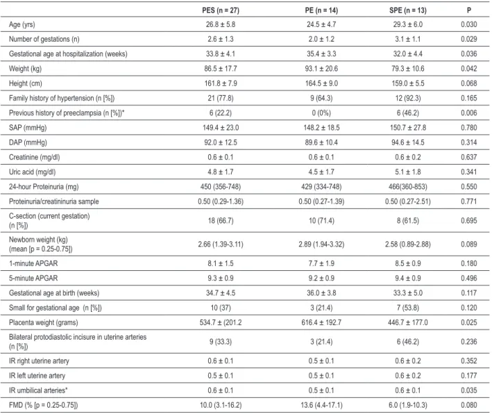
Table 1 - Demographic and clinical aspects of the sample of patients with preeclampsia syndrome
PES (n = 27) PE (n = 14) SPE (n = 13) P
Age (yrs) 26.8 ± 5.8 24.5 ± 4.7 29.3 ± 6.0 0.030
Number of gestations (n) 2.6 ± 1.3 2.0 ± 1.2 3.1 ± 1.1 0.029
Gestational age at hospitalization (weeks) 33.8 ± 4.1 35.4 ± 3.3 32.0 ± 4.4 0.036
Weight (kg) 86.5 ± 17.7 93.1 ± 20.6 79.3 ± 10.6 0.042
Height (cm) 161.8 ± 7.9 164.5 ± 9.0 159.0 ± 5.5 0.068
Family history of hypertension (n [%]) 21 (77.8) 9 (64.3) 12 (92.3) 0.165
Previous history of preeclampsia (n [%])* 6 (22.2) 0 (0%) 6 (46.2) 0.006
SAP (mmHg) 149.4 ± 23.0 148.2 ± 18.5 150.7 ± 27.8 0.780
DAP (mmHg) 92.0 ± 12.5 89.6 ± 10.4 94.6 ± 14.5 0.314
Creatinine (mg/dl) 0.6 ± 0.1 0.6 ± 0.1 0.6 ± 0.2 0.637
Uric acid (mg/dl) 4.8 ± 1.7 4.5 ± 1.7 5.1 ± 1.8 0.341
24-hour Proteinuria (mg) 450 (356-748) 429 (334-748) 466(360-853) 0.550
Proteinuria/creatininuria sample 0.50 (0.29-1.36) 0.50 (0.27-1.39) 0.50 (0.27-2.51) 0.771
C-section (current gestation)
(n [%]) 18 (66.7) 10 (71.4) 8 (61.5) 0.695
Newborn weight (kg)
(mean [p = 0.25-0.75]) 2.66 (1.39-3.11) 2.89 (1.94-3.32) 2.58 (0.89-2.88) 0.089
1-minute APGAR 8.1 ± 1.5 7.7 ± 1.9 8.5 ± 0.9 0.180
5-minute APGAR 9.3 ± 0.9 9.2 ± 0.9 9.4 ± 0.9 0.496
Gestational age at birth (weeks) 34.7 ± 4.5 36.0 ± 3.8 33.3 ± 5.0 0.117
Small for gestational age (n [%]) 10 (37) 3 (21.4) 7 (53.8) 0.120
Placenta weight (grams) 534.7 ± (201.2 616.4 ± 192.7 446.7 ± 177.0 0.025
Bilateral protodiastolic incisure in uterine arteries
(n [%]) 9 (33.3) 3 (21.4) 6 (46.2) 0.236
IR right uterine artery 0.6 ± 0.1 0.5 ± 0.1 0.6 ± 0.2 0.352
IR left uterine artery 0.5 ± 0.1 0.5 ± 0.1 0.6 ± 0.2 0.177
IR umbilical arteries* 0.6 ± 0.1 0.5 ± 0.1 0.6 ± 0.1 0.035
FMD (% [p = 0.25-0.75]) 10.0 (3.1-16.2) 13.6 (4.4-17.1) 6.0 (1.9-10.3) 0.080
Data presented as means (± standard deviations), medians (IIQ25-75) or n (%). The probability was considered signiicant when < 0.05, when comparing the PE and SPE groups. PE - Pure preeclampsia; SPE - superimposed preeclampsia, PES – preeclampsia syndrome.
The data regarding the FMD, patients’ clinical information, obstetric echography, obstetric Doppler, laboratory assessment and maternal and perinatal outcomes were entered into an Excel spreadsheet. Ninety days after the postpartum, the patients were referred to the Ambulatory of Nephrology of Hospital Sao Lucas of PUCRS in order to conclude the diagnosis of PE or SPE, indicated mainly by the measurement of the arterial pressure and proteinuria.
The data were evaluated by measures of central trend and dispersion (mean + standard deviation, or median, or interquartile interval) for parametric and non-parametric data, respectively, or frequencies and percentiles. The comparisons were carried out using Student’s t or Mann-Whitney test, for parametric and non-parametric variables, respectively. The categorical data were compared by the Chi-Square test. The correlations of parametric data were carried out using Pearson’s correlation test and the non-parametric correlations
were carried out using Spearman’s correlation test. The level of significance was set at α = 0.05. The Microsoft Excel program, using a database registry created for this study and the Statistical Package for Social Sciences (SPSS) program, version 11.5 for Windows, were used in this research.
Results
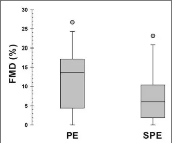
Figure 1 -Distribution of the two groups of pregnant women with preeclampsia
syndrome, according to the measurement of the low-mediated dilatation of the brachial artery < 10.
weight at birth and the Doppler Resistance Index of the umbilical arteries showed a statistically significant difference between the two groups.
Regarding the FMD, its median among patients with SPE was quantitatively lower than in patients with PE; however, the comparison of these values did not show a statistically significant difference (p = 0.080). Considering that endothelial dysfunction was categorized as FMD < 10%, it was observed that the incidence was 69.2% in SPE group and 30.8% in the PE group (p = 0.057). These data are shown in Figure 1.
The PE and SPE groups were not different (p=0.236) when comparing the morphology of the uterine artery waveforms through spectral Doppler analysis. Only three PE patients presented bilateral protodiastolic incisure, whereas 6 patients presented it in the SPE group.
The presence of bilateral protodiastolic incisure was correlated with the FMD, but there was no statistically significance regarding patient distribution, according to the measurement of the vascular response > or < 10%.
The correlation of the FMD with the clinical and perinatal outcomes showed that there is a positive association with the gestational age at the time of the hospitalization (r = 0.42; p = 0.02), newborn gestational age (r = 0.46; p = 0.01) and weight (r = 0.46; p = 0.01).
Discussion
The present study assessed the endothelial function in the differential diagnosis of the preeclampsia syndrome. The median FMD in the SPE group is decreased in comparison with the PE group and when the sample was analyzed with cutoff values < 10%, the following distribution was observed: 1/3 with PE and more than 2/3 with SPE had endothelial dysfunction. A statistically significant difference was not reached due to the sample size. It is evident that the superimposed preeclampsia is associated with a worse endothelial function in comparison with the pure preeclampsia, even though the FMD is not
enough to differentiate both clinical presentations of the preeclampsia syndrome.
Due to its practical characteristics and its applicability, the FMD has become an object of interest for researchers in different areas. Several studies have been carried out with pregnant women aiming at having access to the endothelial function. Several authors have proposed the occurrence of endothelial dysfunction in preeclampsia and have used this indication to carry out their evaluation15-18. This is a practical,
safe and convenient technique to assess pregnant women. The present study seems to be original when it proposes, based on the FMD measurement, to verify whether it is possible to anticipate the differential diagnosis of pregnant women with PE and pregnant women with SPE to the moment of the preeclampsia syndrome manifestation. After the diagnosis had been established, differentiating PE from SPE, the two groups were separated for data analysis. At the moment of data collection, the patients’ diagnosis was not known, as even the patients from the SPE group did not know whether they were hypertensive.
The knowledge that the patient was previously hypertensive was considered an exclusion criterion in order to eliminate a possible observation bias by the examiner, when measuring the FMD.
The data presented in Table 1 regarding the demographic and clinical characteristics, show that the groups are not clearly distinguishable based on the usual clinical measures applied to these patients. Interestingly, the difference was evident in the set of demographic characteristics, which are not routinely employed as high-sensitivity indicators for essential hypertension. The data with higher parity - consequently of also advanced maternal age and previous history of PE in the group of patients with SPE - were expected. These data are in accordance with the proposal that pregnancy is women’s stress test in which the worst outcome demonstrates the lack of capacity for such demand19.
As expected, the SPE group, which can supposedly present previous endothelial damage due to systemic hypertension, presented lower FMD values when compared to the patients from the PE group. However, perhaps due to the sample size, the difference was not statistically significant, although we believe it has a clinical relevance. The fact that both groups presented endothelial dysfunction due to the underlying disease - preeclampsia syndrome - also becomes relevant if one considers that the difference in FMD values was not very high and, therefore, a larger sample size is needed to demonstrate it. Another aspect that must be considered, corroborating this statement, is that two cases from the PE group presented no FMD, thus markedly decreasing the mean in this group. Both cases were categorized as severe preeclampsia (one of them presented HELLP syndrome and another presented eclampsia).
Another difference observed between the two groups of preeclamptic women refers to the disease severity. Preeclampsia presents higher severity when superimposed on previous hypertension12, which can be verified through the
References
1. Roberts JM, Hubel CA. The two stage model of preeclampsia: variations on the theme. Placenta. 2009; 30 (Suppl. A): 532-7.
2. Selligman S, Buyon J, Clancy R, Young B, Abramson S. The role of nitric oxide in the pathogenesis of preeclampsia. Am J Obstet Gynecol. 1994; 171 (4): 944-8.
3. Langenfeld MR, Simmons LA, McCrohon JA, Raitakari OT, Lattimore JD, Hennessy A, et al. Nitric oxide does not mediate the vasodilation of early human pregnancy. Heart Lung Circ. 2003; 12 (3): 142-8.
4. Chavarría ME, Lara-González L, González-Gleason A, Sojo I, Reyes A. Maternal plasma cellular fibronectin concentrations in normal and preeclamptic pregnancies: a longitudinal study for early prediction of preeclampsia. Am J Obstet Gynecol. 2002; 187 (3): 595-601.
5. Pinheiro da Costa BE, Scocco C, Poli de Figueiredo CE, Guimarães JA. Increased serum phosphodiesterase activity in women with pre-eclampsia. BJOG. 2006; 113 (5): 577-9.
6. Mombouli JV, Vanhoutte PM. Endothelial dysfunction: from physiology to therapy. J Mol Cell Cardiol. 1999; 31 (1): 61-74.
7. Celermajer DS, Sorensen KE, Gooch VM, Spiegelhalter DJ, Miller OI, Sullivan ID, et all. Non-invasive detection of endothelial dysfunction in children and adults at risk of atherosclerosis. Lancet. 1992; 340: 1111-5.
8. Faulx MD, Wright AT, Hoit BD. Detection of endothelial dysfunction with brachial artery ultrasound scanning. Am Heart J. 2003; 145 (6): 943-51.
9. Moens AL, Goovaerts I, Claeys MJ, Vrints CJ. Flow-mediated vasodilation: a diagnostic instrument, or an experimental tool? Chest. 2005; 127: 2254-63.
10. Dorup I, Skajaa K, Sorensen KE. Normal pregnancy is associated with enhanced endothelium-dependent flow-mediated vasodilation. Am J Physiol. 1999; 276 (3 Pt 2): H821-5.
11. Cockell AP, Poston L. Flow-mediated vasodilatation is enhanced in normal pregnancy but reduced in preeclampsia. Hypertension. 1997; 30 (2 Pt 1): 247-51.
12. Gifford RW, August PA, Cunningham G, Green LA, Lindheimer MD, McNellis D. Report of the National High Blood Pressure Education Program Working Group on High Blood Pressure in Pregnancy. Am J Obstet Gynecol. 2000; 183: S1-S22.
13. Regattieri NAT, Leite SP, Koch HA, Montenegro CAB. Dilatação fluxo-mediada da artéria braquial: desenvolvimento da técnica, estudo em pacientes de risco para aterosclerose e em um grupo controle. Rev Bras Ultrason.. 2006; 9: 9-13.
14. Quyyumi AA. Prognostic value of endothelial function. Am J Cardiol. 2003; 91(Suppl): 19H-24H.
15. Sader MA, Celermajer DS. Endothelial function, vascular reactivity and gender differences in the cardiovascular system. Cardiovasc Res. 2002; 53 (3): 597-604.
16. Veille JC, Gorsuch L, Weeks W, Zaccaro D. Hyperemic response of the brachial artery during the second half of pregnancy. J Soc Gynecol Investig. 1998; 5 (1): 38-43.
17. Takase B, Gotto T, Hamabe A, Uehata A, Kuroda K, Sattomura K, et al. Flow-mediated dilation in brachial artery in the second half of pregnancy and prediction of pre-eclampsia. J Hum Hypertens. 2003; 17 (10): 697-704.
18. Sierra-Laguado J, Garcia RG, Lopez-Jaramillo P. Flow-mediated dilatation of the brachial artery in pregnancy. Int J Gynaecol Obstet. 2006; 93 (1): 60-1.
19. Poli de Figueiredo CE, Pinheiro da Costa BE, Antonello IC, Valdes G, Germain AM. Pregnancy: women’s stress test. BMJ [serial online] 2003 Apr [cited 26Apr 2003];326:845 [1 screen]. Available from: URL: http://www.bmj. com/cgi/eletters/326/7394/845#31562.
Conclusion
The data obtained from these patients, for both the clinical routine and for this study, contributed little to the objective of attaining the differential diagnosis of preeclampsia during the gestational period. The majority of the clinical and laboratory variables were not different between the two groups, demonstrating that perhaps they are not good indicators of the difference between them, at least during the time when they were collected.
In this context, it seems that the FMD, although it did not present a statistically significant difference, may be seen as a promising variable, especially if the number of observations in this study is increased, as it can contribute to the clinical assessment of the patient with preeclampsia syndrome.
Among the analyzed variables, the FMD differed between the groups, showing to be a useful tool for the differential diagnosis of pre-eclampsia superimposed on hypertension, as shown in Table 1. The gold-standard for the differential diagnosis between PE and SPE remains the diagnosis achieved 12 weeks after the delivery (a retrospective diagnosis).
The present study shows that the flow-mediated dilatation was not capable of definitely differentiating PE from SPE in the studied sample of patients with preeclampsia syndrome. However, it was observed that the SPE is associated with a worse endothelial function, when compared to the PE.
Potential Conflict of Interest
No potential conflict of interest relevant to this article was reported.
Sources of Funding
This study was partially funded by CAPES, Hospital São Lucasda PUCRS and PUCRS.
Study Association