247
Brachial artery flow-mediated dilatation in women with rheumatoid arthritis
Radiol Bras 2007;40(4):247–250
Original Article
BRACHIAL ARTERY FLOW-MEDIATED DILATATION IN WOMEN
WITH RHEUMATOID ARTHRITIS*
Pedro Teixeira Castro1
, Carlos Antônio Barbosa Montenegro2
, Antonio Carlos Pires Carvalho3 , Jorge Fonte de Rezende Filho4
, Washington Bianchi5
, Dante Valdetaro Bianchi6
, Sandra Pereira Leite7
OBJECTIVE: To evaluate the endothelial function in patients with rheumatoid arthritis by means of brachial artery flow-mediated vasodilation. MATERIALS AND METHODS: Sixty-eight patients, 32 with rheumatoid arthritis and 36 in the control group, underwent brachial artery flow-mediated dilatation (B-mode ultrasound for evaluating cardiovascular risk). Afterwards, the correlation among brachial artery flow-mediated vasodi-lation, C-reactive protein, and disease duration in patients with rheumatoid arthritis was evaluated. RESULTS: The brachial artery flow-mediated dilatation of the brachial artery in rheumatoid arthritis corresponded to 5.6 ± 9.69%, and in the control group, to 23.24 ± 5.65%, with a statistically significant difference (p < 0.00001). C-reactive protein result was 4.8 ± 9.1 mg/l. The correlation among age, brachial artery flow-mediated dilatation, and C-reactive protein in patients with rheumatoid arthritis has not shown a statistically significant result. CONCLUSION: Women with rheumatoid arthritis present a significant endothelial dysfunc-tion when compared with normal women.
Keywords: Rheumatoid arthritis; Endothelial dysfunction; Brachial artery flow-mediated dilatation.
Dilatação fluxo-mediada da artéria braquial em mulheres com artrite reumatóide.
OBJETIVO: Avaliar a função endotelial em pacientes com artrite reumatóide, pela dilatação fluxo-mediada da artéria braquial. MATERIAIS E MÉTODOS: Sessenta e oito pacientes, sendo 32 com artrite reumatóide e 36 do grupo controle, foram avaliadas pela dilatação fluxo-mediada da artéria braquial (método ultra-sono-gráfico, no modo B, de avaliação de risco cardiovascular). Em um segundo tempo, foi avaliada a correlação entre a dilatação fluxo-mediada da artéria braquial, a proteína C reativa e o tempo de doença em pacientes com artrite reumatóide. RESULTADOS: A dilatação fluxo-mediada da artéria braquial na artrite reumatóide foi igual a 5,6 ± 9,69% e no grupo controle foi igual a 23,24 ± 5,65%, diferença estatisticamente signi-ficante (p < 0,00001). A proteína C-reativa teve resultado de 4,8 ± 9,1 mg/l. A correlação entre a idade, a dilatação fluxo-mediada da artéria braquial e a proteína C reativa nas pacientes com artrite reumatóide não mostrou resultado estatisticamente significante. CONCLUSÃO: Mulheres com artrite reumatóide apresen-tam importante disfunção endotelial quando comparadas com mulheres normais.
Unitermos: Artrite reumatóide; Disfunção endotelial; Dilatação fluxo-mediada da artéria braquial. Abstract
Resumo
* Study developed at Clínica de Ultra-sonografia Botafogo, in conjunction with the 39th Infirmary of Santa Casa da Misericór-dia do Rio de Janeiro and Departamento de Radiologia da Fa-culdade de Medicina da Universidade Federal do Rio de Janeiro (UFRJ), Rio de Janeiro, RJ, Brazil.
1. Fellow Master Degree, Teaching Assistant, Faculdade de Medicina da Universidade Federal do Rio de Janeiro (UFRJ), Rio de Janeiro, RJ, Brazil.
2. Titular Member of Academia Nacional de Medicina (Natio-nal Academy of Medicine), Director for Clínica de Ultra-sonogra-fia Botafogo, Rio de Janeiro, RJ, Brazil.
3. Adjunct Doctor Professor, Department of Radiology, Adjunct Coordinator for the Program of Post-Graduation in Radiology, Faculdade de Medicina da Universidade Federal do Rio de Ja-neiro (UFRJ), Rio de JaJa-neiro, RJ, Brazil.
4. Private Docent, Faculdade de Medicina da Universidade de São Paulo (FMUSP), São Paulo, SP, Adjunct Professor of Obs-tetrics, Faculdade de Medicina da Universidade Federal do Rio de Janeiro (UFRJ), Rio de Janeiro, RJ, Brazil.
5. Fellow Master Degree in Clinical Medicine, Faculdade de Medicina da Universidade Federal do Rio de Janeiro (UFRJ), Head for the 39th Infirmary of Santa Casa da Misericórdia do Rio de Janeiro, Rio de Janeiro, RJ, Brazil.
6.MD, Physician, the 39th Infirmary of Santa Casa da Mise-ricórdia do Rio de Janeiro, Rio de Janeiro, RJ, Brazil.
7. MD, Radiologist, Clínica de Ultra-sonografia Botafogo, Rio de Janeiro, RJ, Brazil.
Mailing address: Dr. Pedro Teixeira Castro. Rua Voluntários da Pátria, 445/806, Botafogo. Rio de Janeiro, RJ, Brazil, 22270-903. E-mail: [email protected]
INTRODUCTION
Rheumatoid arthritis is associated with high cardiovascular morbimortality. Obser-vational studies demonstrate a three to 18-year decrease in life expectancy(1) in female
patients with rheumatoid arthritis, with cardiovascular diseases accounting for 50% of mortality in these patients(2). With
a higher incidence in women (2–5/1)(3), the
relative risk of acute myocardial infarct in female patients with rheumatoid arthritis is 2.0(4).
Cardiovascular diseases in rheumatoid arthritis are associated with systemic in-flammation and endothelial lesion. Tradi-tionally, sedentarism, high body mass index
and dyslipidemia are some cardiovascular risk factors, however they do not fully ex-plain the increase in cardiovascular dis-eases incidence(5). The silent onset of
car-diovascular diseases occurs on arterial walls and progresses through well-known mechanisms including endothelial dys-function, inflammation,plaque formation and vascular remodeling, later atheroscle-rotic plaque rupture, and thrombosis(6).
Endothelial dysfunction plays an impor-tant role in the formation of cardiovascu-lar diseases(7) and contributes for the
atheroma plaque rupture that accounts for 70% of acute myocardial infarcts in asymp-tomatic patients(8).
The present study was aimed at evalu-ating the endothelium-dependent function by means of brachial artery flow-mediated dilatation (FMD) in women with rheuma-Received September 3, 2006. Accepted after revision
248
Castro PT et al.
Radiol Bras 2007;40(4):247–250
toid arthritis, comparing them with normal women.
Firstly described in 1992, FMD evalu-ates the endothelial function with basis on the production of nitric-oxide released by the endothelium after ischemic stimulus. This stimulus will cause nitric oxide release and arterial dilatation that may be measured by means of ultrasound(9).
Later in the present study, we have com-pared the endothelial function with other method for evaluating inflammation and cardiovascular risk usually utilized in the clinical practice, the C-reactive protein test. FMD was correlated with C-reactive pro-tein, age and duration of the disease in pa-tients with rheumatoid arthritis.
MATERIALS AND METHODS
Sixty-eight patients (32 with rheuma-toid arthritis and 36 in a control group) were examined. Because of the great FMD variation in results described in the litera-ture(10), 36 patients (control group) have
been included for correlation of endothe-lial function in these two groups. The con-trol group has included 36 women in the age range between 20 and 49 years. All of the patients in this group were normotense, non-tobacco users, non-diabetic, were at menacme, with total cholesterol levels ≤ 200 mg/dl, and with no familiar history of vascular diseases.
Between March and August/2004, 32 patients diagnosed with rheumatoid arthri-tis by the Rheumatology Ambulatory at the 39th Infirmary of Santa Casa da Miseri-córdia do Rio de Janeiro were invited to participate in this study.
Only five patients were male. Consid-ering that FMD results are influenced by the sex of the patient(11), the authors
de-cided to exclude the male patients, limiting the study to the female patients. The re-maining 27 female patients with rheuma-toid arthritis were selected according to the Sociedade Brasileira de Reumatologia (Brazilian Society of Rheumatology) crite-ria(12) for the diagnosis of rheumatoid
ar-thritis. With ages between 26 and 71 years, the patients were non-tobacco users and did not present other autoimmune diseases.
Some of the patients were affected by other comorbidities, but this was not taken
into consideration in the present study, be-cause the objective was to evaluate the in-flammatory status of rheumatoid arthritis. Besides, it is already known that rheuma-toid arthritis typically courses with these diseases. Disease intensity, clinical condi-tion and antirheumatic drug use were not evaluated. FMD is highly influenced by the patient’s sex, height and muscular mass, but not by the age(11).
For analyzing the significance of the en-dothelial function evaluation in these pa-tients, C-reactive protein and FMD were separately compared. All of the patients were simultaneously submitted to FMD and C-reactive protein test.
All of the patients have agreed to par-ticipate in the present study that was ap-proved by the Committee for Ethics in Research of our Institution.
FMD was performed according to the already described technique(13,14), with the
patient in dorsal decubitus, after rest, in a calm and controlled-temperature environ-ment. Medicines and food ingestion was not restricted, considering that the objec-tive of the study was to evaluate the patients in their daily habits. The identification of
the right brachial artery was made with the aid of color Doppler, at 2 to 5 cm above the cubital fold. A Toshiba Nemium equipment with a 14 MHz, high resolution, B-mode, linear transducer was utilized. The brachial artery diameter is measured by calculating the distance between the proximal and dis-tal (DI) intima at diastole. Ischemia is in-duced with pneumatic compression of the brachial artery for five minutes, and the artery measurement is repeated between 60 and 90 seconds after interruption of the compression (D2), in the diastole period, with the aid of pulsatile Doppler. The en-dothelium-dependent function is defined by the formula:
(D2 – D1)/D1 × 100 (Figures 1 and 2).
The higher is the numeric value of the study, the better is the endothelial function. The present study has opted for not numeri-cally comparing the FMD with other stud-ies. This is an operator- and equipment-dependent method; besides, the numerical analysis is subject to errors.
For routine evaluation of the disease, C-reactive protein was tested after 12-hour fasting, with positive and quantitative
re-Figure 1. FMD in a patient with rheumatoid arthritis: basal diameter = 4.3 mm; post-occlusion = 4.3 mm; FMD = 0%, showing poor endothelial function, because nitric-oxide production has not occurred after induction (occlusion).
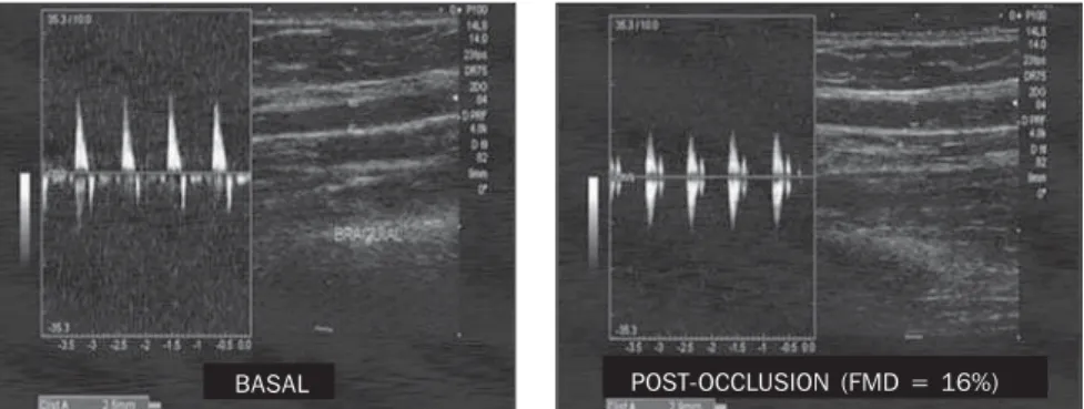
Figure 2. FMD in a patient with a good endothelial function: basal diameter = 2.5 mm; after occlusion = 2.9 mm. FMD = 16%.
Basal diameter = 4.3 mm Post-occlusion diameter = 4.3 mm (FMD = 0%)
249
Brachial artery flow-mediated dilatation in women with rheumatoid arthritis
Radiol Bras 2007;40(4):247–250
sults, in centesimal values above 0.01mg/ dl. For this purpose, the APTEC kit (Selec-tra-Merck; Darmstadt, Germany) was uti-lized — with 0.01 mg/dl sensitivity; RV = 0.0–1.0 mg/dl; intrassay CV = 2.9% and interassay CV = 2.9%.
Results were recorded on average (± standard deviation). The difference be-tween parameter for both groups was evaluated by the Chi-square test. Calcula-tions and statistical analyses were per-formed with the Statistical Package for the Social Sciences (SPSS) for Windows ver-sion 11.0.0 (SPSS, Inc.; Chicago, IL, USA). The difference was considered as statisti-cally significant when p < 0.005.
RESULTS
Mean ages varied significantly between groups. It is important to note that the con-trol group was formed by women at men-acme, considering that menopause is a risk factor for cardiovascular disease. Ages in the control group ranged between 22 and 42 (mean = 28.9 ± 7.1) years. In the rheu-matoid arthritis, the ages ranged between 26 and 76 (mean = 49.9 ± 12.9) years. In this group, ages were significantly (p <
0.000) higher than in the control group (Graph 1).
The control group FMD was 23.24 (± 5.65)%, while in the rheumatoid arthritis was 5.65 (± 9.69)%, with a statistically sig-nificant difference (p < 0.000) (Graph 2).
Five patients failed to demonstrate va-sodilation, with FMD equal or less than zero. Four of these patients were less than 50 years old, demonstrating that the FMD difference between the groups was not af-fected by the ages, as per Graph 3 (p =
0.001).
The C-reactive protein test result was 4.8 (± 9.1) mg/l and disease duration of 6.2 (± 6.5) years. Correlation of age, FMD, C-reactive protein and disease duration dig not show any significant result.
DISCUSSION
The present study demonstrates that pa-tients with rheumatoid arthritis present endothelial dysfunction, and FMD does not correlate with C-reactive protein, patient´s age and disease duration.
Early stages of atherosclerosis are usu-ally found in immunological and inflam-matory diseases where cardiovascular com-plications resulting from the formation and rupture of atherosclerotic plaques represent a relevant cause of morbimortality(15).
Endothelial dysfunction is the earliest event in the atheromatous lesions forma-tion, and its evaluation is a promising tool for estimating coronary prognosis, espe-cially in patients with chronic inflamma-tion, considering that a persistent dysfunc-tion is a reason for an obscure prognosis(16).
Although an ideal method for diagnos-ing endothelial dysfunction has not been
established yet, there are evidences that FMD allows an adequate evaluation of the endothelial behavior and reflects the coro-nary endothelial function and behavior(17).
Age does not affect the endothelial function evaluation with FMD, so the sig-nificant difference in the groups age analy-sis does not affect the data interpretation of age groups. This is corroborated by the dif-ference between patients with less than 50 of age and those in the control group(11).
Like the present study, other studies have found a significant endothelial dys-function in patients with autoimmune dis-eases by means of FMD. The presence of systemic inflammation predisposes to ath-erosclerosis, affecting the endothelial func-tion and decreasing the producfunc-tion of ni-tric oxide by the endothelium. This de-crease in the nitric oxide production leads to a smaller arterial dilatation upon induced ischemia. In a study evaluating the infli-ximab therapy in 11 patients with rheuma-toid arthritis, once the inflammation de-creased, there was an improvement in the FMD(18).
In patients treated for rheumatoid arthri-tis, FMD was related to the HLA-DRB1 (human leukocyte antigen histocompatibil-ity gene) and not to the clinical condition of the patients(19). In a comparison of FMD
results in patients with rheumatoid arthri-tis with control paired by age and sex, pa-tients with low levels of disease activity have demonstrated a significant decrease in
Graph 3. FMD difference between control and rheu-matoid arthritis groups, both with less-than-50-year-old patients.
Rheumatoid arthritis
Control
Graph 2. FMD in the control and rheumatoid ar-thritis groups.
Rheumatoid arthritis
Control
Graph 1. Age of patients (median ± standard de-viation) in the group with rheumatoid arthritis and in the control group.
Rheumatoid arthritis
250
Castro PT et al.
Radiol Bras 2007;40(4):247–250
the endothelial function(20); however, one
study has not found differences between patients with rheumatoid arthritis and con-trol group(14).
FMD and C-reactive protein present correlation in healthy individuals and in those with cardiovascular risk factors.
An investigation evaluating seven car-diovascular risk markers in 2,113 subjects has demonstrated inverse correlation be-tween FMD and C-reactive protein (the higher is the C-reactive protein level, the lower is the nitric oxide production – FMD)(21).
In a study evaluating the endothelium-dependent function in healthy subjects, the C-reactive protein level has presented a correlation with the FMD, but has not pre-sented a closed relation between both meth-ods in the evaluation of an inflammatory process in patients at low cardiovascular risk(22).
In patients with coronary pathology, there was correlation between FMD and C-reactive protein in the 23).
The most surprising data is the non-cor-relation between FMD and C-reactive pro-tein. The present study demonstrates that, even in patients treated for rheumatoid ar-thritis and with normal levels of C-reactive protein, there is presence of systemic in-flammation. The persistent and silent, sys-temic inflammation exposes a clinically managed patient to cardiovascular risk.
Nitrate-mediated endothelium-indepen-dent vasodilation was not utilized. Some-times, this control method is utilized to af-firm that the decrease in arterial dilatation is a result from the endothelial dysfunction, and not an arterial smooth muscle dysfunc-tion.
With disparate results between C-reac-tive protein and FMD, we have concluded that there is endothelial dysfunction in women with rheumatoid arthritis, and that the C-reactive protein level does not pro-vide an adequate evaluation of the cardio-vascular risk in these patients.
Quyyumi(24) affirms that systemic
in-flammation markers like FMD, carotid
media/intimal complex and pulse-wave analysis arise as methods for evaluating the atherosclerosis risk. He recommends the inclusion of these methods, including the biochemical ones, in randomized studies for screening and diagnosis of cardiovas-cular risk. Considering that a high level of systemic inflammation affects the endothe-lial function, inflammation markers like FMD may provide a better evaluation of the cardiovascular risk in patients with rheu-matoid arthritis.
FMD is a promising method for evalu-ation of inflammevalu-ation(25) in the clinical
practice, and may be extended to patients with rheumatoid arthritis for direct mea-surement of vascular physiopathology and preventive therapeutic response.
REFERENCES
1. Sattar N, McCarey DW, Capell H, McInnes IB. Explaining how “high-grade” systemic inflamma-tion accelerates vascular risk in rheumatoid arthri-tis. Circulation 2003;108:2957–2963. 2. DeMaria AN. Relative risk of cardiovascular
events in patients with rheumatoid arthritis. Am J Cardiol 2002;89(6A):33D–38D.
3. Lee DM, Weinblatt ME. Rheumatoid arthritis. Lancet 2001;358:903–911.
4. Solomon DH, Karlson EW, Rimm EB, et al. Car-diovascular morbidity and mortality in women diagnosed with rheumathoid arthritis. Circulation 2003;107:1303–1307.
5. Wang L, Feng G. Rheumatoid arthritis increases the risk of coronary heart disease via vascular en-dothelial injuries. Med Hypotheses 2004;63:442– 445.
6. Cohn JN. Introduction to surrogate markers. Cir-culation 2004;109(25 Suppl 1):IV-20–21. 7. Bugiardini R, Manfrini O, Pizzi C, Fontana F,
Morgagni G. Endothelial function predicts future development of coronary artery disease: a study of women with chest pain and normal coronary angiograms. Circulation 2004;109:2518–2523. 8. Naghavi M, Libby P, Falk E, et al. From
vulner-able plaque to vulnervulner-able patient: a call for new definitions and risk assessment strategies: Part I. Circulation 2003;108:1664–1672.
9. Raitakari OT, Celermajer DS. Flow-mediated di-latation. Br J Clin Pharmacol 2000;50:397–404. 10. Bots ML, Westerink J, Rabelink TJ, de Koning EJ. Assessment of flow-mediated vasodilatation (FMD) of the brachial artery: effects of technical aspects of the FMD measurements on the FMD response. Eur Heart J 2005;26:363–368. 11. Schroeder S, Enderle MD, Baumbach A, et al.
In-fluence of vessel size, age and body mass index on the flow-mediated dilatation (FMD%) of the brachial artery. Int J Cardiol 2000;76:219–225.
12. Laurindo IMM, Pinheiro GRC, Ximenes AC, et al. Consenso brasileiro para o diagnóstico e tra-tamento de artrite reumatóide. Rev Bras Reuma-tol 2002;42:355–361.
13. Corretti MC, Anderson TJ, Benjamin EJ, et al. Guidelines for the ultrasound assessment of en-dothelial-dependent flow-mediated vasodilation of the brachial artery: a report of the International Brachial Artery Reactivity Task Force. J Am Coll Cardiol 2002;39:257–265.
14. Kuscu NK, Kurhan Z, Yildirim Y, Tavli T, Koyuncu F. Detection of endothelial dysfunction in preeclamptic patients by using color Doppler sonography. Arch Gynecol Obstet 2003;268:113– 116.
15. Van Doornum S, McColl G, Jenkins A, Green DJ, Wicks IP. Screening for atherosclerosis in patients with rheumatoid arthritis: comparison of two in vivo tests of vascular function. Arthritis Rheum 2003;48:72–80.
16. Verma S,Wang CH, Lonn E, et al. Cross-sectional evaluation of brachial artery flow-mediated va-sodilation and C-reactive protein in healthy indi-viduals. Eur Heart J 2004;25:1754–1760. 17. Widlansky ME, Gokce N, Keaney JF, Vita JA. The
clinical implications of endothelial dysfunction. J Am Coll Cardiol 2003;42:1149–1160. 18. Hurlimann D, Forster A, Noll G, et al. Anti-tumor
necrosis factor-alpha treatment improves endo-thelial function in patients with rheumatoid arthri-tis. Circulation 2002;106:2184–2187. 19. Gonzalez-Juanatey C, Llorca J, Testa A, Revuelta
J, Garcia-Porrua C, Gonzalez-Gay MA. Increased prevalence of severe subclinical atherosclerotic findings in long-term treated rheumatoid arthri-tis patients without clinically evident atheroscle-rotic disease. Medicine (Baltimore) 2003;82: 407–413.
20. Vaudo G, Marchesi S, Gerli R, et al. Endothelial dysfunction in young patients with rheumatoid ar-thritis and low disease activity. Ann Rheum Dis 2004;63:31–35.
21. Kathiresan S, Gona P, Larson MG, et al. Cross-sectional relations of multiple biomarkers from distinct biological pathways to brachial artery endothelial function. Circulation 2006;113:938– 945.
22. Williams IL, Chowienczyk PJ, Wheatcroft SB, et al. Effect of fat distribution on endothelial-depen-dent and endothelial-indepenendothelial-depen-dent vasodilatation in healthy humans. Diabetes Obes Metab 2006;8: 296–301.
23. Kang SM, Chung N, Kim JY, et al. Relation of vasodilator response of the brachial artery to in-flammatory markers in patients with coronary artery disease. Echocardiography 2002;19:661– 667.
24. Quyyumi AA. Inflamed joints and stiff arteries: is rheumatoid arthritits a cardiovascular risk fac-tor? Circulation 2006;114:1137–1139. 25. Faulx MD, Wright AT, Hoit BD. Detection of