Case Report
Key words
Fontan procedure; bronchitis; protein-losing enteropathies. We report an unusual case of association of plastic bronchitis (PB) to protein-losing enteropathy (PLE) in a girl of 4 years and 9 months of age with double inlet single left ventricle and ventriculoarterial concordance, submitted to total cavopulmonary surgery, with an intracardiac lateral tunnel at the age of three. The elimination of the 10 cm fibrin bronchial mold (PB) and the alpha-1-antitrypsin elevation of 52 mg/g in feces had both become outstanding. Using sildenafil, the thoracic duct ligature and the cardiac transplant were programmed in case of continuity of the process.
Association of Plastic Bronchitis to Protein-Losing Enteropathy after
Fontan Operation
Vanessa Alves Guimarães, Edmar Atik, Jussara Bianchi Castelli, Nana Miura Ikari, Ana Maria Thomaz, Antonio
Augusto Barbosa Lopes
Instituto do Coração (InCor), Hospital das Clínicas da Faculdade de Medicina da Universidade de São Paulo, São Paulo, SP - Brazil
Mailing address: Edmar Atik •
Rua Dona Adma Jafet, 74 conj.73 - Bela Vista - 01308-050 - São Paulo, SP - Brazil
E-mail: [email protected], [email protected]
Manuscript received December 02, 2008; revised manuscript received March 23, 2009; accepted September 01, 2009.
successfully submitted to our services and to the IAC extension by the Rashkind method and, after four days, to the pulmonary trunk bandage and AC closure. At the age of three years and four months, cyanosis and dyspnea were accentuated.
The hemodynamic study indicated mean arterial pressure of 13 mmHg in the pulmonary artery, of 10 mmHg in the pulmonary capillary, with Qp/Qs = 0.9 and pulmonary vascular resistance of 1.7 WU (Wood unit). Afterwards, the patient was submitted to the total cavopulmonary operation with intracardiac lateral tunnel. Three months after the intervention, the patient presented anasarca, ascites and pleural effusion. Serum albumin was of 1.7 mg/dl. Through a hemodynamic study, the obstruction to the venous flow was discarded. The mean pulmonary artery pressure of both the superior and inferior vena cava was of 15 mmHg, the pulmonary capillary pressure was of 7 mmHg and the Qp/Qs was of 0.8.
The research result for alpha-1-antitrypsin in feces was of 52 mg/g, confirming the PLE diagnosis. Through the usual measures - hyperproteic, hypofatty diet, along with me medium-chain triglycerides and anticongestives, such as furosemide, spirinolactone and captopril - there was an improvement in the clinical features, obtaining even alpha-1-antitrypsin normality (less than 3 mg/g). Fourteen months after the operation, there was a new decline in the anasarca picture, as well as incessant coughs, cyanosis and fever peak. Serum albumin was of 2.1 mg/dl and alpha-1-antitrypsin was of 13.2 mg/g. The clinical features showed improvement with the usual anticongestive measures within few days.
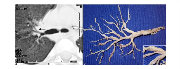
Incidentally, during the respiratory physiotherapy exercises the patient expelled a fibrin bronchial mold about 10-centimeter long (Figure 1). There was perihilar infiltrate in the thorax radiography, and a hypoattenuanting image in bronchial light in the thorax high-resolution CT scan (Figure 1). The anatomopathological report of the bronchial matter was compatible with the PB diagnosis, since it was constituted by compact fibrin combined with numerous lymphoid cells, scarce mucous tissue and permeated by xanthomatous macrophages, besides a focal area with accumulated eosinophils (Figure 2).
Microscopy and immunohistochemistry excluded the infectious causes (bacteria, alcohol-acid resistant bacilli, viruses and fungi). From the diagnosis of the association of PB with PLE, clinical measurements were instituted to fluidify the airways with inhalations and bronchodilators, as well as furosemide, captopril, spirinolactone and sildenafil. In case of continuity of the process, the ligature of the
Introduction
The Fontan operation, though considered the best palliative for univentricular hearts, is accompanied by severe complications, such as plastic bronchitis (PB) and protein-losing enteropathy (PLE), as well as other comorbidities1. It is estimated that PLE occurs in a variable percentage of about 1%-13,4%2,3. The PLE occurrence is very rarely reported4-6, as is the association of these two entities5. Therefore, it is our purpose to report this rare association.
Case report
A female patient, of 4 years and 9 months of age from São Paulo (SP, Brazil), born of a regular term normal birth and daughter of a diabetic mother, has presented early respiratory discomfort and heart murmurs during the first week of life. At that time, the diagnosis was set as a double inlet single left ventricle with ventriculoarterial concordance, interatrial communication (IAC), interventricular communication and arterial channel (AC) persistence, with an important pulmonary hypertension. When one-month-old, the patient was
Case Report
Guimarães et al Plastic bronchitis and PLE after Fontan operation
Arq Bras Cardiol 2010; 94(4) : e48-e50 thoracic duct was programmed, culminating even with a
possible cardiac transplant.
Discussion
Currently, the factors involved in the evolutive prognosis after the Fontan operation consist in the making of an adequate communication between the systemic veins and the pulmonary artery, good ventricular performance, normal
functioning of atrioventricular valves and normal size of the pulmonary arterial tree. The operation has been undergoing great technical modifications and currently consists mainly in the venous pulmonary systemic connection through the use of an extracardiac tube, more than an intracardiac lateral tunnel, with or without fenestration. The final result provides the patient two serial circulations, the systemic venous return being guided directly to the pulmonary artery without the presence of the ventricular pulsating pump. This ‘Fontan
Figure 1 -High resolution thoracic CT scan (a) demonstrating a hypoattenuating image in bronchial light (arrow) corresponding to the mold eliminated from the respiratory tree (b). The detail, in the lower right corner, shows a bright compact tissue, different from the translucent and gelatinous pattern of the mucous ‘corks’ ordinarily observed in airways.
Figure 2 -Microscopic analysis of the bronchial mold, composed mainly of compact ibrin, represented by the pink tissue in (a) and (b), combined with numerous T and B lymphoid cells, observed in (b), (d) and (e), suggesting lymph composition. There are also some areas with scarce mucous tissue (c) and permeated by xanthomatous macrophages, shown in (c) and (f). [a-c: Hematoxylin and Eosin coloration; d-f: Immunohistochemical reactions with CD20 antibodies for the B lymphocyte, anti-CD3 for the T lymphocyte and anti-CD68 for macrophages, in which the brown shade represents positive reaction; objective of 5, 20, 10, 10, 10 and 10, respectively.].
Case Report
Guimarães et al
Plastic bronchitis and PLE after Fontan operation
Arq Bras Cardiol 2010; 94(4) : e48-e50
physiology’ submits a patient to a slower venous flow, thus susceptible to the development of chronic pleural effusion, chylothorax, atrial arrhythmia, PLE and PB4.
As for the PB, it constitutes a rare entity. It is characterized by recurring expectoration of peculiar molds from the tracheobronchial tree. Although its etiopathogeny remains unknown, the central venous pressure elevation and the pulmonary and systemic lymphatic system disorders are probable pathogenic mechanisms5. The same elements are correlated to PLE, in addition to the fact that, chronically, the low cardiac debt would contribute to the increase of mesenteric vascular resistance. This way, the association between mesenteric hypoperfusion and venous congestion would affect the intestinal mucous membrane. In the presented case, the patient was presented with normal pulmonary pressure, thus excluding part of thus pathogenic mechanism.
Usually, patients with PB present dyspnea with an increase in ventilatory work, repetitive episodes of coughing and mold elimination, with a predisposition towards respiratory phenomena, such as infection and atelactasias. Thorax radiography usually reveals a peribronchial infiltrate, as well as atelactasias. The molds of bronchial secretion may cause asphyxia, cardiac arrest and death, the latter being reported in up to 29% of patients with this latent condition6.
In PLE, as a result of the enteric loss of serum proteins, there is edema and poor absorption in the intestinal wall, causing diarrhea. In some patients, this process is more insidious and hypoproteinemia may occur in the absence of diarrheic symptoms. The disease typically initiates with corporal edema, evolving with ascites, pleural and pericardial effusion. Laboratory tests reveal low levels of total serum and albumin protein. The gold standard for the diagnosis is an increased intestinal clearance of alpha-1-antitrypsin, a
protein produced exclusively in the liver and excreted in small amounts throughout the intestine. The decrease of albumin serum levels reduces the calcium transportation, causing tetany and osteopenia. Hypogammaglobulinemia is another possible disorder, causing immunodeficiency. The early stage of this enteropathy may have serious consequences, resulting in the increase of morbidity and mortality. Two studies have described survival in five years, after the diagnosis of only 50% of patients3.
Many times, the treatment of both conditions is merely symptomatic. In PB, it may be of two types: the inflammatory type, secondary to expressive allergic processes, which may be manipulated with inhalating medication (corticosteroids, azithromycin, urokinase); and the acellular type, prevalent after the Fontan operation, which has been benefitting from the circuit fenestration, the use of pulmonary hypotensor medication, the ligature of the thoracic duct or the cardiac transplant4,7-10. In the PLE treatment, there is also another resort: surgically removing the suprahepatic veins. The use of low molecular weight heparin has been described as a remedy to both entities, suggesting it is the same physiopathology2.
Potential Conflict of Interest
No potential conflict of interest relevant to this article was reported.
Sources of Funding
There were no external funding sources for this study.
Study Association
This study is not associated with any post-graduation program.
References
1. Camposilvan S, Milanesi O, Stellin G, Pettenazzo A, Zancan L, D’Antiga L. Liver and cardiac function in the long term after Fontan operation. Ann Thorac Surg. 2008; 86 (1): 177-82.
2. Bhagirath KM, Tam JW. Resolution of protein-losing enteropathy with low-molecular weight heparin in an adult patient with Fontan palliation. Ann Thorac Surg. 2007; 84 (6): 2110-2.
3. Rychik J. Protein-losing enteropathy after Fontan operation. Congenit Heart Dis. 2007; 2: 288-300.
4. Salman S, Shah A, Drinkwater DC, Christian KG. Plastic bronchitis: is thoracic duct ligation a real surgical option? Ann Thorac Surg. 2006; 81 (6): 2281-3. 5. Stiller B, Riedel F, Paul K, Van Landeghem FK. Plastic bronchitis in children
with Fontan palliation: analogue to protein losing enteropathy? Pediatr Cardiol. 2002; 23: 90-4.
6. Brogan TV, Finn LS, Pyskaty DJ Jr, Redding GJ, Ricker D, Inglis A, et al. Plastic
bronchitis in children: a case series and review of the medical literature. Pediatr Pulmonol. 2002; 34 (6): 482-7.
7. Wilson J, Russell J, Williams W, Benson L. Fenestration of the Fontan circuit as treatment for plastic bronchitis. Pediatr Cardiol. 2005; 26 (5): 717-9. 8. Apostolopoulou SC, Papagiannis J, Rammos S. Bosentan induces clinical,
exercise and hemodynamic improvement in a pre-transplant patient with plastic bronchitis after Fontan operation. J Heart Lung Transplant. 2005; 24 (8): 1174-6.
9. Haseyama K, Satomi G, Yasukochi S, Matsui H, Harada Y, Uchita S. Pulmonary vasodilation therapy with sildenafil citrate in a patient with plastic bronchitis after the Fontan procedure for hypoplastic left heart syndrome. J Thorac Cardiovasc Surg. 2006; 132 (5): 1232-3.
10. Nayar S. Treatment of plastic bronchitis. Ann Thorac Surg. 2007; 83 (5): 1884-6.