Original Article
3 5 9 Arq Bras Oftalmol. 2015;78(6):359-62 http://dx.doi.org/10.5935/0004-2749.20150095
INTRODUCTION
Tonometry is fundamental to routine ophthalmological eva-luation, and Goldmann applanation tonometry (GAT) is the gold standard method. Several devices, including the Perkins tonometer, Tono-Pen, Pascal tonometer (dynamic contour), and non-contact to-nometer, can provide reliable values in adults(1-4). However, in general practice, intraocular pressure (IOP) measurement can be diicult in children due to a lack of cooperation.
ABSTRACT
Purpose: High intraocular pressure (IOP) is an important risk factor for a variety of pediatric ophthalmic conditions. The purpose of this study is to evaluate the feasibility, length of examination, and corneal epithelial damage induced by rebound tonometry (RBT) versus Goldmann applanation tonometry (GAT) in school children.
Methods: Healthy children (n=57) participated in a randomized, transversal study with IOP measurement by GAT followed by RBT (study arm 1) or RBT followed by GAT (study arm 2). The number of attempts to acquire a reliable IOP measurement and the length of the examination were quantified. Corneal epithelial damage induced by tonometry was evaluated. Bland-Altman analysis was performed to establish the level of agreement between the two techniques.
Results: The IOP was measured in all children with at least one of the devices. In both study arms, more children failed to be examined with GAT than with RBT (26% vs. 4%, and 16% vs. 6%, p<0.001, in study arm 1 and 2, respectively). The length of examination was shorter for RBT than for GAT (67.81 s ± 35.20 s vs. 126.70 s ± 56.60 s; p<0.0001); IOP measurements with RBT in both study arms were higher than those with GAT (15.20 ± 2.74 mmHg vs. 13.25 ± 2.47 mmHg, p=0.0247 and 16.76 ± 3.99 mmHg vs. 13.92 ± 2.08 mmHg, p=0.003, respectively). No difference was observed between RBT and GAT regarding the corneal epi-thelial damage caused by tonometry.
Conclusion: IOP measurement is feasible in a greater number of children with RBT, and the examination was faster than that for GAT. Compared with GAT, RBT tended to overestimate the IOP. None of the methods induced marked corneal epithelial defects.
Keywords: Glaucoma/diagnosis; Tonometry, ocular; Ocular hypertension; Intraocu-lar pressure; Rebound effect; Visual acuity
RESUMO
Objetivo: A pressão intraocular (PIO) elevada é um importante fator de risco presente em diversas patologias que acometem crianças. O objetivo deste estudo é avaliar a viabilidade, a duração do exame e o dano epitelial corneano induzido pela tonometria de rebote (RBT ) versus a tonometria de aplanação de Goldmann (GAT ) em crianças em idade escolar.
Métodos: Crianças sem comorbidades (n=57) participaram de um estudo ran do-miza do e transversal com medidas da pressão intraocular com GAT seguido de RBT (sequência 1) ou RBT seguido de GAT (sequência 2). O número de tentativas para adqui rir uma medição confiável da pressão intraocular e a duração de exame foi quantificado. Danos epiteliais induzidos pela tonometria foram avaliados. Análise de Bland-Altman foi realizada para estabelecer a concordância entre as duas técnicas.
Resultados: A pressão intraocular foi medida em todas as crianças com pelo menos com um dos dispositivos. Em ambas as sequências do estudo, mais crianças não permitiram o exame com GAT (26% vs. 4% e 16% vs. 6%, p<0,001). A duração exame com RBT foi menor (67,81 ± 35,20 s vs. 126,70 ± 56,60 s; p<0,0001). As medições de pressão intraocular com este tonômetro em ambas as sequências do estudo foram mais elevadas do que as medidas adquiridas com GAT (15,20 ± 2,74 mmHg vs 13,25 ± 2,47 mmHg, p=0,0247 e 16,76 ± 3,99 mmHg vs. 13,92 ± 2,08 mmHg; p=0,003, respec-tivamente). Não foi observada diferença quanto à lesão epitelial corneana induzida pela tonometria com RBT e GAT.
Conclusão: A medição da pressão intraocular foi possível em um maior número de crianças com a tonometria de rebote, além de ser um exame mais rápido do que GAT. A pressão intraocular foi superestimada com RBT em comparação com GAT. Nenhum dos métodos induziu defeito epitelial corneano significativo.
Descritores: Glaucoma/diagnóstico; Tonometria ocular; Hipertensão ocular; Pressão intraocular; Efeito rebote; Acuidade visual
Several conditions, such as childhood glaucoma, uveitis, and ocular trauma, can present elevated ocular pressure. When a routine IOP measurement is not possible, the procedure is often conducted under general anesthesia. However, general anesthesia is associated with a number of complications. Studies have revealed increased mortality associated with IOP measurement under general anesthesia in children compared with that in adults; the procedure may also be associated with long-term adverse neurodevelopmental efects(5-7).
Rebound tonometry versus Goldmann tonometry in school children:
feasibility and agreement of intraocular pressure measurements
Tonometria de rebote versus tonometria de Goldmann em crianças em idade escolar:
viabilidade e concordância entre as medidas da pressão intraocular
Bruno Leonardo Barranco esporcatte1, FLávio siqueira santos Lopes1, camiLa Fonseca netto1, vespasiano reBouças-santos1, diego torres dias1, FáBio igLesias marujo1, christiane roLim-de-moura1
Submitted for publication: May 29, 2015 Accepted publication: October 1, 2015
1. Department of Ophthalmology, Universidade Federal de São Paulo (UNIFESP), São Paulo, SP, Brazil.
Funding: This study was supported by coordenação de aperfeiçoamento de pessoal de nível superior (CAPES).
Disclosure of potential conflicts of interest: None of the authors have any potential conflict of interest to disclose.
Corresponding author: Bruno Leonardo Barranco Esporcatte. Rua Botucatu, 821 - Vila Clementino São Paulo, SP - 04023-062 - Brazil - Email: [email protected]
Rebound tonometry versus Goldmann tonometry in school children: feasibility and agreement of intraocular pressure measurements
360 Arq Bras Oftalmol. 2015;78(6):359-62
The Icare™ tonometer (Icare, Helsinki, Finland) is a portable tono-meter that does not require topical anesthetics and causes minimal discomfort during examination(8). This device is based on the impact and rebound of a probe against the cornea(9). Previous studies have demonstrated a strong agreement between IOP measurements obtained by rebound tonometry (RBT) and GAT. Moreover, the va-riables that inluence reliable measurement are the same for both devices(10,11).
By providing greater comfort during IOP measurement, the Icare tonometer is generally well tolerated by children. Sahin et al. eva-luated the levels of discomfort during IOP examination in school children, with 93.4% reporting no discomfort and 6.6% reporting slight discomfort(12). Flemmons et al. reported that it was possible to acquire a reliable IOP measurement on the irst attempt in over 93% of the examined children(13).
The purpose of the present study was to evaluate and compare the feasibility, length of the examination, and corneal epithelial da-mage induced by tonometry with RBT versus GAT in school children. The level of agreement between the two methods and the corre-lation between IOP and the central corneal thickness (CCT) were analyzed.
METHODS
This protocol was approved by the Ethical Committee of the Fe-deral University of São Paulo and was performed in accordance with the ethical standards laid down in the Declaration of Helsinki and the International Conference on Harmonisation Guidelines for Good Clinical Practice(14). Informed parental consent was obtained for all of the enrolled participants before the study.
Healthy children aged between 6 and 8 years were recruited through a general ophthalmological oice after refractive error screening. Exclusion criteria included logMAR vision worse than 0.7, a history of ocular disease, medication use, and corneal epithelial defects. All children underwent slit-lamp examination, and those found to have corneal pathology were excluded.
The subjects were randomly selected using a sequentially num-bered, opaque sealed-envelope technique(15) to undergo IOP mea-surement by GAT followed by RBT (study arm 1) or RBT followed by GAT (study arm 2). Tonometric examination with the second device was performed 5 min following the end of the irst examination. All of the IOP measurements were conducted by glaucoma specialists with pediatric care experience, and all of the tonometric evaluations and corneal status assessments were performed by three masked examiners in three diferent oices.
Anesthesia was not required for IOP measurement with RBT. The subjects were instructed to look straight ahead, and the instrument was positioned with the tip of the probe at a distance of 4 mm to 8 mm from the corneal apex. For RBT, when the operator activates the tonometer, six measurements are automatically performed, and the mean value is calculated. After topical administration of anes-thesia and luorescein eye drops, GAT measurement was performed using a slit lamp. The length of time taken for each examination was quantiied with a chronometer and included the time taken for administering the drops, explaining the examination procedure, and performing the measurements. The number of attempts with each tonometric method was recorded, with a maximum of three attempts allowed to measure IOP in each eye for each tonometer. Only RBT readings with a high level of reliability were used. After the third attempt, if the subject did not allow the examination to be performed, the test was considered to be unsuccessful. The right eye was measured irst for both tonometers in all subjects.
Corneal epithelial damage was assessed after the irst tonometric method (RBT or GAT) of IOP measurement using biomicroscopic exa-mination of the type and extent of staining. When RBT was performed irst, the examiner instilled anesthetic and luorescein drops after the
measurement to mask the second examiner for the assessment of the corneal status of the eye. The corneal surface was divided into ive zones of equal area (central, nasal, superior, inferior, and temporal). The type of staining in each zone was classiied as absent, micro-punctate, macromicro-punctate, coalescent macromicro-punctate, or patch. The extent of staining in each zone was graded 0 (absent), 1 (1%-15% of the surface), 2 (16%-30% of the surface), 3 (31%-45% of the surface), or 4 (≥46% of the surface).
The CCT was determined using a central ultrasonic pachymeter (Alcon Laboratories Inc., Fort Worth, TX, USA). Under topical anesthe-sia, the seated patient was asked to look at a distant target ahead. The pachymeter probe was aligned perpendicularly and central to the pupil; the mean of ive measurements was calculated.
All statistical analyses were performed using GraphPad Prism version 6.00 for Mac (GraphPad Software, La Jolla, CA, USA). Pooling of data from the right and left eyes during statistical analysis may cause bias as they are not independent variables; therefore, to avoid such bias, only data from the right eyes were analyzed. Moreover, a Bland-Altman analysis was performed to examine the level of agreement between the two devices. The diferences between the category frequencies were evaluated by chi-square tests, and paired analysis of nonparametric measures was performed using the Wil-coxon matched-pairs signed-rank test. Student’s t-tests were used to analyze data with a normal distribution. Correlations between variables were evaluated by Pearson’s correlation coeicients. The level of statistical signiicance was set at p<0.05.
RESULTS
In the present study, 36 (63.2%) out of 57 patients were male and 21 (36.8%) were female. The mean age of the children was 6.76 ± 0.38 years, and the mean uncorrected visual acuity was 0.85 ± 0.11 logMAR.
To achieve a reliable measurement, more attempts were made with GAT than RBT (1.54 ± 0.69 vs. 1.13 ± 0.34, p=0.0021) in both study arms. All children permitted IOP measurements with at least one tonometer. More children did not allow the examination to be performed with GAT than with RBT in both study arms (study arm 1: 26%, n=7 vs. 4%, n=1, p<0.001; study arm 2: 16%, n=5 vs. 6%, n=2,
p<0.001; chi-square test). Examination with RBT was faster than that with GAT (67.81 s ± 35.20 s vs. 126.70 s ± 56.60 s, p<0.0001).
IOP measurements quantified with RBT were statistically diffe-rent from those quantified with GAT in both study arms 1 (15.20
± 2.74 mmHg vs. 13.25 ± 2.47 mmHg, p=0.0247) and 2 (16.76
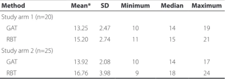
± 3.98 mmHg vs. 13.92 ± 2.08 mmHg; p=0.0003; Table 1). Figure 1 shows the Bland-Altman plots of IOP and the linear regression of these values. A positive correlation between the difference and the mean IOP measurements was observed in study arm 2 (r=0.6347,
p=0.0007; Figure 1 B). No statistically signiicant diference was obser-ved in the degrees of bias in study arms 1 and 2 (1.95 ± 3.58 mmHg vs. 2.84 ± 3.37 mmHg, respectively).
Table 1. Intraocular pressure as measured by GAT and RBT
Method Mean* SD Minimum Median Maximum
Study arm 1 (n=20)
GAT 13.25 2.47 10 14 19
RBT 15.20 2.74 11 15 21
Study arm 2 (n=25)
GAT 13.92 2.08 10 14 17
RBT 16.76 3.98 09 18 24
RBT= rebound tonometry; GAT= Goldmann applanation tonometry; SD= standard deviation. *All recorded values of intraocular pressure are presented in mmHg.
Esporcatte BLB, et al.
361
Arq Bras Oftalmol. 2015;78(6):359-62
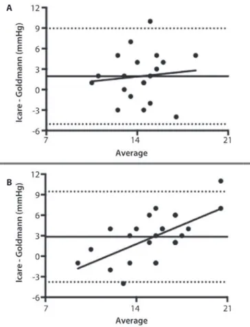
The mean CCT was 558 ± 36 μm, and a positive correlation was found between CCT and IOP for GAT (r=0.3163, p=0.0388) and RBT (r=0.3080, p=0.0445; Figure 2).
The frequency of corneal epithelial damage induced by RBT and GAT was similar (13% vs. 11%; chi-square test), and the most common type of staining was micropunctate on less than 16% of the corneal surface (grade 1). Only one patient who underwent RBT examination presented staining of 16% to 30% of the surface (grade 2), and this was located in the inferior corneal zone.
DISCUSSION
In clinical practice, IOP measurement is often impaired by a lack of cooperation by school children. The present study revealed good tolerance for RBT, with only <6% of the examined children refusing the tonometry. Moreover, the examination was less time consuming, and the number of attempts required was fewer than that with GAT. RBT was better tolerated as it can be performed without adminis-tering anesthesia. Due to the reduction of discomfort, tonometric examination could be successfully performed in more children. The impact of the tonometer tip is extremely gentle in RBT, and frequently does not provoke an eye-blinking rel ex(9).
Although IOP measurements with RBT were statistically signii -cantly higher than those with GAT, this dif erence was not clinically relevant. Several authors have reported a good level of agreement between the two tonometers, with RBT generally overestimating IOP by around 3 mmHg(16-18). The dif erence between the acquired values by the two tested tonometers was higher for high mean IOP values than that for low mean IOP values. Furthermore, in accordance with previous studies, we observed positive correlations between IOP measured by both devices and CCT(19-21).
Despite a small number of children being uncooperative during IOP measurements, neither of the tonometers produced marked cor-neal epithelial defects. This may be because ophthalmologists that conducted the examinations were experienced in pediatric care.
The small number of children enrolled limited our study. As only healthy children were evaluated, these results may not be applicable to patients with childhood glaucoma or other ophthalmic diseases. Further, the corneal applanation induced by GAT may have inl uenced IOP measurements when RBT was performed second (study arm 1). However, only the inl uence of corneal thickness was evaluated in the present study.
CONCLUSION
IOP was successfully measured in all children with at least one of the tonometers. RBT was better tolerated and was faster than GAT examination. RBT did not induce epithelial lesions, although it overes-timated IOP by around 3 mmHg. However, we do not believe this to be a clinically relevant disadvantage of RBT. In routine clinical settings, GAT remains the gold standard for the measurement of IOP; however, RBT may be a useful screening tool for non-cooperative patients, such as school children, and may allow avoidance of the use of general anesthesia for IOP measurement. High RBT measurements should be corroborated by assessment of the clinical presentation, and IOP should be measured by other tonometric methods.
REFERENCES
1. Bradi eld YS, Kaminski BM, Repka MX, Melia M; Pediaric Eye Disease Investigator Group, et al. Comparison of Tono-Pen and Goldmann applanation tonometers for measure-ment of intraocular pressure in healthy children. J AAPOS. 2012;16(3):242-8. 2. Cook JA, Botello AP, Elders A, Fathi Ali A, Azuara-Blanco A, Fraser C, McCormack K,
Margaret Burr J; Surveillance of Ocular Hypertension Study Group. Systematic review of the agreement of tonometers with Goldmann applanation tonometry. Ophthal-mology. 2012;119(8):1552-7.
3. Okafor KC, Brandt JD. Measuring intraocular pressure. Curr Opin Ophthalmol. 2015; 26(2):103-9.
Figure 2. Correlations between central corneal thickness (CCT) and intraocular pressure (IOP) acquired with Goldmann applanation tonometry (GAT) and rebound tonometry (RBT). Positive correlations were observed with GAT (r =0.3163, p=0.0388) and RBT (r=0.3080, p=0.0445).
Figure 1. Bland-Altman analyses of IOP measurements between rebound tonometry (RBT) and Goldmann applanation tonometry (GAT) in study arms 1 (A) and 2 (B). The mean bias (solid line) and 95% limits of agreement (dashed lines) are shown. In study arm 1 (A), the mean bias was 1.95 ± 3.58, and the correlation between the diff erences and the mean values was not signifi cant (r=0.1062, p=0.6558). In study arm 2 (B), the mean bias was 2.84 ± 3.37, and a positive correlation was observed between the diff erences and the mean values (r=0.6347, p=0.0007).
A
Rebound tonometry versus Goldmann tonometry in school children: feasibility and agreement of intraocular pressure measurements
362 Arq Bras Oftalmol. 2015;78(6):359-62
4. Vincent SJ, Vincent RA, Shields D, Lee GA. Comparison of intraocular pressure mea-surement between rebound, non-contact and Goldmann applanation tonometry in treated glaucoma patients. Clin Experiment Ophthalmol. 2012;40(4):e163-70. 5. Chang TC, Cavuoto KM. Anesthesia considerations in pediatric glaucoma
manage-ment. Curr Opin Ophthalmol. 2014;25(2):118-21.
6. DiMaggio C, Sun LS, Ing C, Li G. Pediatric anesthesia and neurodevelopmental im-pairments: a Bayesian meta-analysis. J Neurosurg Anesthesiol. 2012;24(4):376-81. 7. Gonzalez LP, Pignaton W, Kusano PS, Módolo NS, Braz JR, Braz LG. Anesthesia-related
mortality in pediatric patients: a systematic review. Clinics (Sao Paulo). 2012;67(4):381-7. Comment in: Clinics (São Paulo). 2012;67(6):675-6.
8. Grigorian F, Grigorian AP, Olitsky SE. The use of the iCare tonometer reduced the need for anesthesia to measure intraocular pressure in children. J AAPOS. 2012;16(6):508-10. 9. Kontiola AI. A new induction-based impact method for measuring intraocular pressure.
Acta Ophthalmol Scand. 2000;78(2):142-5.
10. Davies LN, Bartlett H, Mallen EA, Wolf sohn JS. Clinical evaluation of rebound tono-meter. Acta Ophthalmol Scand. 2006;84(2):206-9.
11. Munkwitz S, Elkarmouty A, Hof mann EM, et al. Comparison of the iCare rebound tonometer and the Goldmann applanation tonometer over a wide IOP range. Graefes Arch Clin Exp Ophthalmol. 2008;246(6):875-9.
12. Sahin A, Basmak H, Niyaz L, Yildirim N. Reproducibility and tolerability of the ICare rebound tonometer in school children. J Glaucoma. 2007;16(2):185-8.
13. Flemmons MS, Hsiao YC, Dzau J, Asrani S, Jones S, Freedman SF. Icare rebound tono-metry in children with known and suspected glaucoma. J AAPOS. 2011;15(2):153-7.
14. International Conference on Harmonisation of technical requirements for registration of pharmaceuticals for human use. ICH harmonized tripartite guideline: Guideline for Good Clinical Practice. J Postgrad Med. 2001;47(1):45-50.
15. Doig GS, Simpson F. Randomization and allocation concealment: a practical guide for researchers. J Crit Care. 2005;20(2):187-91; discussion 91-3.
16. Beasley IG, Laughton DS, Coldrick BJ, Drew TE, Sallah M, Davies LN. Does rebound to-nometry probe misalignment modify intraocular pressure measurements in human eyes? J Ophthalmol. 2013;2013:791084.
17. Rehnman JB, Martin L. Comparison of rebound and applanation tonometry in the management of patients treated for glaucoma or ocular hypertension. Ophthalmic Physiol Opt. 2008;28(4):382-6.
18. Suman S, Agrawal A, Pal VK, Pratap VB. Rebound tonometer: ideal tonometer for measurement of accurate intraocular pressure. J Glaucoma. 2014;23(9):633-7. 19. Nakamura M, Darhad U, Tatsumi Y, Fujioka M, Kusuhara A, Maeda H, et al. Agreement
of rebound tonometer in measuring intraocular pressure with three types of applana-tion tonometers. Am J Ophthalmol. 2006;142(2):332-4.
20. Rao A, Kumar M, Prakash B, Varshney G. Relationship of central corneal thickness and intraocular pressure by iCare rebound tonometer. J Glaucoma. 2014;23(6):380-4. 21. Salim S, Du H, Wan J. Comparison of intraocular pressure measurements and assessment
of intraobserver and interobserver reproducibility with the portable ICare rebound tonometer and Goldmann applanation tonometer in glaucoma patients. J Glaucoma. 2013;22(4):325-9.