Clinicoradiological Session
Case 6/2009 – 13 Years Old Youngsters with Major Atrial and
Ventricular Septal Defects
Edmar Atik
Hospital Sírio-Libanês de São Paulo - Brazil
Mailing address: Edmar Atik •
InCor - Av. Dr. Enéas Carvalho de Aguiar, 44 - 05403-000 - São Paulo, SP - Brasil E-mail: [email protected]
Key words
Heart defects, congenital / surgery; atrial septal defect; ventricular septal defect; heart septal defects.
Clinical data
Heart murmur had been observed upon birth and tiredness on stress, besides palpitations at rest, has been observed by the patient for some years. The patient had taken Digoxin and Furosemide up to 4 years of age. Good physical development occurred.
Physical examinations found the patient eupneic, ruddy and with normal pulses. Weight was 71.8 lbs, 4.79 ft of height, heart rate of 82 bpm and blood pressure of 105/65 mm Hg. The aorta was not palpated on the suprasternal notch. There were clear impulsions on the precordium and the left sternal edge was diffusely palpated. The heart sounds were accentuated in intensity. The second heart sound was splitted, with both components of equal intensity. Intense and rude systolic thrill and holosystolic murmur, +++ of intensity, were found in the lower left sternal edge, irradiating to the right sternal edge and to the mitral area. Liver was not palpated.
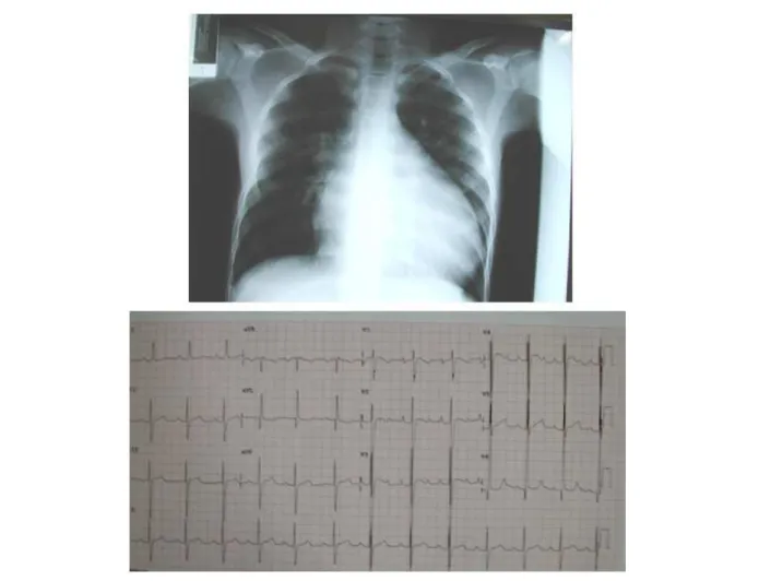
Electrocardiogram tests (fig.1) showed signs of diastolic left ventricular overload with broad qR complexes in the left precordial leads, R wave of 30 mm in V6 and T positive wave, besides blocking on the left antero-superior division. AQRS was at -40o, AP a +30o and AT at +80o.
Radiographic image
Shows cardiac area highly increased by dilation of left cardiac cavities with double atrium arch to the right and ventricular arch elongated and deviated to the left. Arteries were slightly increased and the medium arch elongated and rectified (Figure 1).
Diagnostic impression
The radiographic image is compatible to congenital acianogenic cardiopathy with high volume overload of left cardiac cavities, followed by an increased pulmonary arterial circulation, such as in conditions involving blood from left to the right, such as in ventricular septal defect.
Differential diagnosis
Due to the higher increase of left cardiac cavities, this image
may also lead to a diagnosis of left atrioventricular valve failure. In addition to the acianogenic cardiopathies with obstructed flow to the left of the heart, such as aortic stenosis and aortic coarctation, in cases of left ventricular failure. However, in these abnormal conditions, there will be greater pulmonary venocapillar congestion.
Diagnosis confirmation
Clinical data led to the diagnosis of ventricular septal defect, of repercussion, despite an isolated diastolic left ventricular overload observed in the ECG. Echocardiogram (Figure 2) confirmed this abnormal condition in perimembranous position with extension to the ventricular inlet, with 8 mm of diameter. There was also atrial septal defect with 20 mm of extension. Cardiac cavities measured: Left atrium: 31; Aorta: 24, Right ventricular diastolic diameter: 29, Left ventricular diastolic diameter: 59, Left ventricular sistolic diameter: 38 mm, Shortening ejection fraction: 36%, Ejection fraction: 64%.
Management
Upon the surgery, at extracorporeal circulation of 70’, we found atrial septal defect, ostium secundum, of 40 mm and ventricular septal defect of 8 mm, closed with bovine pericardial patch. A good initial evolution was observed and after 5 months from the surgery, no residual defects were observed.
Comments
Unexpected clinical exteriorization associated to major septal defects in which, despite the large atrial septal defect (ASD), almost a single atrium, there was a higher repercussion of the LV volumetric overload caused by the ventricular septal defect (VSD), with no RV potential. This explains the difficulty in finding a diagnosis for the ASD, well-established by echocardiogram examinations, although at an underestimated size. Such exteriorization is uncommon, as the ASD always works as a escape route in the presence of the other defects.
Clinico Radiological Session
AtikArq Bras Cardiol 2009; 93(3) : 416-417
Figure 2 -Echocardiogram shows a signiicant increase of left cardiac cavities with considerable atrial septal and ventricular septal discontinuity on the inlet way, four-chamber parasternal views. Additionally, in the same cut, in colorDoppler, a clear low from the left to the right through ventricular septal defect.
Figure 1 -Radiographic image shows an increased cardiac area, mainly in the left cavities, with rectiication of the medium arch and pulmonary arteries slightly increased. Electrocardiogram show a considerable increase of the LV electric potentials, of diastolic type, with qR complex at V6 and T positive wave.