1
The name “acute ischemic syndrome” corresponds to a wide range of clinical manifestations including unstable angina, myo-cardial infarction initially without elevated ST segment, myomyo-cardial infarction with ST segment elevation, and sudden death.
The physiopathology of acute ischemic syndrome is the same in all its presentations. The rupture of the plaque and thrombosis are responsible for the change of stable coronary artery disease to unstable coronary artery disease. The intensity of clinical manifes-tations is related to the vessel caliber, to the rupture of the plaque, to the intensity of thrombosis, and to the presence of collateral circulation 1. Vessel occlusion depends on several factors in addition
to the plaque rupture, such as vessel diameter, shape of lesion, distal vasoconstriction, platelet aggregability, and balance between homeostatic and thromboembolic endogenous factors. Coronary obs-truction is usually total in acute myocardial and partial in angina pectoris and myocardial infarction without ST segment elevation 2.
Increased plasma concentration of low-density lipoprotein choles-terol (LDL-C) is directly related to the development of coronary artery disease3 and the low plasma concentration of high-density
lipoprotein cholesterol (HDL-C) has been regarded as one of the strongest independent risk factors for coronary atherosclerotic di-sease4. New evidence indicates that small elevations in triglycerides
increase the risk of coronary events and the development of coro-nary artery disease; it also leads to the formation of new lesions5-7.
Patients with previous myocardial infarction, high levels of total cholesterol, increased LDL-C, and low levels of HDL-C, have an increased risk of reinfarction, death from coronary disease, and death from all causes 8-12. Randomized clinical trials 13-17,
meta-analysis of previous clinical studies 18 and angiographic studies 19-20
have reported the benefits of decreasing LDL-C in patients with coronary artery disease.
Advances in understanding of the physiopathology of atheros-clerosis have revealed the essential role of inflammation throughout the stages of the disease, from the onset to the advanced compli-cations, such as rupture and thrombosis of the plate. Dyslipidemias are associated with a greater inflammatory activity; however, clas-sical risk factors for atherosclerotic disease, such as diabetes and blood hypertension, seem also to be associated with inflammation. More recently, it became evident that inflammation markers may identify high-risk individuals for adverse outcomes that cannot be predicted by risk-factor or lipid profile analysis alone 21. Elevated
levels of highly sensitive C-reactive protein (HSPCR) have been associated with future cardiovascular events 22. Some studies have
demonstrated that HSPCR levels were predictors of early and late mortality in patients with acute coronary syndrome 23-26.
Original Articles
Inflammatory, Lipid, and Metabolic Profile in
Acute Ischemic Syndrome. Correlation with
Hospital and Posthospital Events
Elizabeth da Rosa Duarte, Lucia Campos Pellanda, Vera Lúcia Portal
Porto Alegre, RS - Brazil
Instituto de Cardiologia do Rio Grande do Sul/Fundação Universitária de Cardiologia
Mailing address: Vera Lúcia Portal - Av. Princesa Isabel, 370 Epidemiologia - Cep 90620-001 - Porto Alegre, RS, Brazil E-mail: [email protected]
Received for publication: 12/09/2003 Accepted for publication: 03/17/2004
Objective
To associate the markers of lipid profile, inflammatory profile (high-sensitivity C-reactive protein [HSCRP] and fibrinogen), and metabolic profile (glucose determination) with hospital and post-hospital events in patients with acute ischemic syndrome (AIS) and to describe the predictors of mortality in this population.
Methods
A cohort study with 199 patients with AIS (unstable angina, acute myocardial infarction (AMI) with or without ST segment elevation) admitted to the intensive care unit (ICU) of a university cardiology Hospital, from March to November 2002. The previous diseases, the medication in use, and the coronary risk factors were recorded. The clinical events considered in the hospital were reinfarction, angina, heart failure (HF), ventricular fibrillation, and death; the posthospital events considered (30 days after hospital discharge) were reinfarction, angina, HF, death, and admittance for percutaneous procedures (PTCA) or for revascula-rization (MRS).
Results
HSCRP and altered glycemia were significantly associated with hospital events (P = 0.03 and P < 0.01, respectively); however, they were not associated with posthospital events (P = 0.19 and P = 0.61, respectively). Lipid profile and fibrinogen did not have a statistically significant association in any of the times assessed. Using multiple logistic regression, age (P = 0.04), previous AMI (P = 0.04), myocardial infarction with ST segment elevation (P = 0.008) or without ST segment elevation (P = 0.048), and altered glycemia (P = 0.002) were predictors of hospital mortality.
Conclusion
Increased HSCRP and altered glycemia were associated with a greater number of hospital events, whereas age, previous AMI, AMI with or without ST segment elevation, and altered glycemia were predictors of hospital mortality.
Key words
2
Several clinical and epidemiologic studies have associated fi-brinogen levels with cardiovascular diseases 27-29.
In diabetic patients, cardiovascular events account for 80% of the causes of mortality, and about 75% of the hospital admissions are due to disease complications 30. The presence of diabetes is
particularly harmful in women, especially in those with low levels of HDL-C. They have a greater risk of coronary artery disease than do men with the same condition 31.
The objective of our study was to assess the possible correlation of inflammation (HSCRP and fibrinogen), lipid profile, and metabolic profile (glycemia) with hospital and posthospital events in patients with acute ischemic syndrome, in the intensive care unit (ICU) at a university hospital. The predictors of mortality during hospital admission will also be described.
Methods
A cohort study was performed in the ICU of a cardiology referral center, from March to November 2002.
Patients no more than 10 hours after the onset of acute ischemic syndrome admitted to the ICU were included in the study. These patients fasted for 12 hours to provide samples for laboratory tes-ting, and they all gave written consent to participate in the study that had been approved by the Ethics Committee of the hospital. Exclusion criteria were onset of acute ischemic syndrome lasting longer than 10 hours, absence of 12-hour-fasting, and the presence of chronic inflammatory pathology.
Two hundred patients with acute ischemic syndrome were assessed and followed up during their hospital stay. One patient was excluded from the study at admittance because he was diag-nosed with neoplasia.
Regarding the clinical profile, data were obtained from anam-neses and physical examination on admittance. Variables studied were age, sex, skin color, weight, and height (indicated by the patient), when admitted to the ICU. Body mass index (BMI) was assessed using the height/weight formula2. Previous diseases, the
medication in use, risk factors for coronary disease, and the the-rapeutics adopted were recorded.
Risk factors investigated were familial history of ischemic heart disease, smoking, diabetes mellitus, dyslipidemia, sedentary lifes-tyle, systemic blood hypertension, and alcohol consumption. Posi-tive familial history included those that had first-degree relaPosi-tives (age<55 years old in males and<65 years old in females) with a diagnosis of coronary artery disease or another type of atherosclerotic disease. Those patients who smoked regularly were considered smokers, and those who had quit smoking for at least one year were considered former-smokers. Patients who had a previous diag-nosis of diabetes, those using hypoglycemic medication, and those with a fasting glycemia >126 mg/dL, in previous examinations or during admission were considered diabetic. Patients with systolic blood pressure >140 mm Hg and diastolic blood pressure > 90 mm Hg, or with a diagnosis of systolic blood hypertension before hospital admission, and those using antihypertensive medication were considered as having systemic blood hypertension. Those patients who did not exercise regularly were considered sedentary individuals. Dyslipidemia was determined by the presence of in-creased serum levels of LDL-C or low serum levels of HDL-C or a serum increase in triglycerides (LDL-C >130 mg/dL, HDL-C <40 mg/dL, and TG >150 mg/dL), or all of these. Patients with BMI
>25 kg/m2 were considered overweight. Regarding the use of
al-cohol, the investigation concerned its regular use.
The diagnosis of hospital and posthospital complications was based in the patients’ charts. All the complications described in the charts were recorded; however, only the following were con-sidered hospital events: acute myocardial re-infarction, chest an-gina, heart failure, ventricular fibrillation (recovered cardiac and respiratory arrest), and death. After the first month of hospital discharge, the patients were called, and the charts were reviewed for posthospital events. Posthospital events analyzed were acute myocardial infarction, angina, HF, and readmission for procedures (PTCA and MRS).
Five percent of the sample was lost to follow-up after hospital discharge (11 patients without telephone numbers, and without data for new appointments in the chart).
Blood samples were collected and assessed in the clinical analysis laboratory. Plasma cholesterol and triglycerides were
as-sessed 3 times, using enzymatic kits (Boehringer Mannheim
Diag-nostics). HDL-C was determined, using the heparine-2M Mncl2 method and assessed with the same enzymatic kit used for total plasma cholesterol. LDL-C and VLDL-C were estimated using Fried-wald’s formula in mg/dL. When triglycerides were over 400, LDL
was assessed using the enzymatic method, in a Hitachi 902
appliance. From the lipid and proteic variables, TC/HDL-C ratio and LDL/HDL-C were calculated. Non-HDL cholesterol was cal-culated with the formula NON-HDL= total cholesterol - HDL-C.
Glucose was dosed using commercially available kits (
Boeh-ringer Mannheim Diagnostics). Fibrinogen was assessed with a
CA 500 automated coagulation analyzer. Highly sensitive
C-reac-tive protein was dosed using the Behring BN ll Nephelometer
All laboratory specimens were collected in a single sample, after a 12-hour fast within the first 24 hours of the onset of an ischemic coronary event. Considering that most of the population studied was admitted to the hospital with ischemic syndrome of < 6 hours evolution, the mean time for specimen collection was 18 hours, and the maximum time was 22 hours.
Sample size was estimated with the EPI-INFO 6.0 program, in 182 patients, with an alpha error of 0.05%, power of 80% and an incidence of events of 40% in the exposed population an RR of 2.0. The statistical analysis was performed using the Statistical Package for Social Sciences (SPSS) 10.0 software. Numerical variables are described as mean and standard deviation, or medians and interquartile intervals (25-75%). Categorical variables are des-cribed as proportions. Chi-square tests were used for categorical variables and the Student t test or Mann Whitney’s test for nume-rical variables. For the comparison between more than 2 groups (clinical syndromes), ANOVA and Kruskal-Wallis were used. For all comparisons, a 5% significance level was considered. Additio-nally, a multivariate analysis was performed with multiple logistic regression to assess the predictor factors of events and mortality, including the variables with P < 0.10 in the bi-variate analysis considered in the theoretical model. HF and AVC variables were discharged because of the low frequency of these diagnoses.
Results
3
Regarding the type of acute ischemic syndrome, 17% (34) were admitted due to unstable angina, 12% (23) due to acute myocardial infarction without ST segment elevation, and 71% (142) due to acute myocardial infarction with ST segment elevation. The most common types of acute myocardial infarctions were extensive anterior infarction in 21% (42) of the patients, and dorsal or poste-rior myocardial infarction in 19% (37) of the patients.
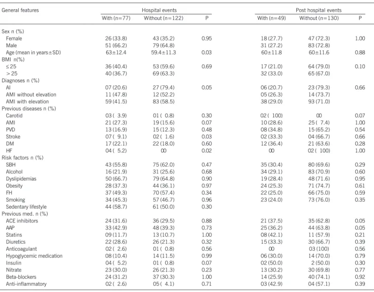
Table I demonstrates the general characteristics of patients at the beginning of the study, the analysis of the possible association of these characteristics, and the occurrence of hospital and pos-thospital events. Of the characteristics assessed (sex, age, AMI, type of AIS, previous cardiovascular disease, coronary risk factors, and medications used), age (P = 0.03), diagnosis of acute myo-cardial infarction with or without ST segment elevation (P= 0.05), and the previous diagnosis of stroke (P = 0.03) and HF (P=0.02) were associated with the development of hospital events. None of the general characteristics assessed demonstrated a statistically significant association with posthospital outcome.
It is important to note that 60% (118) of the patients from the population studied were hypertensive, 60.5% (129) were dys-lipidemic, 42% (84) were smokers, and 55% had AMI above 25
kg/m2. Forty percent of the patients were diagnosed with multiple
metabolic disorders (tab. I).
Regarding lipid profile, 39.5% (79) of the patients had a nor-mal lipid profile, 21.5% (43) had an increase in the total cholesterol, 17.0% (34) had hypertriglyceridemia, 11.5% (23) had low HDL-C, and 10.5% (21) had mixed dyslipidemia. Mean total cholesterol was 192 mg/dL, that of LDL-C was 118 mg/dL, that of HDL-C 46 mg/dL, and that of triglycerides was 137 mg/ dL. Mean non-HDL cholesterol was 146 mg/dL, and the total cholesterol/HDL-C ratio was 4.36. No statistically significant difference was noted between the lipid variables and hospital and posthospital events (tab. II). Only 11% of the patients were taking statins before the onset of acute ischemic syndrome (tab. II)
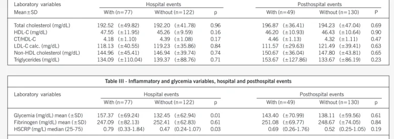
Metabolic (glycemia) and inflammatory (fibrinogen and HSCRP) variables are presented in table III.
Regarding glycemia, 19.6% of the patients had a previous diagnosis of diabetes. In our sample, 42.5% (85) had glycemia > 126 mg/dL, with a 142 mg/dL mean. Altered glycemia was signi-ficantly associated with hospital events (P < 0.01); however, it was not associated with posthospital events (P = 0.61). Among the patients with glycemia >126 mg/dL, 57% (48 patients) had
Table I - General characteristics of hospital and porthospital events
General features Hospital events Post hospital events With (n=77) Without (n=122) P With (n=49) Without (n=130) P
Sex n (%)
Female 26 (33.8) 43 (35.2) 0.95 18 (27.7) 47 (72.3) 1.00
Male 51 (66.2) 79 (64.8) 31 (27.2) 83 (72.8)
Age (mean in years±SD) 63±12.4 59.4±11.3 0.03 60±11.8 60±11.6 0.88 BMI n(%)
≤ 25 36 (40.4) 53 (59.6) 0.69 17 (21.0) 64 (79.0) 0.10
> 25 40 (36.7) 69 (63.3) 32 (33.0) 65 (67.0)
Diagnoses n (%)
AI 07 (20.6) 27 (79.4) 0.05 06 (20.7) 23 (79.3) 0.66
AMI without elevation 11 (47.8) 12 (52.2) 05 (26.3) 14 (73.7) AMI with elevation 59 (41.5) 83 (58.5) 38 (29.0) 93 (71.0) Previous diseases n (%)
Carotid 03 (03.9) 01 (00.8) 0.30 02 (0100) 00 0.07 AMI 21 (27.3) 19 (15.6) 0.07 10 (28.6) 25 (07.4) 1.00
PVD 13 (16.9) 15 (12.3) 0.48 08 (34.8) 15 (65.2) 0.54
Stroke 07 (09.1) 02 (01.6) 0.03 02 (33.3) 04 (66.7) 0.66
DM 17 (22.1) 22 (18.0) 0.60 12 (36.4) 21 (63.6) 0.28
HF 04 (05.2) 00 0.02 00 02 (0100) 1.00
Risk factors n (%)
SBH 43 (55.8) 75 (62.0) 0.47 35 (30.4) 80 (69.6) 0.29
Alcohol 16 (21.9) 31 (25.6) 0.68 34 (29.1) 83 (70.9) 0.60 Dyslipidemias 50 (66.7) 79 (64.8) 0.90 19 (28.4) 48 (71.6) 0.95 Obesity 28 (37.3) 44 (36.1) 0.97 24 (25.3) 71 (74.7) 0.61
FH 37 (49.3) 70 (57.4) 0.34 22 (25.0) 66 (75.0) 0.59
Smoking 34 (45.3) 57 (46.7) 0.96 23 (24.0) 73 (76.0) 0.35 Sedentary lifestyle 44 (58.7) 61 (50.0) 0.30
Previous med. n (%)
ACE inhibitors 24 (31.6) 36 (29.5) 0.88 21 (37.5) 35 (62.8) 0.05
AAP 33 (42.9) 48 (39.3) 0.73 25 (36.2) 44 (63.8) 0.05
Statins 09 (11.7) 13 (10.7) 1.00 08 (42.1) 11 (57.9) 0.21 Diuretics 22 (28.6) 26 (21.3) 0.32 15 (33.3) 30 (66.7) 0.39 Anticoagulant 02 (02.6) 01 (00.8) 0.56 00 03 (100) 0.56 Hypoglycemic medication 08 (10.4) 14 (11.5) 0.99 06 (30.0) 14 (70.0) 0.79 Insulin 04 (05.2) 01 (00.8) 0.07 02 (50.0) 02 (50.0) 0.30 Nitrate 23 (30.0) 26 (21.3) 0.23 13 (30.2) 30 (69.8) 0.77 Beta-blockers 24 (31.2) 37 (30.3) 1.00 14 (25.9) 40 (74.1) 0.92 Anti-inflammatory 02 (02.6) 05 (04.1) 0.71 03 (42.9) 04 (57.1) 0.39
4
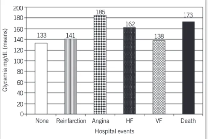
hospital events (4 acute myocardial reinfarction, 21 HF, 7 deaths) (fig.1), and 19% (15 patients) had posthospital events, 14 were hospital admittances (2 acute myocardial reinfarction, 4 HF, and 5 angina). Of the patients who died in the hospital, 70% had elevated glucose.
Median HSCRP was 0.55 mg/L (0.24-1.84), demonstrating a statistically significant association with hospital events (P=0.03), the same did not occur with posthospital events (P=0.19). Figu-re 2 shows the ratio between HSCRP levels and the incidence of hospital events where the same values occurred in patients with angina, heart failure, and those who died. However, in a multiva-riate analysis, HSCRP was not a predictor of hospital mortality.
Fibrinogen was > 277 mg/dL in 41% of patients, with a mean of 251 mg/dL. The difference for the association of fibrinogen with the incidence of hospital and posthospital events was not significant. (tab. III).
The most frequent events during hospital admittance were heart failure in 23.1% (46) of patients, angina in 8.5% (17) of patients, ventricular fibrillation in 11.6% (23) of patients, and reinfarction in 5.5% (11) of patients.
At hospital discharge, 76% (152) of patients were asympto-matic, 19% (38) were clinically stable, and 5% (10) died. Of these patients, 71.4% (142) underwent PTCA, 54% underwent primary PTCA, 2.5% (5) underwent PTCA and MRS, and 5% (10) of the patients underwent MRS.
Of the 48 patients who experienced events 1 month after hospital discharge, 36 were admitted again, 11 patients (5.5%) because of angina, 7 patients (3.5%) because of heart failure, and 5 patients (2.5%) because of acute myocardial re-infarction. Additionally, 5 patients were admitted again to the hospital for procedures (MRS/PTCA). One sudden death occurred at home.
In multiple regression logistics, the factors that remained as predictors of hospital mortality were age, previous acute myocar-dial infarction with or without ST segment elevation, and altered glycemia (tab. IV).
Discussion
The ratio between lipid profile and risk of cardiovascular diseases has been well demonstrated in clinical and observational studies32-34.
These studies have demonstrated that the decrease in cholesterol, especially in LDL-C, helped to prevent coronary artery disease and to reduce coronary events, both in primary (WOSCOPS, AFCAPS/ TexCAPS), and in the secondary prevention (4S,CARE, LIPID and HPS) 13-17,35.
Although in this study mean serum levels of total cholesterol (192 mg/dL), of LDL-C (118 mg/dL), HDL-C (46 mg/dL), and triglycerides (137 mg/dL) were not considered increased, it is important to note that 60.5% of patients had some alteration in lipid levels (21.5% had increased total cholesterol, 17% had hypertriglyceridemia, 11.5% had low HDL-C, and 10.5% had mixed dyslipidemia). Additionally, this is a high cardiovascular risk population with several risk factors as follows: 60% were hypertensive, 42% were smokers, 53.8% had a familial history of atherosclerotic disease, 42.5% had altered glycemia, and 55% had BMI above 25 kg/m2. These data reinforce those of the
lite-rature and call attention to the importance of associating risk factors to determine the risk of coronary events of an individual rather than assessing one isolated risk factor. Several studies have indicated that cholesterol levels decrease during acute myocardial infarction 36-41.Lipid and lipoprotein variation after acute myocardial
infarction occur within 24-48h after the onset of precordial pain and are made evident by decreases in total cholesterol (24% to 70% baseline), in LDL-C (31%), HDL-C (12-18%), and by increases in triglyceride levels (25%). In this study, samples were collected before the acute ischemic syndrome had evolved for 24 hours (mean time was 22h), to eliminate the possibility of an acute-phase reaction. A point to be considered to explain the results obtained is the current knowledge that individuals with comparable serum levels of LDL-C and HDL-C can also have fairly different levels of risk for coronary artery disease because of the differences in subclass distribution of these lipoproteins; however, this was not the objective of our study 42. Patients with a predominance of
small/dense LDL and/or small HDL had a very atherogenic profile. Clinical studies have demonstrated that systemic inflammation markers are strong predictors of clinical events in coronary artery disease 43. CRP is a predictor of cardiovascular events. Liuzzo et
al 24 studied patients with stable angina, unstable angina, and
acute myocardial infarction, using a cut point for normal/high
Table II - Mean of lipid variables and presence or absence of hospital and posthospital events
Laboratory variables Hospital events Posthospital events
Mean±SD With (n=77) Without (n=122) p With (n=49) Without (n=130) P
Total cholesterol (mg/dL) 192.52 (±49.82) 192.20 (±41.78) 0.96 196.87 (±36.41) 194.23 (±47.04) 0.69 HDL-C (mg/dL) 47.55 (±11.95) 45.26 (±9.59) 0.16 46.20 (±10.93) 46.43 (±10.64) 0.90 CT/HDL-C 4.18 (±1.10) 4.39 (±1.08) 0.17 4.46 (±1.13) 4.32 (±1.11) 0.47 LDL-C calc. (mg/dL) 118.13 (±40.55) 119.23 (±35.86) 0.84 111.57 (±29.63) 121.49 (±39.41) 0.63 Non-HDL cholesterol (mg/dL) 144.96 (±45.41) 146.94 (±39.74) 0.74 150.67 (±36.04) 147.80 (±43.81) 0.65 Triglycerides (mg/dL) 134.09 (±110.04) 139.37 (±88.76) 0.71 153.67 (±127.86) 133.67 (±86.19) 0.23
Table III - Inflammatory and glycemia variables, hospital and posthospital events
Laboratory variables Hospital events Posthospital events
With (n=77) Without (n=122) p With (n=49) Without (n=130) p
Glycemia (mg/dL) mean (±SD) 157.37 (±69.24) 132.45 (±62.94) 0.01 143.40 (±70.99) 138.11 (±59.56) 0.61 Fibrinogen (mg/dL) mean (±SD) 247.09 (±82.13) 252.41 (±62.83) 0.61 251.08 (±69.77) 248.67 (±74.05) 0.84 HSCRP (mg/L) median (25-75) 0.79 (0.33-1.84) 0.47 (0.24-1.07) 0.03 0.69 (0.26-1.76) 0.52 (0.25-1.05) 0.19
5
HSCRP at 3 mg/L. Patients with unstable angina had greater serum concentrations of HSCRP than those patients with stable angina, more ischemic episodes, and greater risk of death than those patients with low HSCRP (4.8±2.5/1.8 ±2.4; P=0.02). In patients with acute myocardial infarction, more increased levels of HSCRP are correlated with a greater area of myocardial necro-sis44,45.In a retrospective study of 37 patients with acute myocardial
infarction, HSCRP ≥2 mg/L correlated with a greater risk of myo-cardial rupture 46. In patients with unstable angina, HSCRP
corre-lates with a greater risk of coronary events (acute myocardial infarction, need for angioplasty or myocardial revascularization surgery, or sudden death) 47-49.
Our study reinforces the data from the literature. HSCRP was significantly associated with a greater incidence of hospital events. No significant difference occurred between HSPCR levels and the incidence of posthospital events.
Glycemia mg/dL (means)
200
None 180 160 140 120 100 80 60 40 20 0
Reinfarction Angina HF VF Death Hospital events
133 141 185
162 138
173
Fig. 1 - Glycemia and incidence of hospital events. HF: heart failure; VF: ventricular fibrilation.
HSCRP mg/L (median)
1.4
None 1.2
1
0.8
0.6
0.4
0.2
0
Reinfarction Angina HF VF Death Hospital Events
0.47 1.15
0.57
Fig. 2 - HSCRP and incidence of hospital events.HF: heart failure; VF: ventricular fibrilation.
0.82
0.46 0.98
Table IV - Hospital mortality predictors
Variable P Odds Ratio Confidence Interval (95%)
Age 0.04 1.03 1.00 – 1.06 Previous AMI 0.04 2.3 1.04 – 5.0 AMI with ST segment elevation 0.008 5.7 1.60 – 2.04 AMI without ST segment elevation 0.48 2.7 1.01 – 7.4 Glycemia 0.04 2.5 1.04 – 6.1 (>111 and <126mg/dL)
Glycemia >126mg/dL 0.002 3.5 1.60 – 7.9
CRP levels increased after acute myocardial infarction, reflecting the level of tissue lesion. Kushner et al 50 found a small increase
in CRP after acute myocardial infarction, twice as much as the mean in 8h and a peak at 2-4 days, returning to baseline after 3-4 weeks. In acute ischemic syndrome, CRP may increase modes-tly,51 and this increase seems to be restricted to patients with
tissue necrosis demonstrated by the levels of troponin 52.An isolated
measure of CRP, after infarction, is not predictive of future events53,
unless it is found right after the onset of symptoms and before the acute phase reaction 54. Definition of increased levels of HSCRP
has varied in different studies. These differences are because of the patients’ characteristics, including age, obesity, smoking, DAC extension, regional differences (prevalence of C. pneumoniae or positivity for cytomegalovirus), use of medications that can affect CRP levels (aspirin, statins, estrogens) 55. In the classification of
risk for future cardiovascular events, HSCRP levels have been considered low when they were < 1 mg/L; mild when they were 1 to 3 mg/L; and increased when they were > 3 mg/dL. In our study, HSCRP was between 0.05 and 2.90 mg/L in approximately 90% of patients. Despite being an acute-phase marker, CRP has an individual variability similar to that associated with cholesterol tracking. Several investigators have recommended 2 measures of CRP using the mean or the lower value to determine vascular risk. This is a practice that is in accordance with cholesterol evaluation21.
In the present study, HSCRP was assessed within the first 24h of the onset of the picture and by only one determination.
Fibrinogen, an acute-phase marker, plays an important role in
platelet compliance and aggregation. ECAT (European Concerted
Action on Thrombosis and Disabilities Angina Pectoris Study) 27
and PROCAM (Prospective Cardiovascular Münster) studies 56
de-monstrated an interaction between total cholesterol, LDL-choles-terol, and fibrinogen. In the ECAT study, the presence of moderate and high levels of fibrinogen increased the cardiovascular risk in individuals with hypercholesterolemia, whereas low levels of fibri-nogen identified patients with smaller risk of events even in the presence of increased concentrations of cholesterol. In the PROCAM study, a greater risk of events in individuals with increased fibrinogen and LDL-C was observed. Becker et al 29 verified that a fibrinogen
level in the plasma >300 mg/dL is associated with an increase in death, acute myocardial infarction and spontaneous ischemia in patients with angina and infarction without ST segment elevation. In our study, a statistically significant difference did not occur regarding the level of fibrinogen between events and the absence of events, in both hospital and posthospital events. Mean fibrinogen found in unstable angina was 262 mg/dL, in acute myocardial infarction it was 245 mg/dL, and in infarction with ST segment elevation it was 262 mg/dL. Patients experiencing hospital events had, on average, 242 mg/dL of fibrinogen, and patients with pos-thospital events had 251 mg/dL of fibrinogen.
In the study performed, 42% of patients had altered glycemia (>110 mg/dL), and only 19% had a previous diagnosis of diabe-tes. Among the patients with altered glycemia, 86% (72 patients) had infarction as an acute ischemic syndrome manifestation. Mean glycemia during hospital events was 157 mg/dL, and during pos-thospital events it was 143 mg/dL. Glycemia was significantly associated with hospital events (P = 0.01) and with hospital mortality (P = 0.002). Among the deaths occurring in the hospi-tal phase, 70% of the patients had elevated glucose.
6
diabetes mellitus present have a relative risk for cardiovascular disease increased 2 to 4 times, when compared with individuals without diabetes. Some studies show that, in patients with dia-betes, the risk of cardiovascular disease increases with the elevation of glucose concentration in the plasma. Patients with uncontrolled diabetes, increase in fasting glycemia, or increase in glycated hemoglobin, have an increased risk of cardiovascular disease and mortality, when compared with individuals with controlled glyce-mia. 58-60. Haffner et al 61 assessed cardiovascular mortality for an
8-year period in diabetic and nondiabetic patients, with or without previous acute myocardial infarction; they concluded that the mor-tality of a diabetic individual without previous infarction is equal to the mortality of a nondiabetic individual with previous acute myocardial infarction. Wahab et al62 determined that blood glucose
is an independent risk factor of mortality in acute myocardial infarc-tion, in the thromboembolic age, in 1664 patients. They concluded that hyperglycemia is associated with a worse evolution of acute myocardial infarction even in patients without a previous diagnosis of diabetes and that hyperglycemia added greater risk, regardless of body mass index or history of increased glucose, suggesting that its status per se may contribute to an adverse outcome or may be a key marker. Several studies 63-65 suggest that hyperglycemia, in
nondiabetic patients is, in fact, undiagnosed diabetes. Furthermore, studies suggest a role of stress in the increase of glucose in acute myocardial infarction; however, it is not clear whether this leads to a worse evolution or whether it is only a marker of a worse prognosis. Stress hyperglycemia may be a marker of extensive myocardial damage 66. Hyperglycemia, both acute and chronic, is also related
to involvement of endothelial function67,68, catecholamine release,
decrease in insulin sensitivity 69 and osmotic diuresis. The last leads
to a decrease in myocardial contractility 70.
In our study, hospital mortality was 5%, and posthospital mor-tality was 0.5% in 60 days. In the logistic regression, the mormor-tality predictors were age, previous acute myocardial infarction with or without ST segment elevation, and altered glycemia.
Our data reinforce the importance of differentiating the role of risk factors for a disease and that of the factor predictive of events (prognostic factor). We have studied a population with several classic risk factors for coronary disease, such as dyslipidemia, smoking, systemic blood hypertension, positive familial history of ischemic heart disease, but these risk factors do not contribute to worsen the prognosis of this group of patients. The increase of inflammatory markers, such as HSCRP, and metabolic markers, such as glycemia were related to a greater incidence of hospital events. Regarding increased glycemia, it also contributed to a greater risk of hospital mortality. Therefore, casual factors are not necessarily correlated to a worse prognosis of acute ischemic syndrome.
In conclusion, this study did not find any association between lipid profile and fibrinogen with the hospital and posthospital out-come in patients with acute ischemic syndrome. High sensitivity C-reactive protein and altered glycemia were associated with a greater incidence of hospital events but were not significantly associated with posthospital events. The mortality predictors were age, previous acute myocardial infarction with or without ST seg-ment elevation, and altered glycemia.
1. Fuster V, Lewis A. Conner memorial lecture: Mecanisms leading to myocardial in-farction: insights from studies of vascular biology. Circulation 1994;90:21. 2. Lima VC. Síndromes isquêmicas agudas. Arq Bras Cardiol 1999;72:109-23. 3. Rudel LL, Kesainiemi A. Low-density lipoprotein particle composition: what is the
contribution to atherogenicity? Curr Opin Lipidol 2000;11:227-8.
4. Miller GL, Miller NE. Plasma high-density lipoprotein concentration and the deve-lopment of ischaemic heart disease. Lancet 1975;I:16-9.
5. Assmann G, Schulte H. Role of triglycerides in coronary artery disease: lessons from the prospective cardiovascular munster study. Am J Cardiol 1992;70:10H-13H. 6. Austin MA. Plasma triglyceride and coronary heart disease. Arterioscler Thromb
1991;11:2-14.
7. Hokanson JE, Austin MA. Plasma triglyceride level is a risk factor for cardiovascu-lar disease independent of high-density lipoprotein cholesterol level: a meta-ana-lysis of population based prospective studies. J Cardiovasc Risk 1996;3:213-219. 8. Rossouw JE, Lewis B, Rifkind BM. The value of lowering cholesterol after
myocar-dial infarction. N Engl J Med 1990;1112-19.
9. Wong ND, Wilson PWF, Kannel WB. Serum cholesterol as a prognostic factor after myocardial infarction: The Framingham Study. Ann Intern Med 1991;115:687-93. 10. Frost PH, Verter J, Miller D. Serum lipids and recurrent cardiac events. Am Heart J
1987;1356-64.
11. Ulvenstam G, Bergstrand R, Johansson S, et al. Prognostic importance of choles-terol level after myocardial infarction. Prev Med 1984;13:355-66.
12. Malach M, Quinley J, Imperato PJ, et al. Improving lipid evaluation and manage-ment in medicare patients hospitalized for acute myocardial infarction. Arch Intern Med 2001;161:839-844.
13. Shepherd J, Cobbe SM, Ford I, et al. Prevention of Coronary heart disease with pravastatin in men with hypercholesterolemia. The West of Scotland Coronary Prevention Study (WOSCOPS). N Engl J Med 1995;333:1301-07
14. Downs JR, Clearfield M, Weis S, et al. Primary prevention of acute coronary events with Lovastatin in men and women with average cholesterol levels. Results of AFCAPS/TexCAPS. JAMA 1998;279:1615-22
15. The Long-term intervention with pravastatin in ischaemic disease (LIPID) Study group. Prevention of cardiovascular events and death with Pravastatin in patients with coronary heart disease and a broad range of initial cholesterol levels. N Engl J Med 1998;339:1349-57.
Referências
16. Pedersen TR, Kjekshus J, Berg K, et al. Randomised trial of cholesterol lowering in 4,444 pacients with coronary heart disease: The Scandinavian Simvastatin Sur-vival Study (4S). Lancet 1994;344:1383-9.
17. Sacks FM, Pfeffer MA, Move LA et al. The effect of pravastatin on coronary events after myocardial infarction in patients with average cholesterol levels. The choles-terol and recurrent events trial (CARE). N Engl J Med 1996;335:1001-09. 18. LaRosa JC, He J, Vupputuri S. Effect of statins on risk of coronary disease: a
meta-analysis of randomized trials. JAMA1999;282:2340-6.
19. Brensike JF, Levy RI, Kelsey SF et al. Effects of therapy with cholestyramine on pro-gression of coronary arteriosclerosis: results of the NHLBI Type II Coronary Inter-vention Study. Circulation 1984;69:313-24.
20. Blankenhorn DH, Nessim S A, Johnson RL, et al. Beneficial effects of combined co-lestipol-niacin therapy on coronary atherosclerosis and coronary venous bypass grafts. JAMA 1987;257:3233-40.
21. Ridker PM. Clinical application of C-Reactive Protein for cardiovascular disease de-tection and prevention. Circulation 2003;107:363.
22. Idker PM, Cushman M, Stampfer MJ, et al. Inflammation, aspirin, and risks of cardiovascular disease in apparently healthy man. N Eng J Med 1997;336;973-9. 23. Morrow DA, Rifai N, Antman EM et al. C-reactive protein is a potent predictor of mortality independently of and in combination with troponin T in acute coronary syndromes: a TIMI 11 A substudy. Trombolysis in miocardion infarction. J Am Coll Cardiol 1998;31:1460-5.
24. Liuzzo G, Biasucci LM, Gallimore JR et al. The prognostic value of C-reactive pro-tein and serum amyloid a propro-tein in severe unstable angina. N Engl J Med 1994;331:417-24
25. Haverkate F, Thompson SG, Pyke SD et al. Production of C-reactive protein and risk of coronary events in stable and unstable angina: European concerted action on thrombosis and disabilities angina pectoris study group. Lancet 1997;349:462-6. 26. Mueller C, Buettner HJ, Hodgson JD, et al. Inflammation and long-term mortality after non-ST-elevation acute coronary syndrome treated with a very early invasive strategy in 1,042 consecutive patients. Circulation 2002;105:1412-5. 27. Thompson SG, Kienast J, Pyke SDM, et al. Hemostatic factors and the risk of
7
28. Annel WB, D’Agostinho RB, Belanger AJ. Fibrinogen, cigarette smoking and risk of cardiovascular disease: insights from the Framingham study. Am Heart J 1987; 113:1006-10.
29. Becker RC, Cannon CP, Bovill EG et al. Prognostic value of plasma fibrinogen con-centration in patients with unstable angina and non-Q-wave myocardial infarc-tion. Am J Cardiol 1996;78:142-7.
30. I Diretriz da Sociedade Brasileira de Cardilogia para o Tratamento do Infarto Agudo do Miocárdio. Arq Bras Cardiol 2000; 74(Supl II):1-46.
31. III Diretrizes Brasileiras sobre dislipidemias e diretriz de prevenção da aterosclerose do Departamento de Aterosclerose da Sociedade Brasileira de Cardiologia. Arq Bras Cardiol 2001;77(Supl III):1-48.
32. Sytrowski PA, Kannel WB, D’agostino RB. Changes in risk factors and the decline in mortality from cardiovascular disease. N Engl J Med 1990;322:1635-40. 33. Müller C. Xanthomata, hypercholesterolemia, angina pectoris. Acta Med Scand
1938;89:75-84.
34. Keysa ED. Coronary heart disease in seven countries. Circulation 1970;41 (suppl.I):1.
35. MRC/BHF Heart Protection Study of cholesterol lowering with sinvastatin in 20,536 high-risk individuals: a randomized placebo-controlled trial. Heart Protec-tion Study Collaborative Group. Lancet 2002; 360:7-22.
36. Rosenson RS. Myocardial injury: The acute phase response and lipoprotein meta-bolism. J Am Coll Cardiol 1993;22:933-40.
37. Mainard F, Ozanne P, Madec Y. Variation in lipoproteins, hormones and blood glu-cose during the early acute phase of myocardial infarction. Atherosclerosis 1988; 69:225-31.
38. Gore JM, Goldberg RJ, Matsumoto AS, Castelli WP, Mcnamara PM, Dalen JE. Va-lidity of serum total cholesterol level obtained within 24 hours of acute myocardial infarction. Am J Cardiol 1984;54:722-5.
39. Jackson R, Scragg R, Marshall R et al. Changes in serum lipid concentrations during first 24 hours after myocardial infarction. Br Med J 1987;294:1588-9. 40. Cabana VG, Siegel JN, Sabesin SM. Effects of the acute phase response on the
concentration and density distribution of plasma lipids and apolipoproteins. J Lipid Res 1989;30:39-49.
41. Henkin Y, Crystal E, Goldberg Y et al. Usefulness of lipoprotein changes during acute coronary syndromes for predicting postdischarge lipoprotein level. Am J Cardiol 2002;89:7-11.
42. Otvos JD. Measurement of lipoprotein subclass profiles by NMR spectroscopy. In: Rifai N, Warnick R, Dominiczak M, eds. Handbook of lipoprotein testing. Washing-ton DC: AACC Press, 1997:497-508.
43. Winter RJ, Bholasingh R, Lijmer JG et al. Independent prognostic value of C-reac-tive protein and troponin I in patients with unstable angina or non-Q-wave myo-cardial infarction. Cardiovasc Res 1999; 42: 240-5.
44. de Beer FC, Hind CR, Fox KM, Allan RM, Maseri A, Pepys MB. Measurement of serum C-reactive protein concentration in myocardial ischaemia and infarction. Br Heart J 1982;47(3):239-43.
45. Pietilä KO, Harmoinem AP, Hermens WP et al. Serum C-reactive protein and inf-arct size in myocardial infinf-arct patients with a closed versus an open infinf-arct-related coronary artery after thombolytic therapy. Eur Heart J 1993;14:915-9. 46. Ueda S, Ikeda U, Yamamoto K et al. C-reactive protein as a predictor of cardiac
rupture after myocardial infarction. Am Heart J 1996;131:857-60.
47. Toss H, Lindahl B, Siegbahn A, Wallentin L (for The Frisc Study Group). Prognostic influence of increase fibrinogen and C-reactive protein levels in unstable coronary artery disease. Circulation 1997;96:4204-10.
48. Haverkate F, Thompson SG, Pyke SDM et al. Production of C-reactive protein and risk of coronary events in stable and unstable angina. Lancet 1997;349:462-6.
49. Berk BC, Weintraub WS, Alexander RW. Elevation of C-reactive protein in “active” coronary artery disease. Am J Cardiol 1990;65:168-72.
50. Kushner I, Broder ML, Karp D. Control of the acute phase response. Serum C-reac-tive protein kinetics after acute myocardial infarction. J Clin Ivest 1978;61:235-42. 51. Ferreiros ER, Boissinnet CP, Pizarro R et al. Independent prognostic value of
ele-vated C-reactive protein in unstable angina. Circulation 1999;100:1958-63. 52. Benamer H, Steg PG, Benessiano J et al. Comparison of the prognostic value of
C-reactive protein and troponin I in patients with unstable angina pectoris. Am J Cardiol 1998;82:845-50.
53. Zebrack JS, Anderson JL, Maycock CA, Home BD, Bair TL, Muhlestein JB; Inter-mountain Heart Collaborative(IHC) Study Group. Usefulness of high-sensitivity C-reactive protein in predicting long-term risk of death or acute myocardial infarction in patients with unstable or stable angina pectoris or acute myocardial infarction. Am J Cardiol 2002;89(2):145-9.
54. Tommasi S, Carluccio E, Bentivoglio M et al. C-reactive protein as a marker for car-diac ischemic events in the year after a first, uncomplicated myocardial infarction. Am J Cardiol 1999;83:1595-9.
55. Le Jacq Communicatons. Role of Inflammation in Cardiovascular Disease. Prog Cardiovasc Nurs 2002;17(4):174-85.
56. Heinrich J, Schulte H, Balleisen L, Assmann G, Van De Loo J. Predictive value of haemostatic variables in the Procam Study. J Thromb Haemostas 1991;65:8. 57. Kannel WB, Mcgee DL. Diabetes and cardiovascular disease.The Framingham
Study. J Am Med Assoc 1979;241:2035-8.
58. Moss SE, Klein R, Klein Bek, Meuer SM. The association of glycemia and cause-specific-mortality in a diabetic population. Arch Intern Med 1994;154:2473-9. 59. Andersson DKG, Svardsudd K. Long-term glycemic control relates to mortality in
type Ii diabetes. Diabetes Care 1995;18:1534-43.
60. Gerstein HC. Glucose: a continuous risk factor for cardiovascular disease. Diabetic Med 1997;14:S25-S31
61. Haffner SM, Lehtos S, Ronnemaa T, Pyorala K, Laasko M. Mortality from Coro-nary Heart Disease in subjects with type 2 diabetes and in nondiabetic subjects with and without prior myocardial infarction. N Engl J Med 1998;339:229-34. 62. Wahab NN, Cowden EA, Pearce NJ, Gardner MJ, Merry H, Cox JL. Is blood
glu-cose an independent predictor of mortality in acute myocardial infarction in the thrombolytic era? J Am Coll Cardiol 2002;40:1748-54.
63. Sala J, Masiá R, Gonzalez de Molina FJ et al. Short-term mortality of myocardial infarction patients with diabetes or hyperglycemia during admission. J Epidemiol Community Health 2002;56:707-12.
64. Malmberg K, Norhammar A, Wedel H, Rydén L. Glycometabolic state at admission: important risk marker of mortality in conventionally treated patients with diabetes mellitus and acute myocardial infarction. Circulation 1999;99:2626-32. 65. Norhammar A, Tenerz A, Nilsson G et al. Glucose metabolism in patients with acute
myocardial infarction and no previous diagnosis of diabetes mellitus: a prospective study. Lancet 2002;359:2140-44.
66. Tansey MJ, Opie LH. Plasma glucose on admission to hospital as a metabolic index of the severity of acute myocardial infarction. Can J Cardiol 1986;2:326-31. 67. Williams SB, Goldfine AB, Timimi FK et al. Acute hyperglicemia attenuates
endothe-lium-dependent vasodilation in humans in vivo. Circulation 1998;97:1695-701. 68. Williams SB, Cusco JA, Roddy MA, Johnstone MT, Creager MA. Impaired nitric
oxide-mediated vasodilation in patients with non-insulin dependent diabetes mellitus. J Am Coll Cardiol 1996;27:576-74
69. Leor J, Goldbourt U, Reicher-Reiss H and the APRINT Study Group et al. Cardiogenic shock complicating acute myocardial infarction in patients without heart failure on admission: incidence, risk factors, and outcome. Am J Med 1993;94:265-73. 70. Holubarsch C, Ruf T, Goldstein DJ, et al. Existence of the Frank-Starling mechanism