TEMAS LIVRES FREE THEMES
1 Faculdade de Odontologia de Piracicaba, Universidade Estadual de Campinas. Av. Limeira 901, Areião. 13414-903 Piracicaba SP Brasil. [email protected]
Prevalence of suggestive images of carotid artery calcifications
on panoramic radiographs and its relationship
with predisposing factors
Prevalência de imagens sugestivas de calciicações
da artéria carótida em radiograias panorâmicas
e sua relação com fatores predisponentes
Resumo As radiografias panorâmicas (RP) podem exibir imagens radiopacas sugestivas de ateromas calcificados na artéria carótida em pa-cientes assintomáticos. O objetivo deste estudo foi avaliar a prevalência destas imagens na RP e sua relação com hipertensão, obesidade, idade, sexo e tabagismo. Foram avaliadas RP de 505 pacientes acima de trinta anos, que realizaram esse exame por diversos motivos clínicos. Seu índice de massa corpórea foi calculado; sua circunferência abdo-minal também foi considerada. Informações sobre hipertensão e tabagismo foram obtidas. Os obser-vadores avaliaram as RP para a presença de mas-sas radiopacas na região das vértebras cervicais C3-C4, confirmadas por meio de uma radiografia ântero-posterior (AP). Os resultados mostraram prevalência de 7,92% de imagens sugestivas de calcificações em RP e na radiografia AP. A razão de probabilidade (OR) ajustada mostrou associa-ção com idade e tabagismo. O risco para as pes-soas mais velhas aumenta até cerca de nove vezes quando comparado com aqueles mais jovens, en-quanto para os fumantes, o risco é o dobro, quan-do comparaquan-do com não fumantes. Na população estudada, 7,92% de todos os sujeitos apresenta-ram imagens sugestivas de aterosclerose carotídea em RP e houve associação com idade e tabagismo.
Palavras-chave Radiografia panorâmica,
Hiper-tensão, Obesidade, Acidente vascular cerebral
Abstract Panoramic radiographs (PR) can dis-play radiopaque images suggestive of calcified atheroma in the carotid artery in asymptomatic patients. The aim of this study was to evaluate the prevalence of these images on PR and their link-age with hypertension, obesity, link-age, gender and smoking habits. PR of 505 patients were evaluat-ed. They were older than 30 years old and their PR had been taken for different clinical reasons. Their body mass index was calculated; their waist cir-cumference was also taken into consideration. In-formation about smoking habits and hypertension was obtained. The observers analyzed the presence of radiopaque mass in the region of the cervical vertebrae C3-C4 through the PR, confirmed by an antero-posterior (AP) radiograph. The results showed a 7.92% prevalence of suggestive images of calcifications on PR and on AP radiograph. The adjusted Odds Ratio showed association with age and smoking habits. The calcification process is almost nine times higher for the elderly when compared to the young. As far as smokers are con-cerned, this process is twice worse when compared to no smokers. In conclusion, 7.92% of the group studied presented suggestive images of carotid atherosclerosis on PR, which is directly associated with the age and smoking habits.
Key words Panoramic radiograph, Arterial
hy-pertension, Obesity, Stroke
Ana Caroline Ramos de Brito 1
Helena Aguiar Ribeiro Nascimento 1
Rafaela Argento 1
Thamara Beline 1
Glaucia Maria Bovi Ambrosano 1
B
rit
o A
Introduction
Cardiovascular diseases continue to be the main cause of morbidity and mortality worldwide. Cerebrovascular accidents are responsible for killing or disabling more than half a million Americans every year1. In Brazil, about 68,000
deaths per year are reported to be caused by stroke. This disease is the leading cause of death and disability in the country, generating signif-icant economic and social impact2. People over
55 are at a great risk of having a stroke. This risk increases as they get older3.
Some studies have linked the presence of the carotid artery calciications to systemic diseas-es such as obdiseas-esity (body mass index and waist circumference), hypercholesterolaemia, hyper-tension, diabetes, renal stones, dental infections, prior atherosclerosis and with increasing age4-12.
Panoramic radiograph is one of the most re-quested complementary exam by dentists. Along with the evaluation of dental and maxillofacial hard tissues, panoramic radiograph can be used to spot soft tissue calciications including cal-ciied carotid artery atheroma13. The presence
of calciied carotid artery plaque on panoramic images is an indicator of the risk of future ad-verse cardiovascular events5. Studies show that
the prevalence of theses calciications on pan-oramic radiographs taken for oral health reasons in the general dental outpatient groups, ranges from 2% to 5%14. It is important to highlight that
calciications may not imply signiicant stenosis and not all atherosclerotic lesions are calciied. Despite that, the calciications present on dental radiographs could be quite often associated with signiicant carotid diseases. This inding can be considered a cost-effective incentive for the use of Doppler sonography13.
When dentists are suspicious about the pres-ence of carotid artery atheromas in panoramic radiographs, they play an important role for their patients’ lives, as they guide and immediately re-fer their patients to doctors for adequate medical treatment15.
In recent years, a number of publications have detected calciications on panoramic radio-graphs5,7-12,16-18. Although the diagnosis by
pan-oramic radiography is reported, when it comes to the Brazilian population, no studies have ana-lyzed the calciication prevalence and its associa-tion with risk factors.
The purpose of the present study is, there-fore, to determine the prevalence of radiopacities suggestive of calciied atherosclerotic plaques on
carotid artery on panoramic radiographs. Their association with hypertension, obesity, age, gen-der, smoking habits and history of cardiovascular diseases will also be studied.
Methods
The study was approved by the local ethics com-mittee. Besides, formal consent was given by each subject. 505 patients agreed to take part in the research, all of them over their thirties (199 males and 306 females). The average of age being 50.1 years old. All patients were attended at the Department of Oral and Maxillofacial Radiol-ogy, Piracicaba Dental Scholl, during the years 2013-2014. PR were taken for different purposes regardless of this study.
Information about the lifestyle, such as smoking habits and medical history (hyperten-sion, own cardiovascular disease or family histo-ry of cardiovascular disease, stroke or medication use) of each patient was assessed through a stan-dardized questionnaire applied by a trained ob-server. Weight was measured to the nearest 0.1 kg while the subjects were dressed in light clothing. Height was measured to the nearest 0.1 cm with the subject in stocking feet.
Body mass index (BMI, kg/m2) was
calculat-ed by dividing the body weight (kg) by square of height (m2), according to OMS, 200919:
1 - Malnourished: < 18.5; 2 - Normal: 18.6 - 24.9; 3 - Overweigth: 25.0 - 29.9; 4 - Obesity: > 30.0
Abdominal circumference was mea-sured with the subject standing, at the midway between the lower rib margin and the iliac crest. The abdominal circumference was classiied ac-cording to OMS, 200919:
Female: 1 – Ideal: ≤ 80.0
2 – Increased: 80.0 – 93.9 3 – Substantial: ≥ 94.0
Blood pressure was measured after at least 5 min of rest in the sitting position using a mer-cury sphygmomanometer. Notwithstanding the individuals who reported a state of hypertension previously diagnosed by a doctor and who were under medication to control blood pressure, were directly considered hypertensive.
All panoramic radiographs were obtained with an ORTHOPANTOMOGRAPH® OP100D (Instrumentarium Dental, Tuusula, Finland). The unit was operated with different parameters,
Male:
1 – Ideal: ≤ 88.0
e C
ole
tiv
a,
21(7):2201-2207,
2016
depending on the patient’s estimated jaw size. Two oral and maxillofacial radiologists in-terpreted all the panoramic radiographs. The presence of carotid artery calciications appeared as heterogeneous radiopacities in a verticolinear orientation adjacent to the hyoid bone, epiglottis and cervical vertebrae either at, above or below the intervertebral space between C3 and C4. Each observer interpreted each panoramic radiograph individually, this followed a discussion of each image between the two observers. If the two ob-servers agreed, a positive diagnosis was made.
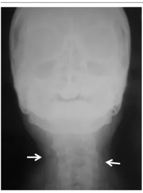
The patients whose cases were deined as pos-itive by the two examiners (Figure 1) were asked to undergo a modiied anterior-posterior (AP) radiograph (with chin elevated) (Figure 2) in or-der to conirm or refute the suggested indings on the panoramic radiograph. If the AP conirmed the calciication, the individual was referred to the cardiologist evaluation and if possible to re-alize carotid Doppler ultrasound.
The data analysis was performed by SAS (9.1.3 version; SAS Institute Inc., Cary, NC, USA). The crude logistic regression was performed to analyze the association between presence and lat-erality of calciication and different factors stud-ied (gender, age, BMI, abdominal circumference, hypertension, smoking and own or family med-ical history of cardiovascular diseases). Variables with p < 0.05 in the crude analysis were selected for initial inclusion in the multivariate logistic regression to determine the independent predic-tors of the presence of calciication.
Results
The prevalence of suggestive images of calciica-tions on PR, conirmed by AP radiograph, was 7.92% in the total of individuals; of these, 32.5% were male and 67.5% were female. In the male population, there was a 6.53% prevalence, while in the female group it was 8.82%.
The presence of calciications according to the predisposing factors studied is presented in Table 1. Subjects with calciications were older and had a higher BMI and abdominal circumfer-ence. They also were hypertensive, had a history of cardiovascular disease and smoking habits. However, BMI, abdominal circumference and history of cardiovascular disease were not signii-cantly associated with calciications (p > 0.05). The brute regression showed that the presence of calciication was associated with age, hyperten-sion and smoking habits.
Table 2 shows the distribution of uni and bi-lateral cases of calciications detected on PR and the factors studied; there was no correlation be-tween the factors and laterality.
Figure 1. Panoramic radiograph of a 63-year-old male patient with heterogeneous radiopacities (arrows), in the left and right sides, adjacent to the hyoid bone and intervertebral space between C3 and C4, posterior to the angle of the mandible.
Figure 2. Modiied anterior-posterior (AP)
B
rit
o A
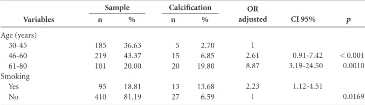
When these same parameters were adjusted, the strength of the association was attenuated for hypertension, but remained statistically signii-cant for age and smoking habits (Table 3). Con-cerning to age, it was observed that the risk for older people increases up to almost nine times when compared to young. For smokers, the risk is twice when compared with no smokers.
Discussion
Atherosclerotic tend to accumulate, in form of irregular plaques, within the bifurcation of the common, external, and internal carotid arteries. However, not all atherosclerotic lesions are calci-ied; furthermore, the presence of calciication in this location is not a deinitive indicator of vascu-lar disease14. Because of its anatomical position,
this region can be viewed on panoramic radio-graphs, which is a radiographic modality
com-monly requested in dentistry. As atherosclerosis may be an asymptomatic condition, it may be an incidental inding on routine panoramic exam-ination of the patient.
As other calciications can appear in the same region of atheroma on panoramic radiograph, which may be anatomic (hyoid bone, cartilage triticeous, epiglottis, etc.) or pathologic (calciied lymph nodes, tonsilloliths, phleboliths, etc.)3, it is
necessary another radiographic (AP) or exam for the diagnosis to be conirmed.
The gold standard for the detection of ather-oma is ultrasonography with Doppler; the pan-oramic radiograph cannot be considered an ac-curate test for stenosis, but a inding on radiogra-phy requires referral to a specialist for diagnosis, because there is a connection between indings in panoramic radiography and cardiovascular events16. Thus, the panoramic radiograph may
be a risk marker for subsequent cardiovascular events12; moreover, previous studies have found
Variables Gender Male Female Age (years) 30-45 46-60 61-80 BMI (Kg m2)
Normal Overweight Obesity Abd circumf. Ideal Increased Substantial Hypertension Yes No Smoking Yes No Pat PH Yes No Fam PH Yes No
Table 1. Brute logistic regression for presence of calciication.
BMI: Body mass índex; Abd circumf.: Abdominal circumference; Pat PH: Patient previous history of cardiovascular disease; Fam PH: Family previous history of cardiovascular disease; OR: Odds ratio; CI: Conidence interval.
e C ole tiv a, 21(7):2201-2207, 2016
a correlation between calciications found in the panoramic radiograph with the indings on ul-trasonography6,13,15,20,21 and others also used the
panoramic radiograph to evaluate the prevalence of atheroma4,16.
In this study, the differential diagnosis were made by the AP radiograph, a method also used by Henriques et al.22 and Almog et al.3. This
aimed the diagnosis, because radiopacities sug-gestive of calciied atherosclerotic plaques on ca-rotid artery is displayed laterally to the vertebra, while the images of other structures of average body region, as calciication in the triticeous cartilage, which is the greatest cause of errone-ous diagnosis of atheroma23, are overlapping to
the spine. This was the method available in the Radiology Clinic where the study was conducted; so the patient could not perform an ultrasound of the neck immediately for conirmation of the presence of atheroma and, when the radiopaque image had been viewed, the patient was referred to an appropriate doctor.
The population studied was in their thirties. In younger patients, only a minor number of cases with carotid calciications can be found16.
Despite this, the accumulation of calcium in the aorta starts to occur after the person is twenty years old. That is why it was decided to include younger patients, so almost all age groups with possibility of having calciication in the carotid artery could be analyzed.
The results of this study demonstrate that 7.92% of the subjects showed a calciied carot-id artery atheroma, which was conirmed by AP radiograph. Previous studies have found a prevalence of carotid calciication on panoram-ic radiographs close to this study’s results, rang-ing from 5.6% to 6.8%9,15,18, while other studies
obtained lower values of 1.6% to 4.8%8,13,16,17,24.
However, in some speciic conditions, such as pa-tients with primary hyperparathyroidism, kidney stones and cardiovascular disease symptoms, the prevalence was higher4,8,18.
Variables Gender Male Female Age (years) 30-45 46-60 61-80 BMI (Kg m2)
Normal Overweight Obesity Abd circumf. Ideal Increased Substantial Hypertension Yes No Smoking Yes No Pat PH Yes No Fam PH Yes No
Table 2. Brute logistic regression for laterality of calciication.
BMI: Body mass índex; Abd circumf.: Abdominal circumference.; Pat PH: Patient previous history of cardiovascular disease; Fam PH: Family previous history of cardiovascular disease. n 10 15 3 10 12 8 8 9 2 11 12 13 12 8 17 2 23 6 19 % 76.92 55.55 60.00 40.00 60.00 66.67 53.33 69.23 50.00 73.33 57.14 68.42 57.14 61.54 62.96 66.67 62.16 46.15 70.37 Unilateral n 3 12 2 5 8 4 7 4 2 4 9 6 9 5 10 1 14 7 8 % 23.08 44.45 40.00 60.00 40.00 33.33 46.67 30.77 50.00 26.67 42.86 31.58 42.86 38.46 37.04 33.33 37.84 53.85 29.63 Bilateral p 0.3377 0.7866 0.9597 0.7705 0.6376 0.7751 0.5188 0.6827 0.7937 0.6419 0.2572 Variables Age (years) 30-45 46-60 61-80 Smoking Yes No
Table 3. Multivariate logistic regression analysis for presence of calciication.
B
rit
o A
Gender, age, obesity, waist circumference, hypertension, previous history of cardiovascular disease and patient’s family history and smoking habits were the risk factors studied, but only the association between calciication, age and smok-ing habits were signiicant. Other studies had also showed a correlation with age13,18,25. In fact, in a
Turkey population, in addition to the age, it was found correlation with gender (male), family his-tory of cardiovascular disease and dyslipidemia; moreover, there was no correlation with hyper-tension and smoking habits24. These
discrepan-cies in the predictors factors could be attributed to differences in the populations’ studies. Indeed, the authors analyzed symptomatic patients of a Cardiologic department whereas this study ana-lyzed asymptomatic patients who were referred to a panoramic radiograph for dental treatment, representatives of as small portion of the Brazil-ian population (very heterogeneous), in the city of Piracicaba/SP with a medium income and without speciic diseases. When the relationship was studied according to speciic conditions, such as in patients with primary hyperparathyroid-ism, despite the prevalence being of 40%, there was no correlation with atherogenic pattern (age, body mass index, hypertension, diabetes,
hyper-lipidemia)3. Postmenopausal Women over 50
years old, for its part, showed 11% of prevalence of carotid calciication, also not correlated with hypertension, past history of myocardial infarc-tion and hypercholesterolemia10.
It is crucial for the dental practitioners to know the characteristics and prevalence of ca-rotid artery calciications to perform a thorough evaluation of areas of panoramic radiograph, which can identify such calciications. Further-more, the professionals should differentiate these calciications from the others when it is possible to perform an AP radiography26. Consequently,
when their presence is suspected or conirmed, the patient has to be informed of the indings and its implications. He has also to be referred to a medical evaluation. To this end, patients with smoking history require special attention.
Conclusions
The study came to the conclusion that, among the factors studied, only the age and smoking habits were correlated to the presence of suggestive im-ages of atheroma in PR. Besides, this prevalence was of 7.92% of all the individuals assessed.
Collaborations
e C
ole
tiv
a,
21(7):2201-2207,
2016
Johansson EP, Ahlqvist J, Garoff M, Karp K, Jäghagen EL, Wester P. Ultrasound screening for asymptomat-ic carotid stenosis in subjects with calciasymptomat-ication in the area of the carotid arteries on panoramic radiographs: a cross-sectional study. BMC Cardiovascular Disorders
2011; 11(44):1-9.
Bayer F, Helfgen EH, Bos C, Kraus D, Enkling N, Mues S. Prevalence of indings compatible with carotid ar-tery calciications on dental panoramic radiographs.
Clin Oral Invest 2011; 15(4):563-569.
Sisman Y, Ertas ET, Gokce C, Menku A, Ulker M, Ak-gunlu F. The prevalence of carotid artery calciication on the panoramic radiographs in Cappadocia region population. Eur J Dent 2007; 1(3):132-138.
Sunman H, Yorgun H, Canpolat U, Hazırolan T, Kaya EB, Ateş AH, Dural M, Aytemir K, Tokgözo lu L, Ka-bakçı G, Akata D, Oto A. Association between family history of premature coronary artery disease and cor-onary atherosclerotic plaques shown by multidetector computed tomography coronary angiography. Int J
Cardiol 2013; 164(3):355-358.
Associação brasileira para o estudo da obesidade e da síndrome metabólica (ABESO). Diretrizes brasileiras de
obesidade 2009/2010. 3ª ed. Itapevi: ACFarmacêutica;
2009.
Romano-Sousa CM, Krejci L, Medeiros FMM, Gracio-sa-Filho RG, Martins MFF, Guedes VN, Fenyo-Pereira M. Diagnostic agreement between panoramic radio-graphs and color doppler images of carotid atheroma. J
Appl Oral Sci 2009; 17(1):45-48.
Friedlander AH, Garrett NR, Chin EE, Baker JD. Ultra-sonographic conirmation of carotid artery atheromas diagnosed via panoramic radiography. J Am Dent Assoc
2005; 136(5):682-683.
Henriques JCG, Kreich EM, Baldani MH, Luciano M, Castilho JCM, Moraes LC. Panoramic radiography in the diagnosis of carotid artery atheromas and the asso-ciated risk factors. Open Dent J 2011; 5:79-83. Kamikawa RS, Pereira MF, Fernandes A, Meurer MI. Study of the localization of radiopacities similar to calciied carotid atheroma by means of panoramic ra-diography. Oral Surg Oral Med Oral Pathol Oral Radiol
Endod 2006; 101(3):374-378.
Bayram B, Uckan S, Acikgoz A, Müderrisoglu H, Aydinalp A. Digital panoramic radiography: a reliable method to diagnose carotid artery atheromas?
Dento-maxillof Radiol 2006; 35(4):266-270.
Horsley SH, Beckstrom B, Clark SJ, Scheetz JP, Khan Z, Farman AG. Prevalence of carotid and pulp calci-ications: a correlation using digital panoramic radio-graphs. Int J Cars 2009; 4(2):169-173.
MacDonald D, Chan A, Harris A, Vertinsky T, Farman AG, Scarfe WC. Diagnosis and management of calciied carotid artery atheroma: dental perspectives. Oral Surg
Oral Med Oral Pathol Oral Radiol 2012;
114(4):533-547.
Artigo apresentado em 22/05/2015 Aprovado em 22/08/2015
Versão inal apresentada em 24/08/2015 15.
16.
17.
18.
19.
20.
21.
22.
23.
24.
25.
26. References
American Heart Association. Heart and stroke statistical
update. Dallas: American Heart Association; 2002.
Portal Brasil. Saúde. Acidente vascular cerebral (AVC). [cited 2015 Apr 1]. Available in: http://www.brasil.gov. br/saude/2012/04/acidente-vascular-cerebral-avc. Almog DM, Tsimidis K, Moss ME, Gottlieb RH, Carter LC. Evaluation of a training program for detection of carotid artery calciications on panoramic radiographs.
Oral Surg Oral Med Oral Pathol Oral Radiol Endod
2000; 90(1):111-117.
Friedlander AH, Aghazadehsanai N, Chang TI, Hara-da N, Garret NR. Prevalence of calciied carotid artery atheromas on panoramic images of individuals with primary hyperparathyroidism. Dentomaxillofac Radiol
2013; 42(8):20130118.
Friedlander AH, Golub MS. The signiicance of carotid artery atheromas on panoramic radiographs in the di-agnosis of occult metabolic syndrome. Oral Surg Oral
Med Oral Pathol Oral Radiol Endod 2006;
101(1):95-101.
Friedlander AH, Sung EC, Chung EM, Garret, NR. Ra-diographic quantiication of chronic dental infection and its relationship to the atherosclerotic process in the carotid arteries. Oral Surg Oral Med Oral Pathol Oral
Radiol Endod 2010; 109(4):615-621.
Friedlander AH, Garrett NR, Norman DC. The preva-lence of calciied carotid artery atheromas on the pan-oramic radiographs of patients with type 2 diabetes mellitus. J Am Dent Assoc 2002; 133(11):1516-1523. Üstun I, Inci M, Demirtas A, Sisman Y, Gökçe C, Tarim Ertas E. Prevalence of carotid artery calciication on panoramic radiographs in patients with renal stones.
Turk Med Sci 2013; 43(5):706-710.
Lee JS, Kim OS, Chung HJ, Kim YJ, Kweon SS, Lee YH, Shin MH, Yoon SJ. The prevalence and correlation of carotid artery calciication on panoramic radiographs and peripheral arterial disease in a population from the Republic of Korea: the Dong-gu study. Dentomaxillofac
Radiol 2013; 42:29725099.
Taheri JB, Moshfeghi M. Prevalence of calciied carotid artery on panoramic radiographs in postmenopaus-al women. J Dent Res Dent Clin Dent Prospect 2009; 3(2):46-51.
Tanaka T, Morimoto Y, Ansai T, Okabe S, Yamada K, Taguchi A, Awano S, Kito S, Takata Y, Takehara T, Ohba T. Can the presence of carotid artery calciication on panoramic radiographs predict the risk of vascular dis-eases among 80-years-olds? Oral Surg Oral Med Oral
Pathol Oral Radiol Endod 2006; 101(6):777-783.
Cohen SN, Friedlander AH, Jolly DA, Date L. Carotid calciication on panoramic radiographs: an import-ant marker for vascular risk. Oral Surg Oral Med Oral
Pathol Oral Radiol Endod 2002; 94(4):510-514.
Imanimoghaddam M, Rooh MR, Hashemi EM, Blou-ri AJ. Doppler sonography conirmation in patients showing calciied carotid artery atheroma in panoram-ic radiography and evaluation of related risk factors. J
Dent Res Dent Clin Dent Prospect 2012; 6(1):6-11.
Almog DM, Horev T, Illig KA, Green RM, Carter LC. Correlating carotid artery stenosis detected by pan-oramic radiography with clinically relevant carotid artery stenosis determined by duplex ultrasound. Oral
Surg Oral Med Oral Pathol Oral Radiol Endod 2002;
94(6):768-773. 1.
2.
3.
4.
5.
6.
7.
8.
9.
10.
11.
12.
13.