https://doi.org/10.1590/0004-282X20170124
ARTICLE
Could infarct location predict the long-term
functional outcome in childhood arterial
ischemic stroke?
¿Puede la localización del infarto predecir el resultado funcional a largo plazo en el ictus
isquémico arterial pediátrico?
Mauricio López-Espejo1, Marta Hernández-Chávez1
Arterial ischemic stroke (AIS) is the most prevalent cerebro-vascular disease encountered in children beyond the newborn period, with an estimated annual incidence of 2.4/100,000 per-sons1. Even though the developing brain possesses remarkable
structural and functional plasticity in response to injury, at least
half of the children who have sufered an AIS have persistent
neurological impairment2,3,4,5. Additionally, the mortality rate
has remained unchanged for at least the past 15 years (7–28%)
and is associated with signiicant age and ethnic variations2,5,6.
Research concerning prognosis after childhood AIS is mainly focused on short-term outcome, and only a few stud-ies have examined the impact of clinical features on the
development of long-term neurologic impairment. he initial
stroke severity and one-year functional status were the stron-gest predictors of long-term outcome in two larger series5,7,
but the possibility that radiological features in MRIs may be risk factors for functional and survival outcomes has not
been evaluated suiciently in children8.
1Pontiicia Universidad Católica de Chile, Escuela de Medicina, Unidad de Neurología, División de Pediatría, Chile.
Correspondence: Mauricio López-Espejo; Unit of Neurology. Division of Pediatrics. School of Medicine. Pontiicia Universidad Católica of Chile. Lira 85, Postal: 6510273 Santiago Chile; E-mail: [email protected]
Conflict of interest: There is no conlict of interest to declare.
Received 31 March 2017; Received in inal form 30 June 2017; Accepted 17 July 2017.
ABSTRACT
Objective: To explore the inluence of infarct location on long-term functional outcome following a irst-ever arterial ischemic stroke (AIS) in non-neonate children. Method: The MRIs of 39 children with AIS (median age 5.38 years; 36% girls; mean follow-up time 5.87 years) were prospectively evaluated. Infarct location was classiied as the absence or presence of subcortical involvement. Functional outcome was measured using the modiied Rankin scale (mRS) for children after the follow-up assessment. We utilized multivariate logistic regression models to estimate the odds ratios (ORs) for the outcome while adjusting for age, sex, infarct size and middle cerebral artery territory involvement (signiicance < 0.05). Results: Both infarcts ≥ 4% of total brain volume (OR 9.92; CI 1.76 – 55.9; p 0.009) and the presence of subcortical involvement (OR 8.36; CI 1.76 – 53.6; p 0.025) independently increased the risk of marked functional impairment (mRS 3 to 5). Conclusion: Infarct extension and location can help predict the extent of disability after childhood AIS.
Keywords: stroke; disability; cerebrovascular disorders; cerebral infarction
RESUMEN
Objetivo: Para explorar la inluencia de la localización del infarto sobre los resultados funcionales a largo plazo después de un primer ictus isquémico arterial (IIA) en niños posterior a la edad neonatal. Métodos: Se evaluaron de forma prospectiva imágenes por RM de 39 niños con IIA (mediana de edad: 5,38 años; 36% niñas; seguimiento promedio: 5,87 años). La localización del infarto fue clasiicada como ausencia o presencia de compromiso subcortical. El resultado funcional fue medido utilizando la escala modiicada de Rankin (mRS) para niños en una evaluación al inal del seguimiento. Utilizamos modelos de regresión logística multivariada para estimar los odds ratios (ORs) para el resultado ajustado para la edad, sexo, tamaño del infarto y compromiso del territorio vascular de la arteria cerebral media (signiicancia < 0,05). Resultados: Tanto el tamaño del infarto > 4% del volumen encefálico total (OR 9,92; IC 1,76–55,9; p 0,009) como la presencia de compromiso subcortical (OR 8,36; IC 1,76–53,6; p 0,025) incrementaron independientemente el riesgo de presentar marcado compromiso funcional (mRS 3 a 5). Conclusión: La extensión y localización del infarto pueden ayudar a predecir la magnitud de la discapacidad posterior a un IIA durante la niñez.
While studies of newborns with ischemic stroke have revealed the extraordinary value of the site and size of the lesion for motor and cognitive outcome9,10, research in older children
exploring the inluence of infarct characteristics on the
long-term functional outcome is lacking and, except for the size of the lesion, these associations have not been fully established11,12,13.
Our purpose in the present study was to explore the inlu
-ence of the infarct location, particularly subcortical involve-ment, on functional outcome in a cohort of children who were followed for at least four years after the occurrence of
a irst, isolated, supratentorial AIS. We hypothesized that the inluence of stroke location on the outcome mentioned above is signiicant.
METHODS
Study design and participants
We undertook a prospective study of a cohort involving 39
consecutive patients who sufered a irst, isolated, supratento
-rial AIS during childhood. he participants were enrolled in the study while hospitalized at the Pontiicia Universidad Católica of Chile’s Clinical Hospital between January 2003 and July 2012. We included all patients with a neurologic deicit of acute
onset, and MRI showing an isolated parenchymal infarct con-forming to known arterial territory and corresponding to clini-cal manifestations, experienced between the ages of 29 days and 18 years14. To avoid factors possibly confounding the
prog-nosis, we excluded patients with bilateral, multiple, infraten-torial or watershed infarcts, previous cerebrovascular disease (including, presumed perinatal stroke and cerebral sinovenous thrombosis15), concomitant hypoxic-ischemic
encephalopa-thy, associated disorder with neurologic impairment at the time of the stroke, and functional impairment before the acute
event. his study was approved by the institutional ethics com
-mittee and written informed parental consent was obtained.
Data collection
A pediatric neurologist illed out a standardized data col
-lection form during the hospital stay. his document enabled us to collect information about each patient’s clinical picture
at the onset of symptoms, underlying conditions, and neu-rologic features from neuroimages. Outcome data was col-lected with a form evaluating functional impairment
(accord-ing to the modiied Rankin scale (mRS) for children5,16) and
abnormalities on the neurologic examination. hese mea
-sures were applied by a pediatric neurologist during an out-patient appointment or home visit four to eight years after the index stroke. Marked functional impairment, as in a pre-vious study5, was deined as neurologic deicits interfering
with daily life activities (mRS score 3 to 5). All information was entered into a database, corrected according to a review of medical and neuroimaging records, and enrolled accord-ing to the institutional protocols.
Imaging analysis
he MRI studies were performed in all patients within
36 hours after symptom onset and were repeated if new neu-rologic signs or symptoms appeared. Vascular imaging was available in 21 children and was performed within three
weeks. All MRI sequences (difusion weighted image,
luid-attenuated inversion recovery, double inversion recovery and T1 with gadolinium, using 1.5 T with 5 mm thick slices, and 2.5 mm separation between cuts) were evaluated by a ologist and subsequently reviewed by two physicians (a radi-ologist, and a pediatric neurologist) together.
Lesion and whole brain (cerebral hemispheres, brain-stem, cerebellum, and ventricles) volumes were measured
by manual segmentation using “NIH Image J” public domain
software available on the National Institute of Health homep
-age. his method was applied to difusion-weighted images obtained at the onset of the illness. he lesion size was expressed
as a percentage of total brain volume, to adjust for changes in
the brain volume with age, and was classiied into large infarct: ≥ 4% of total brain volume, and small infarct: < 4% of total brain
volume, in order to compare with previous studies13.
he infarct location was classiied into two categories
based on the absence or presence of a subcortical involvement infarct (basal ganglia, thalamus, internal capsule and centrum
semiovale) with or without cortical ischemia. he involve
-ment of the anterior cerebral artery, middle cerebral artery, and posterior cerebral artery territories were assessed.
Data analysis
he IBM SPSS Statistics version 22 software (IBM Corp., Somers, NY, USA) was used to identify diferences in study
variables between children admitted with AIS with and
with-out subcortical infarcts, applying Fisher’s exact test and the
t-test for categorical and continuous variables, respectively.
hree logistic binary regression models were created to iden
-tify the association between (1) subcortical involvement, (2) large infarcts, and (3) middle cerebral artery infarcts, and
the odds of long-term functional impairment. hese models
were adjusted for age, stroke location, infarct size and middle
cerebral artery territory involvement. A P-value of < 0.05 was considered statistically signiicant.
RESULTS
Sample characteristics
Of 42 children who met the study’s inclusion criteria,
three could not be contacted (7.14%). Among the 39 patients included in the study, 14 were girls (35.9%) while 25 were
boys (64.1%). he median age at the time of stroke was
(two patients), leukemia, primary intracranial tumor
(discov-ered ive months after the stroke) and a systemic infection (one of each). hirty-two children with a follow-up duration
ranging between 4.2 and 8.72 years (mean 5.87 years) were
considered for an outcome assessment. Baseline demograph
-ics, clinical features and radiological variables of the children
are summarized in Table 1. here were no detected cases of
either hemorrhagic transformation of infarction or intracra-nial hemorrhage in the cohort.
Functional outcome
For the neurological evaluation, marked functional impairment (mRS score 3-5) was found in 10 children (25.7%), severe spastic hemiplegia in seven and profound cognitive impairment and mild spastic hemiplegia in three. Seventeen patients had mild spastic hemiplegia (43.6%), which was not
functionally limiting (mRS score 1), and ive children had
normal functionality (12.8%).
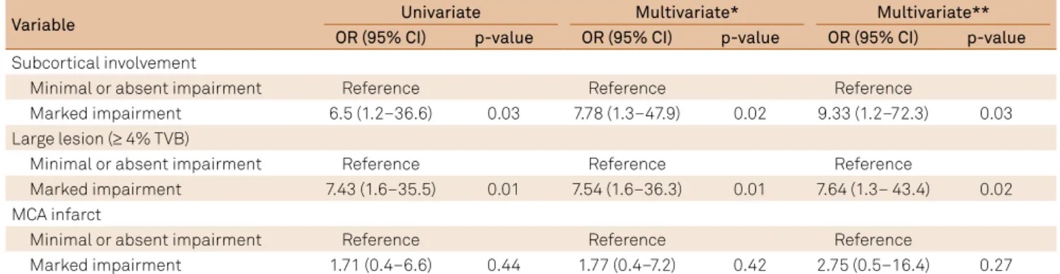
After adjusting for age, arterial distribution and lesion size,
a signiicantly larger proportion of children with marked func
-tional impairment displayed subcortical infarcts (OR 8.36; CI
1.76–53.6; p 0.025) on MRI, compared to the group of survivors
without marked impairment. We also found a positive corre-lation between the lesion size and adverse outcome (OR 9.92;
CI 1.76–55.9; p 0.009). Table 2 summarizes the associations
between radiological variables and the outcomes.
DISCUSSION
he primary goal of this study was to explore whether the
lesion location could predict the occurrence of an adverse functional outcome in pediatric patients beyond the
new-born period following a irst AIS; in this regard, the main ind
-ing of our study was that in this cohort of Chilean children
with an isolated supratentorial infarct, there was an associa-tion between the subcortical locaassocia-tion of the stroke and the presence and severity of long-term functional impairment.
Research in children with perinatal AIS has documented the relationship between basal ganglia and thalamic injury and altered cognitive outcome17, but ours is the irst study
that strongly associates subcortical lesions with functional
impairment in older children. his inding is in contrast to
previous studies, which showed that combined cortical and
Table 1. Demographics, identiied etiologies, and radiological features of children with arterial ischemic stroke with and without an isolated subcortical infarct (Pontiicia Universidad Católica of Chile’s Clinical Hospital: 2003 - 2012).
Variables With subcortical infarct Without subcortical infarct p-value
Overall, n (%) 26 (67) 13 (33)
Demographics and treatment
Female, n (%) 11 (42.3) 3 (23.1) 0.304
Male, n (%) 15 (57.7) 10 (76.9 0.304
Age in years, mean (SD) 6.16 (5.37) 3.83 (4.20) 0.148
Time of follow-up, mean (SD) 4.95 (2.69) 4.93 (1.98) 0.982
Anticoagulant treatment, n (%) 19 (73.1) 10 (76.9) 0.999
Clinical presentation
GCS < 12, n (%) 12 (46.2) 8 (61.5) 0.501
Headache, n (%) 5 (19.2) 2 (15.4) 1.000
Acute seizures, n (%) 9 (34.6) 5 (38.5) 1.000
Focal neurologic deicits, n (%) 20 (76.9) 9 (69.2) 0.704
Stroke etiology
Cardioembolic, n (%) 11 (42.3) 6 (46.2) 1.000
Arteriopathy, n (%) 5 (19.2) 2 (15.4) 1.000
Thrombophilia*, n (%) 2 (7.7) 1 (7.7) 1.000
Other etiology**, n (%) 3 (11.5) 3 (23.1) 0.380
Multifactorial, n (%) 3 (11.5) 0 (0) 0.538
Undetermined, n (%) 5 (19.2) 2 (15.4) 1.000
Stroke-related radiological variables
Right sided, n (%) 15 (57.7) 7 (53.8) 1.000
ACA territory, n (%) 11 (42.3) 3 (23.1) 0.304
MCA territory, n (%) 11 (42.3) 7 (53.8) 0.520
PCA territory, n (%) 4 (15.4) 3 (23.1) 0.666
Large lesion (≥ 4% TBV), n (%) 17 (65.4) 6 (46.2) 0.312
Stroke recurrence, n (%) 3 (13) 1 (8.3) 1.000
subcortical lesions are signiicantly more likely to result in
cognitive impairment in the long term compared to isolated cortical or subcortical infarctions18,19. One study even
associ-ated the presence of isolassoci-ated subcortical lesions with a favor-able outcome20. Although the study criteria could explain
these diferences, the leading cause of these results may lie
in the size of the stroke, since combined lesions are likely to
be the largest, as well. Consistent with research in pediatric
populations, our results showed that patients who had large infarcts were much more likely to develop a long-term
dis-ability. hus, we performed a logistic regression analysis to
adjust for confounding variables, including stroke size, allow-ing us to identify independent predictors of the outcome. In
the results section, we showed that there were no signiicant diferences between children with or without subcortical
involvement with respect to age, sex, underlying conditions, initial clinical manifestations or other radiological features.
he role of the subcortical nuclei in motor regulation and
cognitive functions has been well established21. Subcortical
pathways reciprocally interconnect neuronal activity of an important and diverse set of cerebral cortical areas with the basal ganglia and thalamus. It is noteworthy that the func-tional organization of these circuits changes across devel-opment, allowing the improvement of motor and cognitive behaviors22. However, the molecular mechanism implicated
in these changes is particularly vulnerable to injury in energy failure23. hus, impaired neural plasticity due to ischemia in
the developing brain, particularly during critical periods, may be the pathophysiology basis of functional deterioration in these patients.
It is a unique characteristic of our study that we evalu-ated stroke features in a South American pediatric popu-lation and their impact on long-term functional outcomes.
he results are characterized by a high prevalence of poor
outcomes following pediatric AIS; these data are consis-tent with those of previous studies that included other geo-graphic groups2,18,24,25.
he limitations of this study must be considered when
reviewing these results. Firstly, this is a single center study
with a relatively small sample size. he prevalence of adverse outcomes found in our cohort may not accurately relect our
country’s statistics; thus, future research should include ter
-tiary centers across several regions to obtain results that may be generalized. Furthermore, our study population included many children with heart diseases and relatively few with
CNS arteriopathies; this was probably because the study was
conducted in a referral center for the surgical resolution of complex congenital heart diseases, as well as because there was a relative lack of vascular imaging studies undertaken at the time of diagnosis.
he strengths of the study lie in its clear selection crite
-ria, controlled for confounding variables, prospective
recruit-ment, and uniform evaluation. his study has also contrib
-uted to an increase in the understanding of the long-term outcomes of stroke in children. Finally, the scale used to assess functional outcome does not include any direct mea-sure of cognitive function or self-perceived quality of life; thus, we believe that future research focused on these out-comes may result in the development of a risk scoring system for the development of neurologic disability following an AIS.
In conclusion, early identiication of children at increased
risk for impairment would allow for early interventions and could be useful in reducing permanent disability. In this regard, the size and subcortical involvement of the infarcts are potential predictors of adverse outcome following an iso-lated supratentorial stroke.
Acknowledgment
he authors thank Dr. Isidro Huete, Dr. Escobar–Henríquez (Raúl), Dr. Mesa–Latorre (Tomás), Dr. Núñez–Farias (Alicia), Dr. Beytía–Reyes (María), Dr. Acevedo–Gallinato (Keryma), Dr. Ávila–Smirnov (Daniela), Dr. Samsó–Zepeda (Catalina), and Dr. Arriaza–Ortiz (Manuel) for their valuable contribution to patient recruitment, and Pontiical University Catholic of
Chile’s Clinical Hospital staf and participants for their impor
-tant contributions to this work.
Table 2. Effect of stroke-related radiological features on long-term functional outcome.
Variable Univariate Multivariate* Multivariate**
OR (95% CI) p-value OR (95% CI) p-value OR (95% CI) p-value Subcortical involvement
Minimal or absent impairment Reference Reference Reference
Marked impairment 6.5 (1.2–36.6) 0.03 7.78 (1.3–47.9) 0.02 9.33 (1.2–72.3) 0.03
Large lesion (≥ 4% TVB)
Minimal or absent impairment Reference Reference Reference
Marked impairment 7.43 (1.6–35.5) 0.01 7.54 (1.6–36.3) 0.01 7.64 (1.3– 43.4) 0.02
MCA infarct
Minimal or absent impairment Reference Reference Reference
Marked impairment 1.71 (0.4–6.6) 0.44 1.77 (0.4–7.2) 0.42 2.75 (0.5–16.4) 0.27
References
1. Agrawal N, Johnston SC, Wu YW, Sidney S, Fullerton HJ. Imaging data reveal a higher pediatric stroke incidence than prior Us estimates. Stroke. 2009;40(11):3415-21. https://doi.org/10.1161/STROKEAHA.109.564633
2. Veber GA, MacGregor D, Curtis R, Mayank S. Neurologic outcome in survivors of childhood arterial ischemic stroke and sinovenous thrombosis. J Child Neurol. 2000;15(5):316-24. https://doi.org/10.1177/088307380001500508
3. Ganesan V, Hogan A, Shack N, Gordon A, Isaacs E, Kirkham FJ. Outcome after ischaemic stroke in childhood. Dev Med Child Neurol. 2000;42(7):455-61. https://doi.org/10.1017/S0012162200000852
4. De Schryver EL, Kappelle LJ, Jennekens-Schinkel A, Boudewyn Peters AC. Prognosis of ischemic stroke in childhood: a long-term follow-up study. Dev Med Child Neurol. 2000;42(5):313-8. https://doi.org/10.1017/S0012162200000554
5. Goeggel Simonetti B, Cavelti A., Arnold M., Bigi S, Regényi M, Mattle HP et al. Long-term outcome after arterial ischemic stroke in children and young adults. Neurology. 2015;84(19):1941-7. https://doi.org/10.1212/WNL.0000000000001555
6. Fullerton HJ, Elkins JS, Johnston SC. Pediatric Stroke Belt: geographic variation in stroke mortality in US children. Stroke. 2004;35(7):1570-3. https://doi.org/10.1161/01.STR.0000130514.21773.95
7. Elbers J, Veber G, Pontigon AM, Moharir M. Long-term outcomes of pediatric ischemic stroke in adulthood. J Child Neurol. 2014;29(6):782-8. https://doi.org/10.1177/0883073813484358
8. Greenham M, Gordon A, Anderson V, Mackay MT. Outcome in childhood stroke. Stroke. 2016;47(4):1159-64. https://doi.org/10.1161/STROKEAHA.115.011622
9. Boardman JP, Ganesan V, Rutherford MA, Saunders DE, Mercuri E, Cowan F. Magnetic resonance image correlates of hemiparesis after neonatal and childhood middle cerebral artery stroke. Pediatrics. 2005;115(2):321-6. https://doi.org/10.1542/peds.2004-0427
10. Westmacott R, Askalan R, MacGregor D, Anderson P, Deveber G. Cognitive outcome following unilateral arterial ischaemic stroke in childhood: effects of age at stroke and lesion location. Dev Med Child Neurol. 2010;52(4):386-93. https://doi.org/10.1111/j.1469-8749.2009.03403.x
11. Ganesan V, Ng V, Chong WK, Kirkham FJ, Connelly A. Lesion volume, lesion location, and outcome after middle cerebral artery territory stroke. Arch Dis Child. 1999;81(4):295-300. https://doi.org/10.1136/adc.81.4.295
12. Cnossen MH, Aarsen FK, Akker SL, Danen R, Appel IM,
Steyerberg EW, et al. Paediatric arterial ischaemic stroke: functional outcome and risk factors. Dev Med Child Neurol. 2010;52(4):394-9. https://doi.org/10.1111/j.1469-8749.2009.03580.x
13. Grelli KN, Gindville MC, Walker CH, Jordan LC. Association of blood pressure, blood glucose, and temperature with neurological outcome after childhood stroke. JAMA Neurol. 2016;73(7):829-35. https://doi.org/10.1001/jamaneurol.2016.0992
14. Sébire G, Fullerton H, Riou E, Veber G. Toward the deinition of cerebral arteriopathies of childhood. Curr Opin Pediatr. 2004;16(6):617-22. https://doi.org/10.1097/01.mop.0000144441.29899.20
15. Ichord RN, Benedict SL, Chan AK, Kirkham FJ, Nowak-Göttl U. Paediatric cerebral sinovenous thrombosis: Findings of the International Paediatric Stroke Study. Arch Dis Child.
2015;100(2):174-9. https://doi.org/10.1136/archdischild-2014-306382
16. Bigi S, Fischer U, Wehrli E, Mattle HP, Boltshauser E, Bürki S et al. Acute ischemic stroke in children versus young adults. Ann Neurol. 2011;70(2):245-54. https://doi.org/10.1002/ana.22427
17. Buuren LM, Aa NE, Dekker HC, Vermeulen RJ,
Nieuwenhuizen O, Schooneveld MM et al. Cognitive outcome in childhood after unilateral perinatal brain injury. Dev Med Child Neurol. 2013;55(10):934-40. https://doi.org/10.1111/dmcn.12187
18. Steinlin M, Roellin K, Schroth G. Long-term follow-up after stroke in childhood. Eur J Pediatr. 2004;163(4-5):245-50. https://doi.org/10.1007/s00431-003-1357-x
19. Studer M, Boltshauser E, Capone Mori A, Datta A,
Fluss J, Mercati D, et al. Factors affecting cognitive outcome in early pediatric stroke. Neurology. 2014;82(9):784-92. https://doi.org/10.1212/WNL.0000000000000162
20. Gordon AL, Ganesan V, Towell A, Kirkham FJ. Functional outcome following stroke in children. J Child Neurol. 2002;17(6):429-34. https://doi.org/10.1177/088307380201700606
21. Schroll H, Hamker FH. Computational models of basal-ganglia pathway functions: focus on functional neuroanatomy. Front Syst Neurosci. 2013;7(DEC):122. https://doi.org/10.3389/fnsys.2013.00122
22. Greene DJ, Laumann TO, Dubis JW, Ihnen SK, Neta M, Power JD et al. Developmental changes in the organization of functional connections between the basal ganglia and cerebral cortex. J Neurosci.
2014;34(17):5842-54. https://doi.org/10.1523/JNEUROSCI.3069-13.2014
23. Johnston MV. Clinical disorders of brain plasticity. Brain Dev. 2004;26(2):73-80. https://doi.org/10.1016/S0387-7604(03)00102-5
24. Christerson S, Strömberg B. Stroke in Swedish children II: long-term outcome. Acta Paediatr. 2010;99(11):1650-6. https://doi.org/10.1111/j.1651-2227.2010.01948.x