Vascular Reactivity Response to Mental Stress in Pregnant
Women with Mitral Stenosis
Walkiria Samuel Avila, Osmar Araújo Calil, Ivani C. Trombetta, Carlos Eduardo Negrão, Max Grinberg, Marcelo Zugaib, José Antonio Franchini Ramires
Instituto do Coração do Hospital das Clínicas – FMUSP - São Paulo, SP - Brazil
M a i l i n g A d d r e s s : W a l k i r i a S a m u e l A v i l a • R u a A p i a c á s , 6 8 8 / 3 1 - 0 5 0 1 7 - 0 2 0 – S ã o P a u l o , S P - B r a z i l
E-mail: [email protected] Received on 11/18/05 • Accepted on 01/12/06
O
BJECTIVETo study vascular reactivity according to the analysis of blood fl ow and peripheral vascular resistance at rest and during mental stress in pregnant women with mitral stenosis.
M
ETHODSTwenty two women with mitral stenosis, 13 of whom were pregnant (PS) and 9 were non-pregnant (MIS), and 9 healthy pregnant women (NP) were studied. During gestation, 9 out of the 13 patients of the PS group required a beta-blocker (PSB) and the remaining 4 progressed without medication (PSWB). Plethysmography at rest and during mental stress analyzed muscle blood fl ow, peripheral vascular resistance (PVR), mean arterial pressure (MAP) and heart rate (HR) during gestation and puerperium.
R
ESULTSDuring gestation of PSWB, muscle blood fl ow and HR were higher in 1.6% and 20.5% (p = 0.05), and PVR and MAP were lower in 19.3% and 4.4%, respectively, in comparison to the puerperium; during mental stress, the muscle blood fl ow increased by 55.9%, HR decreased by 30.2% and PVR and MAP were similar. In PSB, muscle blood muscle blood fl ow and HR were greater in 5.9% and 14.9% (p= 0.001) and MAP and PVR were lower in 10.3% and 9.1%, respectively, when compared to the puerperium. During mental stress, muscle blood fl ow and MAP increased by 69.8% and 174.1%, respectively. HR was similar and PVR decreased by 53.7%. The comparative study showed that in the NP group the muscle blood fl ow was higher, PVR was lower, and MAP and HR were similar in relation to the PS group, and that the PS, NP, MIS groups had a similar response to mental stress.
C
ONCLUSIONSVascular reactivity in pregnant women with mitral stenosis was preserved and the analysis of measurements showed lower values of muscle blood fl ow and higher values of PVR when compared to those of healthy pregnant women.
K
EY WORDSThe cycle of pregnancy and puerperium provokes signifi cant and transient cardiocirculatory changes1 in
women’s bodies. As of the seventh day of fertilization2,
adaptations in cardiac output and peripheral vascular resistance to metabolic and hormonal demands and to uteroplacental circulation occur in order to maintain an adequate fetal development.
In light of current knowledge, variations in peripheral vascular resistance (PVR) during pregnancy are not caused only by alterations in the uterine plexus, but also by hormones such as estrogen, prolactin, and circulating prostaglandins, which are responsible for the reduction in vascular response to angiotensin3.
A decrease in the synthesis of prostaglandins or an increase in their metabolism is also known to result in an increase in vascular reactivity to angiotensin II4, a characteristic that is observed, for instance, in
hypertensive syndromes during pregnancy5.
Another clinical situation that has aroused interest is the study of vascular reactivity at rest and during stress in the identifi cation of prognostic markers in patients with congestive heart failure (CHF). In a controlled study of forearm blood fl ow using a plethysmograph, and of muscular reactivity using electromyography during mental stress in patients with CHF, Middlekauff et al6 observed
a signifi cant increase in the sympathetic activity both at rest and during mental stress when compared to healthy volunteers, as well as a reduction of PVR in both groups, although more intense in the control group.
Since we are aware of these data, it would be desirable to apply them to patients with heart diseases, given the differences in the phases of the natural history of heart diseases and PVR adaptations during pregnancy. The literature, however, lacks studies on the behavior of PVR in pregnant women with heart diseases, with emphasis on mitral stenosis, which accounts for most of the heart valve diseases in women of childbearing age and is associated with high percentages – that range from 5% to 30% - of cardiovascular complications7,8, among which pulmonary
congestion is the major cause of mortality.
Given the lack of knowledge on hemodynamic parameters that could correlate with the progression and prognosis of pregnant women with mitral stenosis, we chose the study on the behavior of PVR to collect information that could be applied in the daily clinical practice.
O
BJECTIVETo study the vascular response in pregnant women with mitral stenosis by analyzing muscle blood fl ow and PVR at rest and during mental stress.
M
ETHODSThirty one women of childbearing age followed at
the outpatient service of InCor (Instituto do Coração) were prospectively studied. They were divided into three groups: PS group composed of thirteen pregnant women with isolated mitral stenosis; NP group composed of nine normal pregnant women; and MIS group composed of nine non-pregnant women with isolated mitral stenosis.
All patients were in functional class (FC) I/II, according to the New York Heart Association (NYHA), and had a sinus rhythm recorded by the electrocardiogram (ECG). Those with mitral stenosis used penicillin G benzathine at prophylactic dose for rheumatic disease and had a mitral valve area lower than 1.5 cm2, as estimated by Doppler
echocardiogram (ECHO). In the NP and PS groups, the study started between sixteen and 24 weeks of gestation. All received thorough information on the protocol and gave their written informed consent before the beginning of the study. The study was approved by the Ethics Committee for Analysis of Research Projects (CAPPesq) of the Clinical Board of Hospital das Clínicas and of Faculdade de Medicina da Universidade de São Paulo .
Theexclusion criteria were: age under eighteen or above forty years; NYHA-FC IV; atrial fi brillation, concomitant structural heart lesion; past history of thromboembolism, peripheral vascular disease or pulmonary disease; high blood pressure, diabetes mellitus and smoking.
Clinical protocol -The patients underwent a monthly medical assessment when the NYHA-FC was analyzed, at the third trimester of pregnancy and at the puerperium in the groups of pregnant women, and at the first medical visit in the MIS group. The following events were considered clinical endpoints: congestive heart failure, thromboembolic accident, cardiac arrhythmia, and infective endocarditis. Electrocardiograms and Doppler echocardiograms were performed at admission to the protocol with the purpose of studying changes in heart rhythm and measurements of mitral valve area (MVA), calculated by the pressure half-time method.
Hemodynamic protocol - The hemodynamic parameters studied were: forearm muscle blood fl ow and peripheral vascular resistance, mean arterial pressure and heart rate recorded at restand during mental stress and in two stages for the PS and NP groups, that is, between the 28th and 32nd weeks of gestation and in puerperium, and, for the MIS group, only at admission to the protocol.
calculated using the ratio between mean arterial pressure (mmHg) and muscle blood fl ow (ml/min/100 ml) in the forearm, as follows: PVR = MAP (mmHg) / Blood Flow (ml/min/100 ml).
During plethysmography a mental stress test was performed with colors (Stroop color-word test), in which the participant should name out loud and fast the color of words that are printed in incompatible colors. This procedure was performed for 3 min of resting period (baseline), and followed by 4 min of color test (stress). At the end, each participant was asked about the level of diffi culty of the color test, according to the levels of diffi culty previously established: 0 = not stressing; 1 = little stressing; 2 = stressing; 3 = very stressing; and 4 = extremely stressing.
Mental stress (MS) behavior was assessed by the difference between the mean of the baseline phase and the value obtained at the fi rst minute of mental stress, considered in our study as the test peak value: MS = Mean of Baseline (MB) / fi rst moment of mental stress (M1).
Systolic blood pressures (SBP), diastolic blood pressures (DBP) and mean (MBP) were continuously measured during the test with a cuff connected to an Ohmed 2300 Finapress monitor, and the heart rate (HR) was obtained in a computer-recorded electrocardiographic tracing, all analyzed in specifi c programs (AT/CODAS).
Statistical analysis - Calculation of means and standard deviation, analysis of variance (ANOVA) for the comparison between groups (PS, NP, MIS) and phases (pregnancy and puerperium), analysis of covariance (ANCOVA) for the comparison of stress behavior parameters (mean of baseline minus valueat the fi rst moment of stress) for the following factors: groups, phases and their interaction, and further independent t- tests, and paired t-test for the comparison between group differences (two by two) and between behavior differences during pregnancy and puerperium were used, respectively. The SPSS - Statistical Package Social Science Version 8.0 (1997) was used in these analyses, at a signifi cance
level of 0.05.
R
ESULTSCase series analysis - During gestation, nine out of the thirteen patients of the PS group had therapeutic indication for beta-blocker due to deterioration of FC; therefore, the PS group was subdivided into two distinct subgroups: PSB composed of nine pregnant women on beta-blocker (propranolol at a daily dose ≤ 80 mg), and PSWB composed of four pregnant women not receiving a beta-blocker.
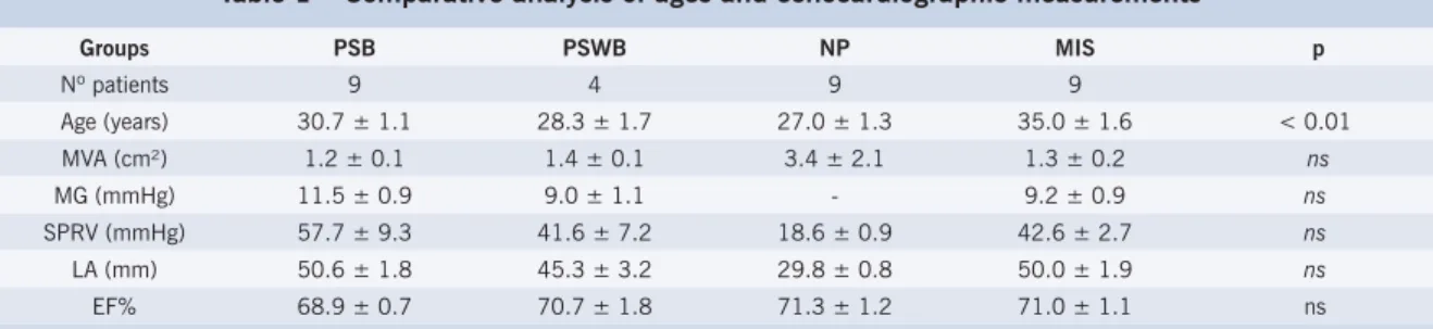
The description and comparison between groups regarding ages and echocardiographic measurements taken at the beginning of the study showed that age in the MIS group was higher than in the NP (Table 1). The analysis of NYHA-FC verifi ed that the nine pregnant women of the NP group remained in FC I/II during gestation and puerperium; among the nine patients of the PSB subgroup, seven progressed to FC III during the study and one patient of the MIS group, the remaining of the PSWB subgroup and of the MIS group remained in FC I/II throughout the study. The statistical analysis between echocardiographic variables was performed only between the groups with mitral stenosis, that is, PS and MIS. Complications such as thromboembolism, infective endocarditis or pulmonary congestion were not recorded during the study.
Comparative analysis of hemodynamic parameters in the groups and subgroups at rest and during stress
Group of normal pregnant women (between gestation and puerperium) - At rest, a signifi cant difference was observed for the following variables: muscle blood fl ow – which increased by 21.3% (p = 0.003); PVR – which decreased by 31.3% (p =0.023), and HR – which increased by 13.1% (p = 0.018) during gestation, whereas MAPremained similar in the two study phases (Table 2). During the stress phase, an increase by 34.4% (p = 0.489) and 348% (p = 0.048) in muscle blood fl ow and MAP, respectively, was verifi ed during gestation, whereas a reduction in PVR by 42.9% (p = 0.280) was observed during puerperium, and HR was similar in both study phases (Table 2).
Table 1 – Comparative analysis of ages and echocardiographic measurements
Groups PSB PSWB NP MIS p
Nº patients 9 4 9 9
Age (years) 30.7 ± 1.1 28.3 ± 1.7 27.0 ± 1.3 35.0 ± 1.6 < 0.01
MVA (cm²) 1.2 ± 0.1 1.4 ± 0.1 3.4 ± 2.1 1.3 ± 0.2 ns
MG (mmHg) 11.5 ± 0.9 9.0 ± 1.1 - 9.2 ± 0.9 ns
SPRV (mmHg) 57.7 ± 9.3 41.6 ± 7.2 18.6 ± 0.9 42.6 ± 2.7 ns
LA (mm) 50.6 ± 1.8 45.3 ± 3.2 29.8 ± 0.8 50.0 ± 1.9 ns
EF% 68.9 ± 0.7 70.7 ± 1.8 71.3 ± 1.2 71.0 ± 1.1 ns
Table 2 – Comparison of hemodynamic parameters of the NP group during pregnancy and puerperitum at rest and during stress
Parameters Pregnancy Puerperium % variation p value
At rest
Flow 2.86 ± 0.12 2.25 ± 0.18 21.3 ↑ 0.003
PVR 30.89 ± 2.09 40.55 ± 4.33 31.3 ↓ 0.023
MAP 85.92 ± 3.78 86.04 ± 4.59 0.1 ↓ 0.750
HR 84.23 ± 3.28 73.19 ± 3.34 13.1 ↑ 0.018
During stress
Flow 1.35 1.07 34.4 ↑ 0.489
PVR -5.91 -11.35 42.9 ↑ 0.280
MAP 12.23 2.38 348.0 ↑ 0.048
HR 6.52 6.25 8.4 ↑ 0.917
↑ increase and ↓ reduction, during pregnancy in relation to puerperium; p- statistical signifi cance value; Flow- expressed in ml/min/100 ml; MAP- mean blood pressure in mmHg; HR- heart rate in beats per minute; PVR- peripheral vascular resistance in mmHg/ml/minute/100 ml.
Table 3 – Comparison of parameters of the PSWB and PSB subgroups during pregnancy and puerperium at rest and during stress
Phase Pregnancy Puerperium % variation p value
PSWB PSB PSWB PSB PSWB PSB PSWB PSB
At rest
Flow 2.3 ± 0.5 2.2 ± 0.3 2.2 ± 0.5 2.0 ± 0.2 1.6 ↑ 5.9 ↓ 0.866 0.857
PVR 39.7 ± 4.6 43.1 ± 5.6 47.4 ± 15.6 46.9 ± 6.8 19.4 ↑ 9.1 ↓ 0.647 0.922
MAP 82.5 ± 4.1 81.6 ± 5.2 86.1 ± 4.6 90.0 ± 2.8 4.4 ↑ 10.4 ↑ 0.119 0.531
HR 86.6 ± 2.8 81.6 ± 4.4 68.8 ± 6.4 71.0 ± 5.2 20.6 ↓ 14.9 ↓ 0.057 0.001
Stress
Flow 1.61 1.07 0.71 0.63 55.9 ↑ 69.8 ↓ 0.118 0.436
PVR - 12.07 - 13.23 - 11.91 - 8.61 1.3 ↓ 53.7 ↓ 0.979 0.576
MAP 6.44 4.33 6.10 1.58 5.3 ↓ 174.1 ↓ 0.753 0.571
HR 8.27 2.71 10.7 2.60 30.2 ↑ 4.2 ↓ 0.228 0.847
↑ increase and ↓ reduction. during pregnancy in relation to puerperium; p- statistical signifi cance value; Flow: expressed in ml/min/100 ml; MAP-
mean arterial pressure in mmHg; HR- heart rate in beats per minute; PVR- peripheral vascular resistance in mmHg/ml/minute/100 ml.
Table 4 – Measurements taken at rest and during stress in the MIS group
Group Flow
(ml/min/100ml)
PVR (mmHg/ml/min/100ml)
MAP (mmHg)
HR (btm)
Baseline 2.39 ± 0.23 39.45 ± 3.00 88.74 ± 4.03 73.84 ± 2.46
M1 4.19 ± 0.55 24.41 ± 2.82 92.16 ± 4.73 88.97 ± 4.70
Stress 1.80 -15.04 3.42 15.13
% variation ↑ 75.31 ↓ 38.12 ↑ 3.85 ↑ 20.49
p value 0.001 0.0001 0.011 0.001
Stress- (Baseline-M1); M1- fi rst minute stress; Baseline- mean of three minutes of stress %l = stress percentage; p- signifi cance.
Subgroup of mitral stenosis without use of beta-blocker (gestation and puerperium)- At rest, the muscle blood fl ow increased by 1.6% (p = 0.866), and HR by 20.5% (p = 057), whereas PVR and MAP had a reduction by 19.3% (p = 0.647) and 4.4% (p = 0.119), respectively, in relation to the puerperium. During stress, there was an increase in muscle blood fl ow by 55.9% (p = 0.118) and HR by 30.2% (p = 0.228), and PVR and MAP were similar in both study phases (Table 3).
Subgroup of mitral stenosis on beta-blocker (gestation and puerperium) - at rest, the muscle blood fl ow increased by 5.9% (p = 0.857), and MAP and PVR decreased
by 10.3% (0.531) and 9.12% (0.922), respectively, during gestation; however, a signifi cant increase in HR by 14.9% (p = 0.001) was observed during gestation. During stress, the muscle blood fl ow increased by 69.8% (p = 0.436), HR was similar, and PVR decreased by 53.7% (p = 0.576), and MAP increased by 174.1% (p = 0.571) (Table 3).
Comparative analysis of hemodynamic parameters between groups and subgroups
Between subgroups of pregnant women with mitral stenosis on beta-blocker (PSB) or not (PSWB)- At rest, similar results of muscle blood fl ow, PVR, and MAP variables, and a mild increase in HR in the PSWB group (p = 0.490) were observed during gestation, whereas the subgroups were similar during puerperium. During stress, an increase in HR (p = 0.490) was observed in PSWB, however it was not signifi cant (Table 3).
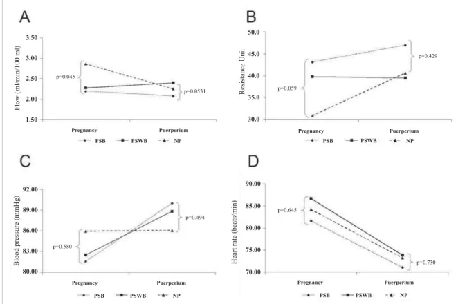
Between the NP group and PSB and PSWB subgroups - At rest, the increase in muscle blood fl ow was higher in the NP group (p = 0.045) in relation to the two subgroups during gestation (Figure 1A); a reduction in PVR, more signifi cant in the NPgroup (p = 0.059) was observed (Figure 1B); and no difference was observed in MAP and HR between the groups and subgroups studied (Figures 1C and 1D). In puerperium, the groups and subgroups were similar as regards the parameters analyzed (Figure 1A, 1B, 1C, 1D).
Between the NP and MIS groups and PSWB and PSB subgroups - (puerperium data) the groups were similar both at rest and during stress for muscle blood fl ow (Figure 2A), PVR (Figure 2B), MAP (Figure 2C), and HR (Figure 2D) parameters. The analysis of covariance
of the parameters in the fi rst phase of mental stress compared with those at rest in the NP group and PSWB and PSB subgroups in gestation and puerperium phases showed a signifi cant difference as regards muscle blood fl ow (p = 0.006), PVR (p = 0.003); MAP (p < 0.001) and HR (p < 0.001).
D
ISCUSSIONThe improvement of knowledge on the physiological aspects of pregnancy and their interaction with the hemodynamic behavior of heart diseases has contributed to the reduction in maternal morbidity and mortality during the cycle of pregnancy and puerperium.
In mitral stenosis, the alarming percentage of 86% of deterioration from class I/II to class III/IV during the second and third trimesters of gestation, as reported by Ávila et al7, shows the clinical importance imposed by
cardiocirculatory overload resulting from hemodynamic alterations during pregnancy.
These data clearly show that pregnancy is a potential factor for clinical deterioration of the mitral lesion during its natural history. Thus, the identifi cation of prognostic variables such as age, functional class, atrial fi brilation, CHF, acute pulmonary edema, pulmonary hypertension, right ventricular overload, and valve area has contributed to the risk stratifi cation of pregnant women with mitral stenosis8,9.
Fig. 1 – Comparison of variables between pregnancy and puerperium at rest, between the PSB and PSWB subgroups and NP group. MS - mental stress; PSB- pregnant woman with mitral stenosis using beta-blocker; PSWB- pregnant woman with mitral stenosis not using beta-blocker, and NP- normal pregnant woman; Flow- ml/min/100 ml; MAP mmHg; PVR mmHg/ml/minute/100; HR- beats/minute.
3.50
3.00
2.50
2.00
1.50
Flow (ml/min/100 ml)
p=0.045
p=0.0531
Pregnancy Puerperium
PSB PSWB NP
50.0
45.0
40.0
35.0
30.0
Resistance Unit p=0.059
p=0.429
Pregnancy Puerperium
PSB PSWB NP
92.00
89.00
86.00
83.00
80.00
Blood pressure (mmHg)
p=0.580
p=0.494
Pregnancy Puerperium
PSB PSWB NP
90.00
85.00
80.00
75.00
70.00
Heart rate (beats/min)
p=0.645
p=0.730
Pregnancy Puerperium
The continuing search for prognostic factors encouraged us to study hemodynamic parameters during pregnancy as compared to post-partum in women with mitral stenosis, with the objective of obtaining clinical and laboratorial support, for a better knowledge of possible complications in this group of pregnant women.
Thus, we included the group of normal pregnant women (NP) and of women with mitral stenosis (MIS) with the purpose of giving support to the analysis of the method used and of the gestation component in the binomial heart disease and pregnancy.
This study considered a period of clinical course - thus subject to therapeutic decisions - and not only a moment of study performance. Thus, the treatment dynamics of each case cannot be predicted in a prospective protocol and its application determined the subdivision of the PS group into two subgroups according to the need or no need of beta-blockers, due to deterioration of the clinical course and/or severity of the valve lesion.
In fact, we observed that seven out of the nine patients in the subgroup of pregnant women with mitral stenosis who required beta-blocker (PSB) progressed to FC III and had a mean valve area of 1.2cm2. These data are
consistent with those of Ávila et al7 study, that signifi cantly
showed a distinct progression of gestation at these limits, that is: mitral valve area greater than 1.2 cm for FC III and lower than this value for FC IV.
The literature lacks studies on the hemodynamic behavior of mitral stenosis and its correlation with clinical aspects during pregnancy. The use of invasive methods available in practice limited the acquisition of knowledge on the behavior of peripheral vascular resistance and muscle blood fl ow and their associations with diseases during pregnancy.
However, non-invasive methods have now been broadly used. Ultrasonography and the venous occlusion plethysmography method are easy and quick to perform at a low cost and have contributed to the study of the endothelial function and vascular reactivity in different clinical situations, including pregnancy10,11.
Edouard et al12, using the plethysmograph, verifi ed
variations in venous tone and blood viscosity of the lower extremities in nine normal pregnant women, when the fi rst and the third trimesters of gestation were compared to the period of three to six months after delivery.
In the present study, the 31% reduction in PVR of normal pregnant women (NP) in relation to the puerperium is consistent with the data verifi ed by Clark et al13, who
used the thermodilution technique and pulmonary artery catheterization in ten normal primiparous pregnant women and observed a 21% reduction in PVR in the third trimester of gestation, when compared with the period between the 11th and 13th week after delivery.
Fig. 2 – Comparison of variables at rest and during stress between the PS, MIS and NP groups in puerperium. MS - mental stress; PS - pregnant woman with mitral stenosis; MIS - non-pregnant woman with mitral stenosis; and NP - normal pregnant woman; Flow- ml/min/100 ml; MAP mmHg; PVR mmHg/ml/minute/100; HR- beats/minute.
5.00
4.00
3.00
2.00
1.00
0,00
Flow (ml/min/100 ml)
p=0.590
p=0.113
Baseline
Interaction
PS MS NP
MIS
p=0.000
Baseline
PS MS NP
MIS
Interaction p=0.000
Resistance Unit
50.00
45.00
40.00
35.00
30.00
25,00
20,00
p=0.499
p=0.112
Baseline
PS MS NP
MIS
Interaction p=0.000
Blood pressure (mmHg)
94.00
92.00
90.00
88.00
86,00
84,00
p=0.786
p=0.807
Baseline
PS MS NP
MIS
Interaction p=0.000
Heart rate (beats/min)
p=0.858
p=0.155
90.00
85.00
80.00
75.00
70,00
The mechanisms of arteriolar dilation during pregnancy have been attributed to estrogenic components3,4, to
prolactin and to the increase in circulating prostaglandin levels (PGE2 and PGI2), a substance that is responsible for the reduction in vascular response to angiotension. Recently, alterations in vascular tone during pregnancy have been shown to be possibly attributed, in part, to changes in the synthesis of vasoactive substances derived from the endothelium, especially the endothelin which is theoretically able to mediate the synthesis of prostaglandins and the nitric oxid which has been related to vasodilation during pregnancy14-16.
PVR also decreased by 19% and 9% during pregnancy of the subgroups with mitral stenosis (PSWB and PSB), respectively, not very differently from the puerperium; however, the values were lower than the percentage of 31% of normal pregnant women. This different PVR behavior at rest in pregnant women with mitral stenosis may be attributed to an abnormal state of vascular response at rest due to the low cardiac output or to an inadequate response to mechanisms of vasodilation during pregnancy.
Similar data were published by Sakai et al17, who,
using a plethysmograph, demonstrated that the severity of pre-eclampsia in pregnant women with high blood pressure was related to an inadequate vascular response, increase in peripheral vascular resistance and reduction in peripheral vascular muscle blood fl ow.
In face of these results, it is reasonable to assume that diseases such as arterial hypertension or mitral stenosis can interfere with vascular reactivity during pregnancy, predisposing to an abnormal PVR response at rest.
The muscle blood fl ow increase by 21% in normal pregnant women (NP), when compared to the post-partum, corroborates Dorup et al’s data18, who reported
a 26% increase in peripheral muscle blood fl ow in normal pregnant women in the fi rst trimester, and 46% at the end of gestation in relation to non-pregnant women, as assessed by ultrasonography.
The fi nding of a small increase by 1.6% and 5.9% in blood muscle blood fl ow in pregnant women of the subgroups of pregnant women with mitral stenosis (PSB and PSWB respectively), in turn, supports the hypothesis that mitral stenosis may be included in the special group of diseases that modify vascular response to muscle blood fl ow during pregnancy. Vascular muscle blood fl ow reduction was also observed to occur in both subgroups, that is, the one with beta-blocker (PSB) and the other without beta-blocker (PSWB).
The signifi cant increase in HR verifi ed during gestation in relation to puerperium in the groups studied is consistent with previous reports19 that have demonstrated
a progressive increase in HR, from the fourth week of gestation up to term. The initial changes in HR seem to be related to the production of chorionic gonadotrophin,
and the late changes to cardiocirculatory alterations that accompany the fetal and placental development.
HR variation between gestation and puerperium was higher (20.6% versus 14.8%) in the subgroup without beta-blocker (PSWB) as expected; however it was not signifi cant probably due to the limited sample size.
Brooks20, assessing the sensitivity of barorefl exes in
the control of heart rate in the hearts of pregnant rabbits, evidenced an increase in baseline sympathetic activity during gestation and, additionally, a reduced action of the parasympathetic nervous system on the heart. Anyway, the similarity of HR between the group of normal pregnant women (NP) and the subgroup (PSB) that had more severe characteristics of mitral stenosis in our case series corroborates the benefi t of beta-blockers in mitral stenosis for the control of HR in pregnancy.
Although non-signifi cant, MAP had a tendency to a reduction during pregnancy in the groups studied when compared to puerperium, consistent with longitudinal studies that have verifi ed a decrease in systemic blood pressure from the beginning to the half time of gestation21,
particularly at the expense of diastolic pressure and further elevation close to pre-gestational values by term.
Rubler et al22 did not verify signifi cant differences in
MAP between pregnant and non-pregnant women at several gestational ages. Controversy on MAP variations during pregnancy may be related to variations in manual measurements in which BP may be underestimated because of the absence of the fi fth Korotkoff sound resulting from the hyperkinetic state, and also because of infl uences of the patient posture.
The similarity found in hemodynamic parameters at rest between the groups and subgroups of pregnant women in the post-partum and those of the patients with mitral stenosis supports the idea that pregnancy represents a transient risk of hemodynamic maladaptation; however, it seems not to interfere in the natural history of the disease.
Another aspect of the originality of this study was the assessment of the response to mental stress of pregnant women with mitral stenosis. The stress testsstimulated in laboratory may be seen as a strategy for investigation of the infl uence of environmental factors on the development of cardiovascular diseases.
The behavior of vascular reactivity during stress may represent a pathogenic mechanism in the development of diseases, that is, a marker of the autonomic central nervous system in the control of the cardiovascular system, or it may demonstrate the alterations of vascular compliance in individuals prone to diseases in the future.
Atterhog et al23, using mental stress tests (Stroop
thirteen healthy men and twelve asymptomatic men with primary electrographic abnormalities of ventricular repolarization and demonstrated higher variations of plasma levels of noradrenalin and adrenalin, of increase in blood pressure (BP) and HR in the group with electrographic abnormality.
The present study, which is perhaps the pioneer in the utilization of mental stress in mitral valve disease, showed a signifi cant variation in all hemodynamic parameters during the test in the group of non-pregnant women with mitral stenosis (MIS), similar to those recorded in normal individuals24.
This behavior is different from that of patients with heart failure whose situation is characterized by vasoconstriction at rest and abnormal vasodilation response during physical or mental stress.
Negrão et al25 showed that both at rest and during
stress test muscular blood muscle blood flow and refl ex vasodilation response are reduced in patients with heart failure, and that the impaired endothelium-mediated vasodilation may not be the major cause of vasoconstriction at rest, and it should be attributed to other mechanisms, such as alterations in the cholinergic and nitric oxide response.
It is reasonable to hypothesize that the adaptation mechanisms of mitral stenosis do not interfere in peripheral vascular reactivity; that is, the endothelium-dependent sympathetic response, at least in adapted patients, is preserved under stress stimulus unlike in other situations where endothelial injury is present, such as those with low cardiac output due to ventricular dysfunction or atherosclerotic ischemic disease26,27.
In the SOLVD study28, the analysis of noradrenalin
and renin levels showed higher values in the group of symptomatic patients with ventricular dysfunction (ejection
fraction lower than 35%) in relation to the asymptomatic patients, suggesting that the neurohormones with a vasoconstrictor effect predominate in relation to those with vasodilator effect in patients with a poorer prognosis.
During pregnancy, studies on vascular reactivity during mental stress are limited to cases of pre-eclampsia, among which we should point out Woisetschlager’s29, who, when
assessing the changes in blood pressure and elevation in heart rate, demonstrated an exacerbation of vascular reactivity in pregnant women with pre-eclampsia when compared to normal pregnant women.
In the present study, the similarity between vascular reactivity during stress in gestation and in the post-partum – a period that is not under the infl uence of gestational hormones, shows that sympathetic and endothelium-dependent responses are preserved both in normal women and in those with mitral stenosis, whether pregnant or not.
The limitation of this study is due to the small number of patients in the subgroups of pregnant women with mitral stenosis according to the need for treatment with a beta-blocker. However, the literature lacks original information, particularly concerning the behavior of vascular reactivity in pregnant women with mitral stenosis.
We concluded that the vascular response to mental stress, as measured by plethysmography, is preserved in pregnant women with mitral stenosis. Additionally, we observed higher values of PVR and lower values of blood muscle blood fl ow in comparison with healthy pregnant women.
Acknowledgements
To Dr Maéve Barros Correa, hemodynamicist, for spell checking.
R
EFERENCES1. Ueland K, Metcalfe J. Circulatory changes in pregnancy. Clin Obstet Gynaecol. 1975; 18: 41-8.
2. Cunningham GF, MacDonald CP, Gant FN. The endometrium and uterine decidua. In: Williams Obstetrics. 8th ed. Dallas, Texas, USA:
Appleton & Lange; 1989: 23-37.
3. Gant NF, Daley GL, Chand S, et al. A study of angiotensina II pressor response throughout primigravid pregnancy. J Clin Invest. 1973; 52: 2682-89.
4. Van Assche FA. The role of prostacyclin and tromboxane in pregnancy. Verh K Acad Geneeskd Belg. 1990; 52: 105-25.
5. Roberts JM, Redman CWG. Pre-eclampsia: more than pregnancy induced hypertension. Lancet. 1993; 341: 1447-51.
6. Middlekauff HR, Nuyen AH, Negrão CE, et al. Impact of acute mental stress on sympathetic nerve activity and regional blood flow in advanced heart failure. Circulation. 1997; 96: 1835-42.
7. Avila WS, Grinberg M, Décourt LV. Evolução clínica de mulheres portadoras de estenose mitral na gravidez e puerpério. Arq Bras Cardiol. 1992; 58: 359-62.
8. Silversides CK, Colman JM, Sermer M, Siu SC. Cardiac risk in pregnant women with rheumatic mitral stenosis. Am J Cardiol. 2003; 91: 1382-85.
9. Siu SC, Sermer M, Colman JM, e al. Prospective multicenter study of pregnancy outcomes in women with heart disease. Circulation. 2001; 104: 515-28.
10. Steptoe A, Vogele C. Methodology of mental stress testing in cardiovascular research. Circulation. 1991; 83(suppl II): II –14- II-24.
11. Falkner B, Onesti G, Angelakos E T, Fernandes M, Langman C. Cardiovascular response to mental stress in normal adolescents with hypertensive parents. Hypertension. 1979; 1: 23-30.
12. Edouard DA, Pannier BM, London GM, Cuche JL, Safar ME. Venous and arterial behavior during normal pregnancy. Am J Physiol. 1998; 274: H1605-12.
13. Clark SL, Cotton DB, Lee W, et al. Central hemodynamic assessment of normal term pregnancy. Am J Obstet Gynecol. 1989; 161: 1439-42.
15. Sladek SM, Magness RR, Conrad KP. Nitric oxide and pregnancy. Am J Physiol. 1997; 272: R441-63.
16. Williams DJ, Vallance PJ, Neild GH, Spencer JA, Imms FJ. Nitric oxide-mediated vasodilatation in human pregnancy. Am J Physiol. 1997; 272: H748-52.
17. Sakai K, Imaisumi T, Maeda H, Tsukimori K, Takeshita A, Nakano H. Venous distensibility during pregnancy. Comparisons between normal pregnancy and preeclampsia. Hypertension. 1994; 24: 461-66.
18. Dorup I, Skajaa K, Sorensen KE. Normal pregnancy is associated with enhanced endothelium-dependent fl ow-mediated vasodilatation. Am J Physiol. 1999; 276: H821-5.
19. Clapp JF. Maternal heart rate in pregnancy. Am J Obstet Gynecol 1985; 152: 659-62.
20. Brooks E M. Altered heart rate barorrefl ex during pregnancy: role of sympathetic and parasympathetic nervous systems. Am J Physiol 1997; 273: 960-66.
21. Christianson RE. Studies on blood pressure during pregnancy I. Infl uence on parity and age. Am J Obstet Gynecol. 1976; 125: 509-13.
22. Rubler S, Pradodhkumar MD, Pinto ER. Cardiac size and performance during pregnancy: estimates with echocardiography. Clin Obstet Gynecol. 1977; 40: 534-40.
23. Atterhog JH, Eliasson K, Hjemdahl P. Sympathoadrenal and
cardiovascular responses to mental stress, isometric handgrip, and cold pressor test in asymptomatic young men with primary T wave abnormalities in the electrocardiogram. Br Heart J. 1981; 46: 311-9.
24. Harris CW, Edwards JL, Baruch A, Rilley WA, Pusser BE, Rejeski WR, et al. Effects of mental stress on brachial artery fl ow-mediated vasodilatation in healthy normal individuals. Am Heart J. 2000; 139: 405-11.
25. Negrão CE, Hamilton MA, Fonarow GC, Hage A, Morigushi JD, Middekauff HR. Impaired endothelium-mediated vasodilation is not the principal cause of vasoconstriction in heart failure. Am J Physiol Heart Circ Physiol. 2000; 278: H168-74.
26. Katz SD, Biasucci L, Strom JA, et al. Impairment of endothelial-mediated vasodilatation in the peripheral vasculature of patients with congestive heart failure. J Am Coll Cardiol. 1992; 19: 918-25.
27. Yeung AC, Vekshein VI, Krantz DS, et al. The effects of atherosclerosis on the vasomotor reponse of coronary arteries to mental stress. N Engl J Méd. 1991; 325: 1551-6.
28. The Solvd Investigators. Effect of enalapril on mortality and development of heart failure in asymptomatic patients with reduced left ventricular ejections fractions. N Engl J Med. 1992; 327: 685-9.