Hemodynamic responses to acute
aortic coarctation in conscious
sinoaortic denervated rats
1Departamento de Fisiologia, Faculdade de Medicina de Ribeirão Preto,
Universidade de São Paulo, Ribeirão Preto, SP, Brasil
2Departamento de Ciências Biológicas, Faculdade de Medicina
do Triângulo Mineiro, Uberaba, MG, Brasil R. Fazan Jr.2,
B.H. Machado1 and
H.C. Salgado1
Abstract
The hemodynamic responses to acute (45 min) partial aortic constric-tion were studied in conscious intact (N = 7) or sinoaortic denervated (SAD) adult male Wistar rats (280-350 g, N = 7) implanted with carotid and femoral arterial catheters, a pneumatic cuff around the abdominal aorta and a pulsed Doppler flow probe to measure changes in aortic resistance. In addition, the hypertensive response and the reflex bradycardia elicited by total (N = 8) vs partial (N = 7) aortic constriction (monitored by maintenance of the pressure distal to the cuff at 50 mmHg) were compared in two other groups of intact rats. Intact rats presented a smaller hypertensive response (26 to 40% above basal level) to partial aortic constriction than SAD rats (38 to 58%). The calculated change in aortic resistance imposed by constriction of the aorta increased progressively only in intact rats, but was signifi-cantly smaller (193 to 306%) than that observed (501 to 591%) in SAD rats. Intact rats showed a significant bradycardia (23 to 26% change in basal heart rate) throughout coarctation, whereas the SAD rats did not (1 to 3%). Partial or total occlusion of the aorta induced similar hypertensive responses (37-38% vs 24-30% for total constriction) as well as reflex bradycardia (-15 to -17% vs -22 to -33%) despite a greater gradient in pressure (97-98 vs 129-140 mmHg) caused by total constriction. The present data indicate that the integrity of the baroreflex in intact rats can cause the hypertensive response to level off at a lower value than in SAD rats despite a progressive increase in aortic resis-tance. In addition, they also indicate that the degree of partial aortic constriction by maintenance of the pressure distal to the cuff at 50 mmHg already elicits a maximal stimulation of the arterial baroreflex.
Correspondence H.C. Salgado
Departamento de Fisiologia FMRP, USP
14040-900, Ribeirão Preto, SP Brasil
E-mail: [email protected]
Research supported by FAPESP, PRONEX, CAPES and CNPq.
Received January 9, 1997 Accepted August 11, 1997
Key words
•Hypertension
•Aortic coarctation
•Doppler flowmetry
•Aortic resistance
•Reflex bradycardia
•Sinoaortic denervation
Introduction
The model of acute aortic coarctation provides remarkable hemodynamic changes such as an increase in carotid pressure, which has been attributed to the sudden increase in the resistance to aortic flow, associated with neurohumoral mechanisms triggered by
a gradual increase in aortic resistance during acute (45 min) aortic constriction as a conse-quence of the overall increase in the imped-ance to blood flow caused by the release of neurohumoral factors (e.g., angiotensin II and vasopressin) triggered by the kidneys (2). In the present study we investigated the hemodynamic responses (changes in carotid pressure, heart rate and aortic resistance to blood flow) of conscious intact and sinoaortic denervated (SAD) rats during the acute (45 min) hypertensive response to partial aortic constriction (pressure distal to the cuff main-tained at 50 mmHg). In addition, we com-pared both the extent of the hypertensive response and the reflex bradycardia obtained with partial vs total occlusion of the aorta in two other groups of intact rats.
Material and Methods
The experiments were performed on con-scious freely moving male Wistar rats (280-350 g) equipped with intra-arterial catheters (carotid and femoral), a pneumatic cuff around the abdominal aorta above the renal arteries, and a miniaturized Doppler flow probe. Two other groups used to compare the hemodynamic responses to partial (N = 7) or total (N = 8) aortic constriction were equipped with vascular catheters and the pneumatic cuff only. Carotid and femoral arterial pressures were recorded continuously with a Statham pressure transducer (Model P23-Db, Hato Rey, PR) attached to a Hewlett-Packard recorder (Model 7848, Palo Alto, CA). The wire leads from the flow probe were connected to an ultrasonic pulsed Dop-pler flowmeter (University of Iowa Bioengi-neering Department, Iowa City, IA) which allows measurement of changes in blood velocity recorded as Doppler shift in kHz. Aortic resistance was calculated as the ratio of mean carotid pressure (MCP) and aortic blood flow Doppler shift and then normal-ized in terms of percentage (7).
Two days before the experiment, under
sodium pentobarbital (40 mg/kg, ip) anes-thesia, both pneumatic cuff and flow probe were placed around the aorta, immediately below the diaphragm, by means of ample laparotomy. This procedure was the same as that used in previous studies (6). Briefly, a 4-6 mm length of abdominal aorta was care-fully isolated below the diaphragm. A pneu-matic cuff, prepared according to a tech-nique developed in our laboratory (8), was placed around the abdominal aorta above the renal arteries, and the miniaturized flow probe secured with 6-0 cotton sutures was placed above the cuff immediately below the dia-phragm. The tubing connected to the pneu-matic cuff and the wires of the Doppler flow probe were tunneled and exteriorized through the animal’s back. Twenty-four hours before the experiment, under ether anesthesia, the animals were submitted to sinoaortic deaf-ferentation (SAD rats, N = 7), according to Krieger’s technique (9), or sham operation (intact rats, N = 8) and the catheters were introduced into the femoral and carotid ar-teries. In animals submitted to partial aortic coarctation the degree of constriction was monitored by reduction of the pressure distal to the occlusion to 50 mmHg. Total occlu-sion of the aorta was monitored by mainte-nance of a maximal gradient of pressure between carotid and femoral arterial pres-sure. The animals did not exhibit any sign of distress with this maneuver. On the day of the experiment the animals had basal MCP and heart rate (HR) measured over a period of 10-15 min followed by acute (45 min) partial or total aortic coarctation. HR was obtained by counting pulses at higher re-corder speed.
curves. When the time course of the curves was different, individual comparisons were performed during each period by the Student
t-test. Differences were considered signifi-cant if P<0.05.
All animals received humane care in com-pliance with the Principles of Laboratory Animal Care formulated by the National Research Council (NCR) in the Guide for the Care and Use of Laboratory Animals prepared by the National Academy of Sci-ences and published by the National Insti-tutes of Health (Publication No. (NIH) 85-23, revised 1985).
Results
Total vs partial occlusion of the aorta
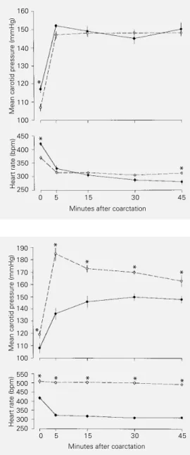
MCP and HR values before and during maintained (45 min) partial and total aortic coarctation induced by the pneumatic cuff are shown in Figure 1. The animals submit-ted to partial occlusion of the aorta presensubmit-ted a basal MCP of 107 ± 2 mmHg. Five min after coarctation, MCP had already reached a plateau (37% above control) and remained close (37 to 38%) to this level throughout the experiment. The animals submitted to total occlusion of the aorta presented a basal MCP of 117 ± 2 mmHg, and five min after the constriction the MCP had also reached a plateau (30% above control) and remained close (27 to 28%) to this level throughout the experiment. During partial constriction of the aorta, the pressure distal to the pneu-matic cuff was maintained precisely at 50 mmHg, whereas during total coarctation the distal pressure (data not shown) oscillated between 12 and 17 mmHg. The basal HR of the rats submitted to partial occlusion was 369 ± 6 bpm and five min after coarctation the reflex bradycardia had already leveled off (305 ± 2 to 314 ± 3 bpm). The animals submitted to total occlusion presented a basal HR of 421 ± 7 bpm which declined progres-sively during total constriction; this reflex
bradycardia reached 329 ± 13 and 280 ± 10 bpm 5 and 45 min after the constriction, respectively.
Intact vs SAD rats
MCP and HR values before and during the maintained (45 min) aortic coarctation in intact and SAD rats are shown in Figure 2. The intact group presented a basal MCP of 108 ± 4 mmHg. Fifteen minutes after coarc-tation, MCP reached a plateau (36% above control) and remained close (36 to 40%) to
Figure 1 - Mean carotid pres-sure (upper panel) and heart rate (lower panel) responses to acute aortic coarctation in con-scious unrestrained intact rats submitted to total (continuous line) or partial (broken line) aor-tic constriction. Data are re-ported as means ± SEM. *P<0.05 compared to partial constriction (Student t-test).
Mean carotid pressure (mmHg)
160
140
130
120
110
100
450
350
300
250 400 150
Heart rate (bpm)
0 5 15 30 45
Minutes after coarctation
*
*
*
Mean carotid pressure (mmHg)
190
160
140
130
120
100 180
170
150
110
550
450 400 350 500
Heart rate (bpm)300250
0 5 15 30 45
Minutes after coarctation
Figure 2 - Mean carotid pressure (upper panel) and heart rate (lower panel) responses to acute aortic coarctation in conscious unrestrained intact (continuous line) and sinoaortic denervated (SAD) (broken line) rats. Data are reported as means ± SEM. *P<0.05 compared to the corre-sponding point in the intact group (Student t-test). *
* *
*
* * * * *
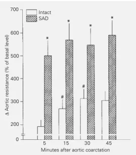
this level throughout the experiment. The SAD group presented a basal MCP of 119 ± 2 mmHg. Five min after constriction, the SAD group presented an increase in MCP almost twice (58%) that observed in intact rats, and remained at a higher level (38-46%) throughout the experiment. The basal HR of the intact group was 417 ± 4 bpm. Five minutes after coarctation the reflex brady-cardia leveled off (323 ± 5 to 309 ± 4 bpm) throughout the experiment. In contrast, the SAD group presented a remarkable tachy-cardia (509 ± 8 bpm) before coarctation and the HR did not change (492 ± 4 to 502 ± 6 bpm) during aortic constriction. Figure 3 shows the calculated changes in aortic resis-tance (%) of the intact and SAD groups during coarctation. The change in aortic re-sistance of the intact group increased pro-gressively from 5 min (193 ± 28%) to 30 min (314 ± 41%) after coarctation when it at-tained a plateau (306 ± 40%). The change in aortic resistance of SAD rats during aortic constriction was significantly higher than that of the intact group. Nevertheless, the change in aortic resistance of the SAD group had already reached a plateau (501 ± 67%) 5 min after constriction, remaining within a relatively narrow range (548 ± 72 to 591 ± 63%) throughout the experiment.
Discussion
Partial aortic occlusion with the mainte-nance of the pressure distal to the cuff pre-cisely at 50 mmHg (8) keeps the renal perfu-sion pressure well below the lower limit of renal blood flow autoregulation (10) provid-ing a powerful stimulus for renin (1,4) and vasopressin release (2,3,6,11,12). In addi-tion, this maneuver induces a marked me-chanical increase in aortic resistance which represents a substantial challenge for circu-latory homeostasis (6). In the present study we found that, despite a greater gradient in pressure (129-133 mmHg) through the cuff caused by total aortic occlusion, the hyper-tensive response did not differ from that of the animals submitted to partial occlusion, but the time course of the reflex bradycardia differed between groups particularly 45 min after constriction.
The finding that the degree of partial occlusion monitored by the maintenance of the mean femoral pressure at 50 mmHg elic-ited a hypertensive response equivalent to that elicited by total occlusion is not surpris-ing. Previous findings by Gupta and Wiggers (13) demonstrated that 85 to 95% constric-tion of the aortic lumen is necessary to re-duce the distal pressure to 50 mmHg in anesthetized dogs. Therefore, the amount of resistance imposed by the maintenance of the pressure distal to the occlusion at 50 mmHg already elicits a maximal hyperten-sive response. This finding validates the use in our protocol of partial aortic occlusion to promote maximal stimulation of the arterial baroreflex in conscious intact rats.
The basal MCP of conscious intact and SAD rats differed. Over the last two decades there has been a debate about whether or not sinoaortic denervation causes sustained hy-pertension (14-16). Currently, there is a gen-eral agreement that sinoaortic denervation causes an extreme lability of arterial pres-sure, not necessarily accompanied by arteri-al pressure elevation (17), but with the SAD Figure 3 - Bar graph showing
change as percentage of basal level (∆%) in calculated aortic re-sistance due to aortic coarcta-tion in conscious unrestrained intact and sinoaortic denervated (SAD) rats. Data are reported as means ± SEM. *P<0.05 com-pared to the corresponding point in the intact group (Student t -test); #P<0.05 compared to the
anterior value in the same group (Student t-test).
∆
Aortic resistance (% of basal level)
700
600
500
400
300
200
0
5 15 30 45
Minutes after aortic coarctation Intact
SAD
* *
* *
rats being more reactive to environmental stimuli (18), e.g., noise (19). In the present study we also observed that the SAD rats presented a remarkable tachycardia when compared to their intact counterparts, which might be attributed to sympathetic overac-tivity (19-22). During acute (45 min) partial aortic coarctation intact rats presented a steady increase in MCP accompanied by a stable reflex bradycardia, indicating a buff-ering effect of the arterial baroreflex due to parasympathetic activation (23). In contrast, during aortic constriction SAD rats exhib-ited both a greater hypertensive response and a greater change in aortic resistance to blood flow than the intact animals. The main-tenance of the MCP of SAD rats 38 to 58% above the basal level during constriction of the aorta, without a change in HR, indicates complete sinoaortic deafferentation. From a hemodynamic point of view, it is noteworthy that in intact rats both MCP and HR re-mained stable throughout the period of co-arctation, whereas the overall resistance to blood flow increased gradually as indicated by the change in aortic resistance. This find-ing might be explained by Green’s peripher-al resistance equation (resistance = pressure/ flow) (24) where the fall in cardiac output would compensate for the increase in overall resistance with the maintenance of a steady MCP. Nevertheless, SAD rats exhibited stable MCP and HR, without a gradual change in aortic resistance to blood flow, presum-ably due to the maintenance of a constant cardiac output and the lack of reflex vasodi-latation throughout the period of coarcta-tion.
Studies from our laboratory on conscious rats demonstrated that, in addition to the mechanical effect of aortic constriction, the vasopressor hormones angiotensin II and vasopressin participate in the physiopatho-genesis of acute (45 min) aortic coarctation hypertension (1-3,11,12). Moreover, a pre-vious study from our laboratory indicated that the release of these neurohumoral
fac-tors increased the overall resistance to blood flow throughout the circulation (6). As dem-onstrated before (6), in the present study there was a gradual increase in aortic resis-tance during coarctation of the aorta in intact rats, which may have been due to an increase in overall resistance to blood flow. In con-trast, the aortic resistance of SAD rats reached a maximum immediately after constriction of the aorta, a finding that might be inter-preted as being due to an already increased sympathetic tone at the beginning of the period of aortic coarctation (20,22). There-fore, the increased sympathetic drive of SAD rats presumably masked the effect of neuro-humoral factors on overall resistance to blood flow as seen in intact animals. On the other hand, the smaller and gradual increase in aortic resistance associated with a sharp bradycardia that leveled off 5 min after aor-tic constriction in the intact rats might sug-gest the already known rapid resetting of the baroreceptors (25,26) that was demonstrated to be present 30 min after the onset of the hypertensive response (27).
overall resistance to blood flow might be explained by a failure of the baroreflex to override the effect of vasopressor hormones and/or by a differential control of HR and vascular resistance during strong activation of the arterial baroreflex.
Acknowledgments
The authors gratefully acknowledge the excellent technical assistance of Jaci A. Castania and Mauro de Oliveira.
References
1. Salgado HC & Krieger EM (1986). Me-chanical and renin-angiotensin system components in acute aortic coarctation hypertension. Hypertension, 8 (Suppl I): I.133-I.136.
2. Salgado HC & Salgado MCO (1989). Acute aortic coarctation hypertension: role of vasopressin and angiotensin II. American Journal of Physiology, 257 (Heart and Cir-culatory Physiology, 26): H1480-H1484. 3. Salgado HC, Skelton MM, Salgado MCO
& Cowley Jr AW (1994). Physiopathogen-esis of acute aortic coarctation hyperten-sion in conscious rats. Hypertension, 23 (Suppl I): I.78-I.81.
4. Yagi S, Kramsch DM, Madoff IM & Hol-lander W (1968). Plasma renin activity in hypertension associated with coarctation of the aorta. American Journal of Physiol-ogy, 215 (Heart and Circulatory Physiolo-gy, 19): H605-H610.
5. Krieger EM, Salgado HC & Michelini LC (1982). Resetting of baroreceptors. In: Guyton AC & Hall JE (Editors), Interna-tional Review of Physiology. Vol. 26. Uni-versity Park Press, Baltimore, 119-146. 6. Fazan Jr R, Machado BH, Salgado MCO &
Salgado HC (1993). Effect of bilateral ne-phrectomy on hypertension produced by acute aortic coarctation. Brazilian Journal of Medical and Biological Research, 26: 765-771.
7. Haywood JR, Shaffer RA, Fastenow C, Fink GD & Brody MJ (1981). Regional blood flow measurements with pulsed Doppler flowmeter in conscious rat. American Journal of Physiology, 241 (Heart and Circulatory Physiology, 2): H273-H278.
8. Maio AA, Moreira ED, Salgado HC & Krieger EM (1981). Cardiovascular re-sponses of conscious rats due to arterial occlusion. Brazilian Journal of Medical and Biological Research, 14: 115 (Abstract). 9. Krieger EM (1964). Neurogenic
hyperten-sion in the rat. Circulation Research, 15: 511-521.
10. Arendshorst WJ, Finn WF & Gottschalk CW (1975). Autoregulation of blood flow in the rat kidney. American Journal of Physiology, 228: 127-133.
11. Salgado HC, Fazan Jr R, Machado BH & Salgado MCO (1992). Mechanical and neuro-humoral factors in acute aortic co-arctation hypertension. Agents and Ac-tions, 36 (Suppl I): 152-163.
12. Fregonese JB, Salgado MCO, Castro e Silva EJ & Salgado HC (1995). Hyperten-sive response to acute aortic coarctation in chronic vasopressin deficient states. Clinical and Experimental Hypertension, 17: 977-988.
13. Gupta TC & Wiggers CJ (1951). Basic he-modynamic changes produced by aortic coarctation of different degrees. Circula-tion, 3: 17-31.
14. Norman Jr RA, Coleman TG & Dent AC (1981). Continuous monitoring of arterial pressure indicates sino-aortic denervated rats are not hypertensive. Hypertension, 3: 119-125.
15. Persson P, Ehmke H, Kirchheim H & Seller H (1988). Effect of sino-aortic denervation in comparison to cardiopulmonary deaf-ferentation on long-term blood pressure in conscious dogs. Pflügers Archives, 411: 160-166.
16. Krieger EM (1984). Neurogenic hyperten-sion in the rat. In: de Jong W (Editor), Handbook of Hypertension. Vol. 4. Exper-imental and Genetic Models of Hyperten-sion. Elsevier Science Publishers, Amster-dam, 305-363.
17. Beloni SNE, Costa RS, Machado BH & Salgado HC (1992). Chemical renal medullectomy and arterial pressure re-sponse to sinoaortic denervation. Hyper-tension, 19 (Suppl II): II.116-II.120. 18. Schreihofer AM & Sved AF (1994). Use of
sinoaortic denervation to study the role of baroreceptors in cardiovascular regula-tion. American Journal of Physiology, 266 (Regulatory, Integrative and Comparative Physiology, 36): R1705-R1710.
19. Trindade AS & Krieger EM (1984). Long term analysis of the hypertension pro-duced by sinoaortic denervation in the rat. Brazilian Journal of Medical and Biological Research, 17: 209-217.
20. Irigoyen MC, Moreira ED, Ida F, Pires M, Cestari IA & Krieger EM (1995). Changes of renal sympathetic activity in acute and chronic conscious sinoaortic denervated rats. Hypertension, 26: 1111-1116. 21. Alper RH, Jacob HJ & Brody MJ (1987).
Regulation of arterial pressure lability in rats with chronic sinoaortic deafferenta-tion. American Journal of Physiology, 253 (Heart and Circulatory Physiology, 22): H466-H474.
22. Barres C, Lewis SJ, Jacob HJ & Brody MJ (1992). Arterial pressure lability and renal sympathetic nerve activity are dissociated in SAD rats. American Journal of Physiol-ogy, 263 (Regulatory, Integrative and Comparative Physiology, 32): R639-R646. 23. Stornetta RL, Guyenet PG & McCarty RC (1987). Autonomic nervous system con-trol of heart rate during baroreceptor acti-vation in conscious and anesthetized rats. Journal of the Autonomic Nervous Sys-tem, 20: 121-127.
24. Green HD & Kepchar JH (1959). Control of peripheral resistance in major systemic vascular bed. Physiological Reviews, 39: 617-686.
25. Salgado HC & Krieger EM (1978). Time course of baroreceptor resetting in short-term hypotension in the rat. American Journal of Physiology, 234 (Heart and Cir-culatory Physiology, 3): H552-H556. 26. Dorward PK, Andresen MC, Burke SL,
Oliver JR & Korner PI (1982). Rapid reset-ting of the aortic baroreceptors in the rab-bit and its implications for short-term and longer-term reflex control. Circulation Re-search, 50: 428-439.
28. Guo GB, Thames MD & Abboud FM (1982). Differential baroreflex control of heart rate and vascular resistance in rab-bits. Circulation Research, 50: 554-565. 29. Mancia G, Ferrari A, Gregorini L, Parati G,
Ferrari MC, Pomidossi G & Zanchetti A (1979). Control of blood pressure by ca-rotid sinus baroreceptors in human be-ings. American Journal of Cardiology, 44: 895-902.
30. Machado BH, Bonagamba LG, Castania JA & Menani JV (1992). Changes in vas-cular resistance during carotid occlusion in normal and baroreceptor-denervated rats. Hypertension, 19 (Suppl II): II.149-II.153.