A
R
TI
G
O
O
R
IG
IN
A
L
/ O
R
IG
IN
A
L
A
R
TI
C
LE
INTRODUCTION
Colorectal cancer is a frequent tumor in western world and it has been increasing its incidence over the past decades(1).It remains as a great source of morbidity and mortality, despite improvement on early diagnosis, surveillance procedures for high risk individuals, surgical techniques and adjuvant treatment(5, 10, 12, 17).
The biologic and clinic behavior of rectal tumor has not been entirely clarified. Although its prognosis is related to the histopathologic aspects such as invasion into the rectal wall or perirectal fat and lymph node involvement, patients with tumors at early stages and operated with a curative intention, may present an undesirable outcome and on the other hand far advanced tumors may do well(1, 10, 12, 18, 23).
New prognostic factors have been studied to better clarify these unpredictable outcomes, including genetic alteration and cell cycle control mechanisms. The establishing of a correlation between genetic damage and prognosis has been the goal of extensive investigations in the last few years, as well as the mechanisms of the cell cycle(18, 25).
p53 is a tumor suppressor gene, located in the short arm of chromosome 17 and produces a protein named p53(2, 14). Its primary function is to control checkpoints at the cell cycle, to promote DNA repair by stimulating other proteins (pRb, p21) and to stimulate apoptosis. Mutations of this gene produce an inactive p53 protein, which accumulates in the tumor cells. The expression of this altered protein is detected in 30% to 70% of rectal tumors and may be related to poor prognosis(2, 14).
This study describes the correlation between p53 protein immunoexpression and clinical and pathologic factors, survival rates and prognosis.
PATIENTS AND METHODS
We evaluated 83 patients with rectal adenocarcinoma submitted to potentially curative surgery at Coloproctology Unit of the “Hospital de Clínicas de Porto Alegre” (HCPA), Porto Alegre, RS, Brazil, from 1985 to 1997.
The clinical and pathologic data was carefully obtained from patient charts through retrospective assessment of the clinical follow-up notes, their respective diagnostic exams and anatomic or pathologic exams of the surgical
EXPRESSION OF THE p53
PROTEIN AND CLINICAL
AND PATHOLOGIC CORRELATION
IN ADENOCARCINOMA
OF THE RECTUM
Márcia Teresinha
JURACH
, Luise
MEURER
and Luis Fernando
MOREIRA
ABSTRACT – Background - p53 is a tumor suppressor gene that has been implicated in the pathogenesis of a wide range
of tumors, including colorectal cancer. Aim - To evaluate the prognostic significance of p53 protein expression, and to correlate with clinical and pathologic variables as well as survival. Patients and Methods - Immunohistochemical staining for p53 protein was performed in a series of 83 patients with rectal cancer with a follow-up of at least 5 years. Results
– Expression of nuclear protein was positive in 36 (43.4%) of the tumors. We found association between positive p53 and death, local recurrence, distant metastasis and overall recurrence and we also found association between p53 and death, local recurrence, metastasis and overall recurrence when analysis was adjusted to Astler-Coller stage and grade of differentiation. Conclusion - p53 immunoexpression has shown to be an independent prognostic factor in these series.
HEADINGS – Protein p53. Rectal neoplasms. Adenocarcinoma.
This work was partially granted by CAPES – PRODOC – 2002.
Department of Surgery, Federal University of Rio Grande do Sul, Porto Alegre, RS, Brazil.
specimens. The stage of the tumor was defined according to the Astler-Coller classification.
The studied variables were age, gender, tumor location, symptoms, type of the surgical procedure, staging, number of metastatic lymph nodes, survival rates, disease-free intervals, tumor aspect and size, neoplasia-free distal margin, loco-regional recurrence and metastasis.
The p53 immunostaining was performed on surgical specimens of 83 patients with rectal adenocarcinoma. The p53 evaluation by the immunostaining technique (IHC) used specific antibodies, anti-p53 human (Pab-1801) and its expression was classified according to the percentage of positive nuclei. For positive p53, a cutoff value of 20% was considered, in other words, when more than 20% of nuclei stained in a microscopic field magnified 400X. The preparation of the slides (IHC and hematoxylin-eosin) and their assessment were performed by the Pathology Unit of the HCPA, using the formaline-fixed paraffin-embedded blocks from the surgical specimens, and analyzed by two independent pathologists with experience in gastrointestinal pathology.
Antibody
The primary antibody used was a mouse mononuclear antibody Pab-1801 (Sigma Biosciences, St Louis, MO, USA) which recognizes both wild type and mutant human p53, but given the extremely short half-life of the wild type protein, staining is likely to represent abnormal accumulation of the mutant type only.
Immunostaining technique
Samples of tumor tissues were processed for p53 protein expression as a routine immunostaining technique at our hospital, using the primary antibody previously described. After dewaxing, rehydration and antigen recovery, inactivating endogenous peroxidase activity, and blocking cross-reaction with normal serum, the sections were incubated for 12 hours at 4oC with a solution of the primary antibody diluted at 1:100. Identification of the primary antibody site was achieved by subsequent application of biotinylated antibody, streptavidin horseradish peroxidase conjugate (LSAB, Dako) and its reaction by the use of diamino-benzidine tetrahydrochloride as chromogen (Kit DAB, Dako). The control staining of the slides was performed with Harris hematoxylin. All the immunostaining section had positive (breast cancer) and negative (absence of primary antibody) controls.
Statistical analysis
The quantitative data were described as average and standard deviation (SD). For categoric variables, absolute and percentage frequencies were used. Overall survival and disease-free interval were initially calculated by Kaplan-Meyer curves. The bivariate association between outcomes of interest and patient characteristics, including determination of the p53, as evaluated by relative risk (RR), confidence interval (CI), and their significance by Chi-square test. The simultaneous influence or multiple variables over mortality, local recurrence and metastasis was evaluated by Cox’s regression method, obtaining the adjusted relative risks and their respective confidence intervals. The level of significance in the bivariate analysis was P = 0.05, and in the multivariate regression analysis was P = 0.10. The data were processed and analyzed in a SPSS version 10.0 software.
RESULTS
We studied 83 cases of rectal adenocarcinoma; 47% of them (39) were male. The mean (SD) age at the time of the operation was 61.8 (SD = 14.3) years. The most frequent symptoms were bleeding, diarrhea, constipation, anal pain and an average weight loss of 9.4 kg. These symptoms were referred in an average of 9 months prior to the diagnosis.
Considering the tumor site, 48% were located in the lower rectum, 34% in the middle rectum and 18% in the upper rectum. The mean (SD) distance between the tumor and the anal verge was 7 (3.9) cm. The surgical procedures performed consisted of Miles operation in 42 patients; low anterior resection with manual anastomosis in 21 patients; stapled anastomosis in 11, coloanal anastomosis in 3 and Hartmann’s procedure in 6 patients. Table 1 shows the clinical and pathologic characteristics of the patients included in the study. The distal margin could be analyzed in 41 (49%) cases. The other 42 cases underwent rectal amputation. The tumor macroscopic appearance was considered elevated, infiltrating or ulcerated, but in most of the cases showed an association between two or the three forms. The exophitic appearance of the lesion was demonstrated in 82.4% of the cases. In eight cases the appearance was unclear.
The staging classification used was the Astler-Coller (Duke’s modified classification, 1954). Since there was no patient in stage A, all cases were divided into four categories only: B1, B2, C1 and C2, as shown in Table 1.
TABLE 1 – Clinical and pathologic characteristics of the patients
Characteristics n Description
Age, years (mean) 83 61.8 (14.3)
Male gender 83 39 (47.0)
Distance from the anal verge (cm) 83 7.0 (3.9)
Distal limit (cm) 43 2.3 (1.5)
Tumor size (cm) 73 5.5 (2.0)
Macroscopic appearance 75
Ulcerated 59 (78.7)
Infiltrating 23 (27.7
Exophitic 61 (82.4)
Astler-Coller classification 83
B1 21 (25.3)
B2 16 (19.3)
C1 10 (12.0)
C2 36 (43.4)
Grade of differentiation 83
Well differentiated 11 (13.3)
Moderately differentiated 61 (73.5)
Undifferentiated 11 (13.3)
Figures are presented as average and standard deviation of frequency
The mean number of lymph nodes examined on each surgical specimen was 15.4 (9.2) and the average of positive lymph nodes was 5.6.
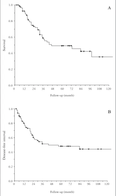
FIGURE 1 – Kaplan-Meier graphic representation of overall survival (A)
and disease-free interval (B) during the follow-up period of 83 patients with rectal adenocarcinoma
1.0 0.8 0.6 0.4 0.2 0.0 1.0 0.8 0.6 0.4 0.2 0.0
0 12 24 36 48 60 72 84 96 108 120
Follow-up (month)
0 12 24 36 48 60 72 84 96 108 120
Follow-up (month)
B A
Survival
Desease-free interval
There was no relationship between p53 and tumor size, symptoms, grade of differentiation, macroscopic tumor appearance, location and gender (P >0.05). Table 3 shows the p53 protein prevalence in relation to the Astler-Coller classification, and Tables 4 and 5, demonstrate the association of p53, Astler-Coller classification and grade of differentiation with mortality, local recurrence, metastasis and overall recurrence.
TABLE 2 – Sites of metastasis presented by the patients
Site n Local recurrence*
Liver 9 (1)
Lungs 5 (1)
Bones 2 (1)
Bones + lungs 3 (1)
Liver + lungs 3 (1)
Axillary + lungs 1 (1)
Total 23 (6)
* Synchronous cases of associated local recurrence
The survival rates were calculated in relation to the disease-free interval and overall survival according to the Kaplan-Meyer method and are displayed in Figure 1. The mean (SD) disease-free interval was 17 (10.6) months.
TABLE 3 – p53 and Astler-Coller classification
Stage p53 immunoexpression
n (negative p53) n (positive p53)
B1 11 10
B2 11 5
C1 6 4
C2 19 17
Total 47 (5.6%) 36 (4.4%)
Cutoff = 20%
DISCUSSION
The prognosis of rectal adenocarcinoma has being determined through histologic stage at the moment of the operation, but some patients are still recurring despite potentially curative procedure and earlier pathologic staging(20, 22).
It has been proposed that genetic alterations acquired during tumor development may predict prognosis. In rectal cancer, the p53 mutation and allelic loss of the short arm of chromosome 17 lead to genetic alterations that inactivate the suppressor function of the p53, i.e., alterations of this gene leads to a non-functioning p53 protein. The association of the p53 through immunostaining study that evaluates the nuclear concentration of the mutated p53 protein has been intensively assessed. The prevalence of the altered protein in rectal tumors varies from 40% to 60%(7). In our trial we demonstrated p53 positive expression in 43% of the cases.
The prognostic meaning of the p53 on the treatment of colorectal cancer remains uncertain. Many authors have found relation between positive p53 and survival improvement, such as BOSARI et al.(3), SOONG et al.(26), SUN et al.(27) and ZENG et al.(28), while others haven failed to show the same relation(2, 6, 13, 19, 21). DIEZ et al.(6) and MAEDA et al.(16) discovered a significant increase in immunopositive tumor and recurrence risk, while no other researchers could confirm these findings(6, 8, 16).
TABLE 5 – Immunoexpression of p53, Astler-Coller classification and grade of differentiation according to local recurrence and metastasis
Bivariate analysis Multivariate analysis
RR CI95% P RR CI90% P
Local recurrence
p53 ≤20%† 1.00 1.00‡
p53 >20% 2.71 1.06-6.84 0.04* 2.64 1.19-5.83 *0.05
Astler-Coller
B1† 1.00 1.00‡
B2 0.63 00.9-4.18 0.68 0.62 0.09-4.26 0.68
C1 2.36 0.61-9.10 0.29 1.88 0.46-7.73 0.46
C2 3.61 1.24-10.4 0.05* 2.69 0.85-8.53 0.16
Histology
WD† 1.00 1.00‡
MD 3.24 0.43-24.50 2.41 0.39-14.90 0.43
PD 6.23 0.55-70.70 3.22 0.36-28.75 0.38
Metastasis
P53≤20%† 1.00 1.00‡
P53>20% 2.54 1.07-6.00 0.03* 2.45 1.18-5.07 0.04*
Astler-Coller
B1† 1.00 1.00‡
B2 1.68 0.32-8.71 0.61 2.11 0.39-11.5 0.47
C1 7.51 2.00-28.2 0.01* 7.01 1.66-30.2 0.03*
C2 5.28 1.50-18.6 0.03* 4.45 1.07-18.4 0.09*
Histology
WD† 1.00 1.00‡
MD 1.33 0.30-5.83 0.64 0.16-2.57 0.60
PD 10.87 2.04-58.01 3.81 0.79-18.28 0.16
RR = relative risk; CI = confidence interval; WD = well differentiated; MD = moderate differentiated; PD = poorly differentiated; * statistical significance; † reference category; ‡ RR adjusted in Cox’s regression model containing the
Astler-Coller terms; + p53 ( [+] >20% vs [- ]≤20%) and histology
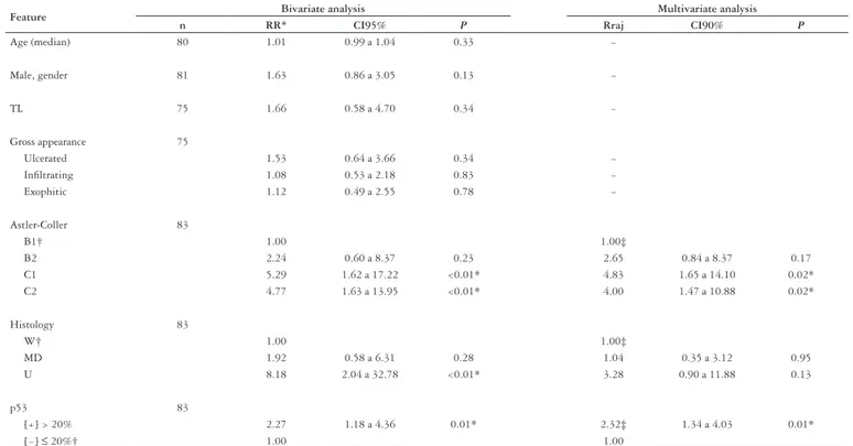
TABLE 4 – Analysis of clinical and pathologic features and mortality in patients with rectal adenocarcinoma
Feature Bivariate analysis Multivariate analysis
n RR* CI95% P Rraj CI90% P
Age (median) 80 1.01 0.99 a 1.04 0.33 −
Male, gender 81 1.63 0.86 a 3.05 0.13 −
TL 75 1.66 0.58 a 4.70 0.34 −
Gross appearance 75
Ulcerated 1.53 0.64 a 3.66 0.34 −
Infiltrating 1.08 0.53 a 2.18 0.83 −
Exophitic 1.12 0.49 a 2.55 0.78 −
Astler-Coller 83
B1† 1.00 1.00‡
B2 2.24 0.60 a 8.37 0.23 2.65 0.84 a 8.37 0.17
C1 5.29 1.62 a 17.22 <0.01* 4.83 1.65 a 14.10 0.02*
C2 4.77 1.63 a 13.95 <0.01* 4.00 1.47 a 10.88 0.02*
Histology 83
W† 1.00 1.00‡
MD 1.92 0.58 a 6.31 0.28 1.04 0.35 a 3.12 0.95
U 8.18 2.04 a 32.78 <0.01* 3.28 0.90 a 11.88 0.13
p53 83
[+] > 20% 2.27 1.18 a 4.36 0.01* 2.32‡ 1.34 a 4.03 0.01*
[−] ≤ 20%† 1.00 1.00
* statistical difference; † reference category; ‡ RR adjusted in Cox’s regression model containing Astler-Coller classification + histology + p53 ([+] >20% vs [−] ≤20%); RR = relative risk; CI = confidence interval; TL = tangencial limit;
The lack of agreement among the studies may be attributed to a short follow-up period, different staging criteria and biases use on patient selection. Moreover, there is no consensus in the literature on criteria for interpreting immunostaining results. The amount of nuclear staining (expression) necessary to consider p53 positive is still unclear. The use of a low cutoff leads to disparity and difficult the establishment of the p53 protein expression either with prognosis, survival rate, loco-regional recurrence and metastasis. The great majority of the researchers use a cutoff of 5%.
Another important fact is to consider colon and rectal cancer patients in the same group. GERVAZ et al.(9) and DIEZ et al.(7) demonstrate that colon molecular biology differs from rectal molecular biology, specially regarding p53 protein, that is why it is important to analyze only rectal cancer cases, as we did in this study.
In our study, we found no association between positive p53 and clinical and pathologic variables, such as gender, symptoms, location, size tumor macroscopic and gross appearance, circumferential tumor limit, which is in agreement with the literature. We also have found no association between these variables with recurrence and survival.
In these series we observed an association between positive p53, in a cutoff of 20% with death (P = 0.01), local recurrence (P = 0.04), distant metastasis (P = 0.03) and overall recurrence (P = 0.01). We also found an association between p53 positive and death (0.01), local recurrence (P = 0.05), metastasis (P = 0.04) and overall recurrence (P = 0.01), when analysis was adjusted to Astler-Coller stage, grade of differentiation proven p53 as an independent prognostic factor.
CONCLUSION
Our analysis demonstrated that immunoexpression of the p53 protein is adequate to define prognosis in patients with rectal adenocarcinoma. The p53 protein is associated to established variables of worst diagnosis such as tumor stage C1 and C2, to slightly differentiated grade of differentiation degree as well as correlates an increased risk for recurrence and decreased survival, as demonstrated in the multivariate analysis. Therefore p53 immunoexpression can be considered as an independent prognostic factor in cancer treated by surgery.
Jurach MT, Meurer L, Moreira LF. Expressão da proteína p53 e relação clínico-patológica no adenocarcinoma de reto. Arq Gastroenterol. 2006;43(1):14-9.
RESUMO – Racional - p53 é um gene imunossupressor tumoral que tem sido implicado na patogênese de vários tumores, incluindo o câncer
colorretal. Objetivo - Avaliar a expressão nuclear da proteína p53 com o valor prognóstico e correlacionar com variáveis clínico-patológicas e sobrevida. Material e Métodos - Foi realizado estudo imunoistoquímico da proteína p53 em 83 pacientes com diagnóstico de câncer retal com seguimento de, no mínimo, 5 anos. A expressão da proteína p53 foi associada com o estádio, com o grau histológico e com a recidiva.
Resultados - A expressão nuclear da proteína p53 foi positiva em 36 (43.4%) dos tumores. Encontrou-se associação entre p53 positivo e óbito, recidiva local, metástases à distância e recidiva total e, também, associação entre p53 e óbito, recidiva local, metástases e recidiva total, quando a análise foi ajustada para Astler-Coller e grau histológico. Conclusão - A expressão da proteína p53 tem demonstrado fator prognóstico independente.
REFERENCES
1. American Cancer Society. Cancer facts and figures. [cited 2002 Sept 30] Available from: http://www.cancer.org/statistcs.
2. Bell SM, Scott N, Cross D, Sagar P, Lewis FA, Blair GE, Taylor GR, Dixon MF, Quirke P. Prognostic value of p53 overexpression and c-ki-ras gene mutations in colorectal cancer. Gastroenterology. 1993;104:57-64.
3. Bosari S, Viale G, Bossi P. Cytoplasmic accumulation of p53 protein: an independent prognostic indicator in colorectal adenocarcinomas. J Natl Cancer Inst. 1994;86:681-7. 4. Bosari S, Viale G, Roncalli M, Graziani D, Borsani G, Lee AKC, Doggi G. p53
gene mutations, p54 protein accumulation and compartmentalization in colorectal adenocarcinoma. Am J Pathol. 1995;147:790-8.
5. The Centers for Disease Control and Prevention. Screening for colorectal cancer: United States, 1997. JAMA. 1999;281:1581-2.
6. Diez M, Medrano M, Muguerza JM, Ramos P, Hernandez P, Villeta R, Martin A, Noguerales F, Ruiz A, Granell J. Influence of tumor localization on the prognostic value of P53 protein in colorectal adenocarcinomas. Anticancer Res. 2000;20(5C):3907-12. 7. Diez M, Enriquez JM, Camunas J. Prediction of recurrence in B-C stages of colorectal
cancer by p53 nuclear overexpression in comparison with standard pathological features. Eur J Surg Oncol. 1995;21:635-59.
8. Gallego MG, Acenero MJ, Ortega S, Delgado AA, Cantero JL. Prognostic influence of p53 nuclear overexpression in colorectal carcinoma. Dis Colon Rectum. 2000;43:971-5. 9. Gervaz P, Bouzourene H, Cerottini JP, Chaubert P, Benhattar J, Secic M, Wexner S, Givvel
JC, Belin B. Dukes B colorectal cancer: distinct genetic categories and clinical outcome based on proximal or distal tumor location. Dis Colon Rectum. 2001;44:364-72. 10. Hertz REL, Deddish MR, Daye E. Value of examination in detecting cancer of the
rectum and colon. Postgrad Med. 1960;27:290-4.
11. Kandioler D, Zwrtek R, Ludwig C, Janschek E, Ploner M, Hofbauer F, Kührer I, Kappel S, Wrba F, Horvath M, Karner J, Renner K, Bergmann M, Karner-Hhanusch J, Pötter R, Jakesz R, Teleky B, Herbst F. TP53 genotype but not p53 immunohistochemical result predicts response to preoperative short-term radiotherapy in rectal cancer. Ann Surg. 2002;235:493-8.
12. Khankhanian N, Mauligit GM, RusseL WO. Prognostic significance of vascular invasion in colorectal cancer of Dukes B class. Cancer. 1997;39:1195-200. 13. Kressner U, Lindmar G, Gerdin B. Immunohistological p53 staining is of limited
value in the staging and prognostic prediction of colorectal cancer. Anticancer Res. 1996;16:951-7.
14. Kressner U, Inganäs M, Bydin S, Blikstad I, Pählman L, Glimelius B, Lindmark G. Prognostic value of p53 genetic changes in colorectal cancer. J Clin Oncol. 1999;17:593-9.
15. Leahy DT, Salman R, Mucahy, Sheahan K, O’Nonoghue DP, Parfrey NA. Prognostic significance of p53 abnormalities in colorectal carcinoma detected by PCR-SSP and immunohistochemical analysis. J Pathol. 1996;180:364-70.
16. Maeda K, Chung YS, Kang SM. Overexpression of cyclin D1 and p53 associated with disease recurrence in colorectal adenocarcinoma. Int J Cancer. 1997;74:310-5. 17. Miller SF, Knigth AR. The early detection of colorectal cancer. Cancer. 1997;40:945-9.Cancer. 1997;40:945-9. 18. Moreira LF, Iwagaki H, Hizuta A, Sakagami K, Orita K. Outcome in patients withOutcome in patients with
early colorectal carcinoma. Br J Surg. 1992;79:436-8.
19. Nathanson SD, Linden MD, Tender P. Relationship among p53, stage and prognosis of large bowel cancer. Dis Colon Rectum. 1994;37:527-34.
20. Olson RM, Perencevich NP, Malcom AW, Caffey JT, Wilson RE. Patterns of recurrence following curative resection of adenocarcinoma of the colon and rectum. Cancer.Cancer. 1980;45:2969-74.
21. Pereira H, Silva S, Julião R, Garcia P, Perpétua F. Prognostic markers of colorectalPrognostic markers of colorectal cancer: expression of p53 and Bcl-2. World J Surg. 1997;21:210-3.
22. Ratto C, Sofo L, Ippoliti M, Merico M, Doglietto GB, Crucitti F. Prognostic factors in colorrectal cancer. Literature review for clinical application. Dis Colon Rectum.Dis Colon Rectum. 1998;41:1033-49.
23. Ribeiro U, Alves VA, Souza PMB, Ribeiro AVS, Rawet V, Nonogaki S, Rodrigues JG, Habr-Gama A. Correlação das proteinas p53 e KI-67 com o prognóstico de pacientes com adenocarcinoma de reto distal. Rev Bras Coloproctol. 2000;20:248-56.Rev Bras Coloproctol. 2000;20:248-56. 24. Rich T, Gunderson II, Lew R, Galdibini JJ, Cohen AM, Donalsdsib G. Patterns of recurrence
of rectal cancer after potentially curative surgery. Cancer. 1983;52:1317-29. 25. Scwandner O, Schiedeck TH, Bruch HP. Apoptosis in rectal cancer: prognostic
significance in comparison with clinical histopathologic, and immunohistochemical variables. Dis Colon Rectum. 2000;43:1227-36.
26. Soong R, Powell B, Elsaleh H, Gnanasampanthan G. Prognostic significance of TP53 gene mutation in 995 cases of colorectal carcinoma. Influence of tumour site stage, adjuvant chemotherapy and type of mutation. Eur J Cancer. 2000;36:2053-60. 27. Sun XF, Carstensen JM, Stal O. Prognostic significance of p53 expression in relation to
DNA ploidy in colorectal adenocarcinoma. Virchows Arch A Pathol Anat Histopathol. 1993;423:443-8.
28. Zeng ZS, Sarkis AS, Zhang ZF. p53 nuclear overexpression: an independent predictor of survival in lymph node-positive colorectal cancer patients. J Clin Oncol.J Clin Oncol. 1994;12:2043-50.