245
Gastroesophageal reflux: contrast-enhanced study and ultrasonography
Radiol Bras. 2009 Jul/Ago;42(4):245–248
Original Article • Artigo Original
Gastroesophageal reflux: a comparative study
of receptiveness and sensitivity of upper
gastrointestinal series and ultrasonography*
Refluxo gastroesofágico: estudo comparativo da receptividade e sensibilidade entre seriografia e ultrassonografiaMakoto Sakate1, Guilherme Lopes Silveira2, Bruno Paulino de Muzio2, Hudson Teigao Junior2, Jorge Guilherme Okanobo Ozaki2, Marcelo Dimas Spadim2, Roberto Cesar Teixeira Dantas2, Nilton Carlos Machado3
OBJECTIVE: Comparative study of receptiveness (cooperation) and sensitivity of upper gastrointestinal series and intraabdominal esophagus ultrasonography in patients with suspicion of gastroesophageal reflux. MATERIALS AND METHODS: The present study included 42 pediatric patients (26 male, with mean age of 33.64 ± 34.33 months, and 16 female, with mean age of 31.02 ± 35.56 months) with suspicion of gastroesophageal reflux, who were initially submitted to upper gastrointestinal series and subsequently to intraabdominal esophagus ultrasonography. RESULTS: The statistical comparative analysis covering sexes and ages suggests no evidence of association with cooperation, both for the upper gastrointestinal series and the intraabdominal esophagus ultrasonography. However, in the classification of the patients’ cooperation, the technique of upper gastrointestinal series presented less than 50% of cooperation, while 80.49% of patients cooperated in the intraabdominal esophagus ultrasonography examinations. As regards the methods sensitivity for the diagnosis of gastroesophageal reflux, the technique of intraabdominal esophagus ultrasonography was significantly superior (85.7%) to the upper gastrointestinal series (47.6%). CONCLUSION: The present study suggests that an intraabdominal esophagus ultrasonography should be performed even if gastroesophageal reflux even if it is not detected at the upper gastrointestinal series.
Keywords: Gastroesophageal reflux; Intraabdominal esophagus; Ultrasonography; Contrast-enhanced study.
OBJETIVO: Estudo comparativo da receptividade (colaboração) e sensibilidade da seriografia do esôfago, estômago e duodeno em relação à ultrassonografia do esôfago intra-abdominal em pacientes com suspeita de refluxo gastroesofágico. MATERIAIS E MÉTODOS: Foram incluídos no estudo 42 pacientes pediátricos (26 masculinos, com idade média de 33,64 ± 34,33 meses, e 16 femininos, com idade média de 31,02 ± 35,56 meses) com suspeita de refluxo gastroesofágico, os quais foram submetidos, inicialmente, a seriografia do esôfago, estômago e duodeno, e posteriormente, a ultrassonografia do esôfago intra-abdominal. RESUL-TADOS: A análise estatística comparativa entre os sexos e as idades sugere não haver evidência de associa-ção com a colaboraassocia-ção, tanto para a seriografia do esôfago, estômago e duodeno como para a ultrassonografia do esôfago intra-abdominal. Entretanto, na classificação quanto ao tipo de colaboração, a técnica de seriografia do esôfago, estômago e duodeno apresentou menos de 50% de colaboração, enquanto 80,49% dos pacien-tes colaboraram com a ultrassonografia do esôfago intra-abdominal. Quanto à sensibilidade do diagnóstico de refluxo gastroesofágico, a técnica de ultrassonografia do esôfago intra-abdominal (85,7%) foi significa-tivamente superior à de seriografia do esôfago, estômago e duodeno (47,6%). CONCLUSÃO: O presente estudo sugere que se proceda a ultrassonografia do esôfago intra-abdominal, mesmo na ausência de refluxo gastroesofágico na seriografia do esôfago, estômago e duodeno.
Unitermos: Refluxo gastroesofágico; Esôfago intra-abdominal; Ultrassonografia; Estudo contrastado.
Abstract
Resumo
* Study developed at the Center of Diagnostic Imaging – Hos-pital das Clínicas da Faculdade de Medicina de Botucatu da Universidade Estadual Paulista (FMB-Unesp), Botucatu, SP, Bra-zil.
1. PhD, Assistant Professor of the Discipline of Radiodiagnosis, Department of Tropical Diseases and Imaging Diagnosis – Faculdade de Medicina de Botucatu da Universidade Estadual Paulista (FMB-Unesp), Botucatu, SP, Brazil.
2. MDs, Residents, Discipline of Radiodiagnosis, Department of Tropical Diseases and Imaging Diagnosis – Faculdade de
Sakate M, Silveira GL, Muzio BP, Teigao Jr H, Ozaki JGO, Spadim MD, Dantas RCT, Machado NC. Gastroesophageal reflux: a comparative study of receptiveness and sensitivity of upper gastrointestinal series and ultrasonography. Radiol Bras. 2009;42(4):245–248.
0100-3984 © Colégio Brasileiro de Radiologia e Diagnóstico por Imagem
Medicina de Botucatu da Universidade Estadual Paulista (FMB-Unesp), Botucatu, SP, Brazil.
3. PhD, Professor of the Department of Pediatrics – Faculdade de Medicina de Botucatu da Universidade Estadual Paulista (FMB-Unesp), Botucatu, SP, Brazil.
Mailing address: Dr. Makoto Sakate. Rua Aleixo Varoli, 651, Jardim Paraíso. Botucatu, SP, Brazil, 18610-295. E-mail: [email protected]
Received September 26, 2008. Accepted after revision May 5, 2009.
INTRODUCTION
Gastroesophageal reflux (GER) is the involuntary passage of the gastric content backwards up into the esophagus. It is a relevant and relatively frequent finding in infants(1,2). In neonates, this disorder may
246
Sakate M et al.
Radiol Bras. 2009 Jul/Ago;42(4):245–248 cases, self-limited and benign(3).
Esoph-ageal reflux occurs because of a defect and/ or immaturity of the lower esophageal sphincter(4,5). The integrity of this
func-tional valvular system is important to avoid the gastric content reflux, considering that the intraabdominal pressure is higher than the intrathoracic pressure. The refluxed material is rapidly returned into the stom-ach by the secondary esophageal peristal-tic waves(6,7).
The gastric content reflux degree may range from mild to severe, and the fre-quency is variable. In most of cases, the gastroesophageal reflux is mild, transitory, bringing no consequence in particular. However, persistent episodes should be considered as pathologic(8,9).
The traditional method for studying GER is the upper gastrointestinal series (UGIS) with barium and, most recently, intraabdominal esophagus ultrasonography (IEUS)(10,11).
Similarly to UGIS, IEUS allows a safe, noninvasive evaluation of structural ana-tomic details and can identify several pathologic processes, including GER, in real time (12) and without ionizing radiation.
The present study is aimed at compar-ing the receptiveness (cooperation) and sensitivity of UGIS in relation to IEUS in pediatric patients with suspicion of GER.
MATERIALS AND METHODS
The research project was presented and received the approval from the Committee for Ethics in Research of the Institution were the study was developed. A term of free and informed consent was signed by the parents of the patients, who had re-ceived information on the study methodol-ogy.
The present study included 42 pediatric patients with suspicion of GER, as follows: 26 boys with ages ranging between 1 and 120 months (mean, 33.64 ± 34.33 months), and 16 girls between 2 and 113 months (mean, 31.02 ± 35.56 months).
All the patients were previously submit-ted to UGIS in accordance with the proto-col of the service of radiodiagnosis, and an additional examination form being filled out. In the present study the additional ex-amination form included the following
data: name, age, sex, weight, height, num-ber of reflux episodes observed, time of intermittent observation with five-minute radioscopy after barium repletion, besides an indication of the behavior of the patient as follows: cooperative (c), partially coop-erative (pc), excited during examination (ede), and very difficult patient (vdp).
The UGIS technique utilized corre-sponded to the protocol of the unit of radi-ology consisting of: after four-hour fast, the patient was placed on the examination table of the x-ray equipment in right lateral de-cubitus position. During the ingestion of diluted barium through a baby bottle, three radiographic views of the esophagus were obtained (initial phase, during and after in-gestion). Subsequently contrast-enhanced images were acquired from the stomach and duodenal arch. Once a complete reple-tion of the stomach was achieved, the pa-tient was placed in horizontal dorsal decu-bitus and a five-minute intermittent radios-copy was performed in an attempt to ob-serve and recording the GER. After the UGIS, all the patients were referred to the ultrasonography department.
The US equipment utilized was a Logiq 400 (General Electric Medical Systems; Milwaukee, USA), with 2–5 MHz semi-convex transducer.
After approximately one-week interval, IEUS was performed by a single sono-graphist who did not know the results of the previous UGIS. The examination was per-formed at morning, after breakfast with breast milk, infant formula or yogurt (for the older children) for GER study.
The patients were placed in horizontal dorsal decubitus, with their head slightly raised. The IEUS was performed with the transducer placed on the left region of the xiphoid appendix(13) for identifying the
intraabdominal esophagus. Real-time con-tinuous observation was developed along five minutes. Similarly to the UGIS form, a IEUS form was filled out with the follow-ing data: age, sex, weight, height, GER presence or absence, number of GER epi-sodes and behavior of the patients during examination. The study was recorded on a videotape for later, detailed analysis of the local anatomic conditions (Figure 1).
Data of both UGIS and IEUS were sta-tistically analyzed for evaluation of intermethod agreement by means of the chi-square test (χ2), and according to the
confidence interval for agreement ratio(14).
RESULTS
In the comparative study considering sex and age of the 42 children submitted to diagnostic imaging for GER, the statistical analysis by the χ2 test suggests that there
is no evidence of association between the patients cooperation both for the UGIS (p = 0.98745) and the IEUS (p = 0.3966) tech-niques. However, in the classification of the patients’ behavior during examination, the UGIS technique presented < 50% of cooperation, while 80.49% of the children cooperated with the IEUS. Practically, all the children who cooperated in the UGIS also did it with the IEUS (94.74%). Among the 22 children who were noncooperative
Figure 1.A: US image of intraabdominal esophagus at rest (arrow) and stomach filled with food (star).
B: US image of dilated intraabdominal esophagus (arrow) because of gastroesophageal reflux (star).
247
Gastroesophageal reflux: contrast-enhanced study and ultrasonography
Radiol Bras. 2009 Jul/Ago;42(4):245–248
nal region of the esophagus for 24-hour continuous recording of the ph values in this region(17). Because of this method
char-acteristics, it is considered as an invasive study.
Scintigraphy is performed in the suspi-cion of GER, after swallowing of a food bolus mixed with a radioactive substance. An increase in the uptake, by the esoph-ageal region, of the radiation coming from the gastric region during a given time, means that GER has occurred(16). This
method requires the utilization of radioac-tive material and highly expensive equip-ment, so its utilization as a routine study is unfeasible.
UGIS is performed with an ionizing ra-diation source (x-rays) both during and after barium ingestion. Intermittent obser-vation of the stomach fundus filled with barium is performed with radioscopy for detecting the contrast medium reflux from the stomach backwards up to the esopha-gus(10).
Currently, the comparative study of IEUS and UGIS has demonstrated that IEUS presents a higher sensitivity in the diagnosis of GER(9,17) and in the evaluation
of the intraabdominal esophagus, with a reduction of the exposure to ionizing radia-tion for these patients(11,18). However, the
determination of reflux degree as well as the evaluation of the intrathoracic esophagus cannot be performed with this method(18,19).
The determination of the GER degree could be done with IEUS, although not as specifically as the UGIS does, by means of a comparative study between these two techniques. The GER degree obtained at UGIS for each patient would be compared with the diameter and the time of opening of the intraabdominal esophagus during the GER observed at the IEUS(20,21) in the same
patient. After this analysis the GER degree could be inferred only with IEUS.
The intrathoracic esophagus could be indirectly evaluated with the utilization of the “esophageal time”(18), that allows the
determination of the typical esophageal transit time for a liquid or pasty food from the moment of deglutition up to its passage through the intraabdominal esophagus. Most of times, the association of “esoph-ageal time” and IEUS could evaluate the intrathoracic esophagus and, indirectly, the
Table 2 Distribution of 42 children submitted to UGIS and IEUS, regarding results in the diagnosis of gastroesophageal reflux. IEUS Negative Positive Total (%) Negative 6 16 22 (52.4) UGIS Positive – 20 20 (47.6) Total (%) 6 (14.3) 36 (85.7) 42 (100)
UGIS, upper gastrointestinal series; IEUS, intraabdominal esophagus ultrasonography.
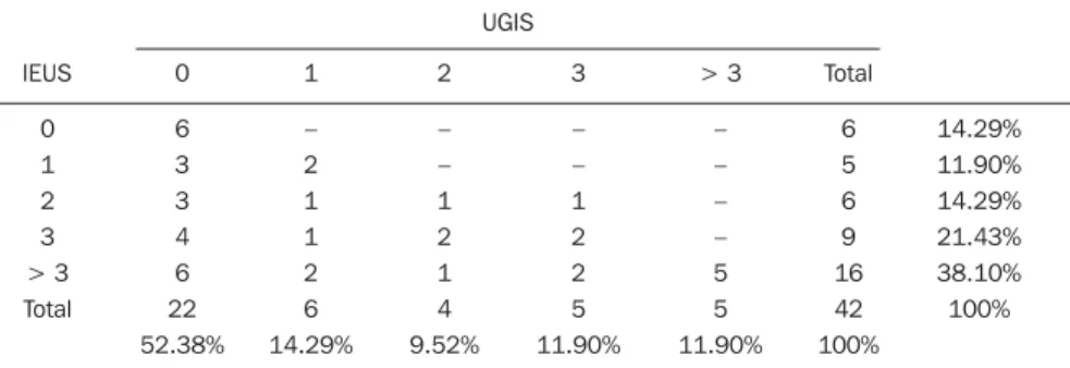
Table 3 Distribution of 42 children submitted to UGIS and IEUS, regarding the number of episodes of gastroesophageal reflux. IEUS 0 1 2 3 > 3 Total UGIS 0 6 3 3 4 6 22 52.38% 1 – 2 1 1 2 6 14.29% 2 – – 1 2 1 4 9.52% 3 – – 1 2 2 5 11.90% > 3 – – – – 5 5 11.90% Total 6 5 6 9 16 42 100% 14.29% 11.90% 14.29% 21.43% 38.10% 100%
UGIS, upper gastrointestinal series; IEUS, intraabdominal esophagus ultrasonography. during UGIS, 68.18% cooperated with the
IEUS technique (Table 1).
Statistically, one can conclude that there is a difference between the UGIS and IEUS technique in terms of rates corresponding to the different levels of cooperation (McNemar’s test: U2 = 12.25;
p = 0.0005). In terms of cooperation, both techniques are positively associated (χ2 = 4.578;
p = 0.0324).
As regards the diagnostic results of both techniques (UGIS and IEUS), IEUS was significantly superior to UGIS (Tables 2 and 3).
Statistically, one can conclude that there is a difference in the rate of positive results for GER between the UGIS (47.6%) and IEUS (85.7%) techniques (McNemar’s test: U2 = 16.0;
p = 0.00006).
In terms of diagnosis for gastroesoph-ageal reflux, both techniques are positively associated (χ2 = 6.3636;
p = 0.01165).
DISCUSSION
Gastroesophageal reflux is a well known entity frequently found in pediatric patients. Several methods are available for evaluation of this condition in children: 24-hour esophageal pH test, radioisotope scin-tigraphy and UGIS, the latter being the method most frequently utilized to confirm the presence of GER and mainly for ruling out obstructive diseases such as vascular compression, congenital esophageal steno-sis, among others(15,16).
Esophageal pH test is performed with the placement of an electrode in the
termi-Table 1 Distribution regarding the patients’ behavior during IEUS and UGIS in the evaluation of gastro-esophageal reflux. IEUS c pc ede vdp Total c 18 1 – – 19 46.34% pc 11 1 – 1 13 31.71% ede 3 2 – – 5 12.20% vdp 1 2 1 – 4 9.76% Total 33 6 1 1 41 100% 80.49% 14.63% 2.44% 2.44% 100% UGIS
248
Sakate M et al.
Radiol Bras. 2009 Jul/Ago;42(4):245–248 GER in the majority of cases, especially in
centers where only ultrasonography units are available. In the presence of obstructive diseases of the intrathoracic esophagus, such as vascular compression, dysphagia and odynophagia will be observed during food ingestion, and with increase in the esophageal time at IEUS.
Different practitioners performed the UGIS (physicians, residents in radiodiag-nosis) and IEUS (a physicians-sonograph-ist), which may imply more or less empa-thy with the children. Even so, it is sug-gested that IEUS is performed in cases where gastroeshophageal reflux is not de-tected by UGIS, considering that more than half (52.4%) of diagnoses by UGIS are negative and, on the other hand, more than 80% of the patients are diagnosed as posi-tive by IEUS. The discrepancy between the diagnoses with the two methods becomes evident in cases of reflux > 3, where the rates achieve 11.9% and 38%, respectively for UGIS and IEUS, similarly to the results reported by Milward(11). However, the
au-thors could not find in the literature, simi-lar studies evaluating and comparing the patients’ receptiveness (cooperation) with these two diagnostic modalities.
Probable limiting factors for UGIS are: examination room illumination, number of outsiders in the room, presence of equip-ment unfamiliar to the child, ingestion of substances with unpleasant flavor (barium), and movement in the room. Ad-ditionally, one must consider the utilization of an appropriate dose-area product of ion-izing radiation(22), but exposing young,
growing patients, whose probability of in-teraction with radiation is higher.
Positive aspects related to the IEUS technique are: calm environment, equip-ment with less frightening appearance with a TV-like display, half-light, ingestion of a substance with pleasant flavor (infant for-mula or yogurt) and few persons in the
room. Additionally, there are other advan-tages of ultrasonography: low-cost, nonin-vasiveness, short examination time, and the functioning of the organ and adjacent struc-tures is not affected (both at short- and long-term). The examination can be re-peated as many times as necessary in even more physiological circumstances, in real-time, allowing the evaluation of normal anatomic structural details or identification of local pathological processes.
CONCLUSION
The comparative study of the sensitiv-ity and children receptiveness (coopera-tion) in relation to the IEUS, with gastric repletion (breast milk, infant formula or yogurt), and UGIS with barium, in the evaluation of GER, has demonstrated the following aspects: first, a higher level of cooperation from the patients at IEUS as compared with UGIS in the evaluation of GER; secondly, IEUS demonstrated higher sensitivity in the diagnosis of GER; thirdly, the present study suggests that, most of times, IEUS can substitute UGIS for indi-rectly evaluating the intrathoracic esopha-gus and GER, especially in centers where only ultrasonography is available.
REFERENCES
1. Costa L, Campobasso P. Gastroesophageal reflux. Pediatr Med Chir. 1999;21(5 Suppl):33–52. 2. Naik DR, Bolia A, Moore DJ. Comparison of
barium swallow and ultrasound in diagnosis of gastro-oesophageal reflux in children. Br Med J (Clin Res Ed). 1985;290:1943–5.
3. Balistreri WF, Farrell MK. Gastroesophageal re-flux in infants. N Engl J Med. 1983;309:790–2. 4. Gomes H, Lallemand A, Lallemand P. Ultrasound of the gastroesophageal junction. Pediatr Radiol. 1993;23:94–9.
5. Boix-Ochoa J, Canals J. Maturation of the lower esophagus. J Pediatr Surg. 1976;11:749–56. 6. Boix-Ochoa J. The physiologic approach to the
management of gastric esophageal reflux. J Pediatr Surg. 1986;21:1032–9.
7. DeMeester TR, Wernly JA, Bryant GH, et al. Clinical and in vitro analysis of determinants of
gastroesophageal competence. A study of the principles of antireflux surgery. Am J Surg. 1979; 137:39–46.
8. Costa CD, Zomignan HP, Rocha JIP, et al. Re-fluxo gastroesofágico em pediatria: estudo radio-lógico. Pediat (S Paulo). 1986;8:136–40. 9. Swischuk LE.Alimentary tract. In: Swischuk LE,
editor. Imaging of the newborn, infant and young child. 3rd ed. Baltimore: Williams & Wilkins; 1989. p. 353–69.
10. Meschan I, Ott D. Oropharynx, laryngopharynx, and esophagus. In: Meschan I, editor. Roentgen signs in diagnostic imaging: abdomen. 2nd ed. Philadelphia: WB Saunders; 1984. p. 487–560. 11. Milward CMMO. A ultrassonografia como mé-todo diagnóstico do refluxo gastroesofágico no paciente pediátrico. Radiol Bras. 2004;37:230.
12. Mádi-Szabó L, Kocsis G. Examination of gastroe-sophageal reflux by transabdominal ultrasound: can a slow, trickling form of reflux be responsible for reflux esophagitis? Can J Gastroenterol. 2000; 14:588–92.
13. Cerri GG. Vísceras ocas. In: Rocha DC, Cerri GG, Prando A, et al. Ultrassonografia abdominal. São Paulo: Sarvier; 1988. p. 169–86.
14. Zar JH. Biostatistical analysis. 3rd ed. New Jer-sey: Prentice-Hall; 1996.
15. Gomes H, Menanteau B. Gastro-esophageal re-flux: comparative study between sonography and pH monitoring. Pediatr Radiol. 1991;21:168–74. 16. González Fernández F, Argûelles Martin F, Rodri-guez de Quesada B, et al. Gastroesophageal scin-tigraphy: a useful screening test for GE reflux. J Pediatr Gastroenterol Nutr. 1987;6:217–9. 17. Schindlbeck NE, Heinrich C, König A, et al.
Op-timal thresholds, sensitivity, and specificity of long-term pH-metry for the detection of gastro-esophageal reflux disease. Gastroenterology. 1987; 93:85–90.
18. Sakate M, Teixeira AS, Yamashita S, et al. Um novo método de avaliação do “tempo esofágico” com ultra-sonografia por abordagem externa. Radiol Bras. 2008;41:309–12.
19. Westra SJ, Derkx HH, Taminiau JA. Symptom-atic gastroesophageal reflux: diagnosis with ul-trasound. J Pediatr Gastroenterol Nutr. 1994;19: 58–64.
20. Lynch CS, Chammas MC, Mansur LL, et al. Bio-mecânica ultra-sonográfica da deglutição: estudo preliminar. Radiol Bras. 2008;41:241–4.
21. Spadotto AA, Gatto AR, Cola PC, et al. Software para análise quantitativa da deglutição. Radiol Bras. 2008;41:25–8.