Article
J. Braz. Chem. Soc., Vol. 22, No. 1, 80-85, 2011. Printed in Brazil - ©2011 Sociedade Brasileira de Química 0103 - 5053 $6.00+0.00
A
*e-mail: [email protected]
Biosynthesis of Aphidicolin Proceeds via the Mevalonate Pathway in the Endophytic
Fungus
Nigrospora sphaerica
Adriana A. Lopes and Mônica T. Pupo*
Departamento de Ciências Farmacêuticas, Faculdade de Ciências Farmacêuticas de Ribeirão Preto, Universidade de São Paulo, 14040-903 Ribeirão Preto-SP, Brazil
Previamente o fungo endofítico Nigrospora sphaerica foi identiicado como um profícuo produtor do diterpeno bioativo aidicolina. Neste artigo foi realizado um estudo a im de se veriicar as melhores condições para a produção do diterpeno em meio Czapek. A fonte de açúcar (glicose e sacarose) e o tempo de incubação (4-15 dias) foram otimizados, para posterior aplicação no estudo biossintético do diterpeno. Os níveis mais elevados da produção da aidicolina foram obtidos no 8° dia com 1% de glicose e no 12° dia com 3% de sacarose nos meios de cultura. A biossíntese da aidicolina foi estudada empregando [1-13C]-D-glicose como precursor, e o padrão de incorporação
foi determinado através de espectroscopia de RMN de 13C quantitativo. A incorporação de [1-13
C]-D-glicose mostrou que as unidades isoprênicas da aidicolina são derivadas da via do mevalonato.
We have previously identiied the endophytic fungus Nigrospora sphaerica as a proliic producer of the bioactive diterpene aphidicolin. Herein we report a study to establish the best conditions for the production of aphidicolin by N. sphaerica in Czapek medium. The sugar source (glucose and sucrose) and the incubation time (4-15 days) were optimized for further application on biosynthetic studies of the diterpene. The highest levels of production of aphidicolin were found on the 8th day
with 1%-glucose and on the 12th day with 3%-sucrose based media. The biosynthesis of aphidicolin
was investigated using [1-13C]-D-glucose as a precursor, and showed that the isoprene units of
aphidicolin are derived from the mevalonate pathway.
Keywords: Nigrospora sphaerica, aphidicolin, biosynthesis, [1-13C]-D-glucose, mevalonate
pathway
Introduction
Terpenoids constitute one of the most diverse groups of natural products in nature. Dimethylallyl pyrophosphate (DMAPP) and isopentenyl pyrophosphate (IPP) are the universal precursors in the biosynthesis of terpenoids. These precursors can be produced by two distinct biosynthetic pathways: the mevalonate pathway (MVA pathway) and methylerythritol pathway (MEP pathway).1
The key regulatory step of the MVA pathway involves reduction of 3-hydroxy-3-methylglutaryl-CoA into mevalonic acid, catalyzed by the enzyme HMG-CoA reductase.2 MVA pathway is of common occurrence
among eukaryotes, including plants, fungi and yeasts, but is also present in some bacteria.3 The MEP pathway
is nearly completely elucidated, and its distribution is now fairly well known.4 Many eubacteria, cyanobacteria,
green algae and higher plants are known to use the MEP pathway.5,6
MVA and MEP pathways can be readily differentiated by the labeling pattern resulting from the incorporation of 13C after feeding with 13C-precursor into isoprenoids.
Studies for the elucidation of the biosynthetic origin of terpenoid metabolites are usually based on [1-13C]-
D-glucose as general precursor.7,8 Isoprenoids biosynthesized
by the MVA pathway and by the MEP pathway show different enrichment pattern into isoprene units. MVA-derived terpenes will be labeled at C-2, C-4 and C-5, while MEP-derived terpenes will be labeled at C-1 and C-5.9,10
Recently, we reported the isolation and remarkable cytotoxic activity against cancer cell lines of the diterpene aphidicolin (1), produced in liquid and solid cultures by the endophytic fungus Nigrospora sphaerica.12 Aphidicolin
(1) was irst isolated from Cephalosporium aphidicola13
as an antiviral agent against Herpes simplex type 1. The diterpene also showed a variety of biological activities such as antitumor, phytotoxic and speciic inhibition of DNA polymerase α. This latter property makes aphidicolin a valuable tool for studies of eukaryotic DNA synthesis and cell cycles.14,15
Considering the biological signiicance of aphidicolin and the absence of biosynthetic studies involving this diterpene, the aim of the present study was to investigate the biosynthetic pathway of aphidicolin (1) in the endophytic fungus N. sphaerica.
Experimental
Procedures
1H and 13C NMR spectra were recorded in CD 3OD
in a Varian INOVA 500 spectrometer. Chemical shift
values were recorded relative to the dH (4.80 ppm) and dC
(49.0 ppm) signals of the predominantly deuterated solvent. The MS system used was a quadrupole time-of-light instrument (UltrOTOF-Q, Brucker Daltonics, Billerica, MA, U.S.A.), equipped with an ESI positive and negative ion source. The analyses were performed with the mass spectrometer in full scan mode. The following settings were applied for the analyses: capillary voltage 3,900 V; dry gas low 4 L h-1; nebulizer gas nitrogen. The instrumentations
for HPLC analysis consisted of a Shimadzu (Kyoto, Japan) HPLC system, coupled of a LC-6AD model solvent pump, system controller SCL 10AVB, a column oven CTO-10ASVP, a Rheodyne model 7725 injector with a 20 μL loop, a SPD-M10AVP diode array detector operating at 190 nm and a software Class VP for data acquisition.
Biological material
The endophytic fungus N. sphaerica SS67 was isolated from the roots of Smallanthus sonchifolius (Asteraceae).12
The fungus has been maintained on potato dextrose agar slants, on mineral oil, on agar plugs in sterile water and on silica-gel (1-4 mm Synth, Brazil), stored at 4 oC at our
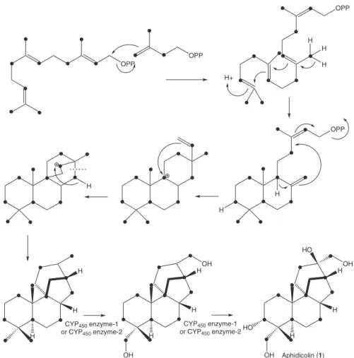
Figure 1. Biosynthesis of aphidicolin (1).
OPP
OPP
OPP
H+
H H
H
H
OPP
H
H
H
H H
H
H H
OH
OH
H
H H
OH
OH
HO
HO CYP450 enzyme-1
or CYP450 enzyme-2
CYP450 enzyme-1
or CYP450 enzyme-2
laboratory, under voucher number SS67. The [1-13C]-
D-glucose (99% isotopic abundance) was purchased from Sigma-Aldrich.
Quantiication of aphidicolin production
Liquid cultures were conducted in two steps. First, the slant stored fungi were inoculated in Petri dishes containing PDA medium and incubated at 30 °C for 7 days. After this period, three agar plugs (0.5 cm diameter) were cut from the N. sphaerica PDA cultures, inoculated in 20 mL of seed medium (2 falcon lasks of 10 mL each, supplemented with 5% of triptone, 10% of dextrose, 3% of yeast extract) and the resulting culture was incubated at 30 ºC for 48 h with shaking at 120 ºC rpm. After cultivation, the resulting mycelia were transferred to 180 mL of Czapek medium (lask of 500 mL) at pH 6.2, 30 °C, 120 rpm. Three different concentrations of sugar were used, 1% glucose, 3% glucose and 3% sucrose, and the cultures were incubated at the same conditions varying the time of incubation (4-16 days). Flasks with no fungal inoculation were incubated at the same conditions for control purposes. The culture broths were extracted by partition with ethyl acetate (3 × 70 mL) at the even days between the 4th and the 16th day. Crude
extracts (from cultures and controls) were analyzed by HPLC. The quantiication of aphidicolin was performed on a Shimadzu CLC-ODS (C-18) column 250 × 4.6 mm, 5 μ particle size protected with a 4-Pack endcapped guard column Agilent Technologies 12.5 × 4.6 mm; solvent, 35% acetonitrile-65% water (using an isocratic mobile phase; 1.0 mL min-1 low rate; detection at 190 nm). Aphidicolin
was detected at 9.6 min, and the amount of aphidicolin was calculated from a calibration curve (prepared in acetonitrile at concentration 1, 2, 4, 6 and 8 mg mL-1) of standard in
respect of the peak area and amount.
Administration of [1-13C]-D-glucose to the fungal culture
N. sphaerica was inoculated onto PDA medium in Petri dishes and incubated at 30 °C for 7 days. After this period, three agar plugs (0.5 cm diameter) were cut and inoculated in 20 mL of seed medium (6 falcon lasks of 10 mL each, supplemented with 5% of triptone, 10% of dextrose, 3% of yeast extract) and the culture was incubated at 30 ºC for 48 h with shaking at 120 ºC rpm. The resulting mycelia (2 falcon lasks) were transferred to 180 mL of Czapek medium (total of 3 lasks of 500 mL with 180 mL of medium each) at pH 6.2, 30 °C, 120 rpm with 1% of labeled sugar ([1-13C]-
D-glucose) and cultures were reincubated at 30 ºC for 8 days at 120 rpm. Fungus in the 8th day of culture was extracted
by the addition of 200 mL of ethanol 95% to the broth
(to cause the death of the microorganism), the resulting suspension was vacuum-iltered and the iltrate submitted to liquid-liquid partition with ethyl acetate (5 × 350 mL). The organic layer was evaporated under reduced pressure to produce the EtOAc crude extract (393 mg).
Extraction and isolation of 13C-labeled aphidicolin from
N. sphaerica
The EtOAc crude extract (393 mg) was fractionated using column chromatography over silica gel (70-230 mesh; Merck, column size 8.5 × 1.5 cm) eluted with EtOAc (isocratic system) to yield ive fractions. Fraction 5 (128 mg) was further puriied by column chromatography over silica gel (70-230 mesh; Merck, column size 4.5 × 1.0 cm) eluted with EtOAc (isocratic system) to yield enriched aphidicolin (43 mg).
Evaluation of the isotopic abundance
The 13C NMR spectra of samples of 1 obtained from 13C at natural abundance and 1-13C-labeled obtained from
incorporation experiments involving [1-13C]-D-glucose
as precursor, respectively, were measured under identical experimental conditions. 13CNMR spectra were acquired
using NS = 65536. The Spin Works 3.0 version 3.1.2.0 was the software used to process the spectra obtained by measuring the integral of the carbons. The relative
13C-isotopic abundances of individual carbon atoms in 1 and
1-13C-labeled were calculated from the integrals obtained
from the labeled sample in comparison with those of the natural abundance sample (Table 1). The values were normalised with reference to an abundance value of 1.1% for the carbon with the lowest possible 13C enrichment.
Results and Discussion
Two different pathways are known for the formation of IPP: the well-known acetyl-CoA/MVA route and MEP pathway starting from glyceraldehyde 3-phosphate and pyruvate. They can be clearly distinguished by incorporation of a 13C-labeled carbon source such as
glucose (especially when they are utilized as single carbon and energy source) and examination of the 13C NMR spectra
of the analyzed isoprenoids.
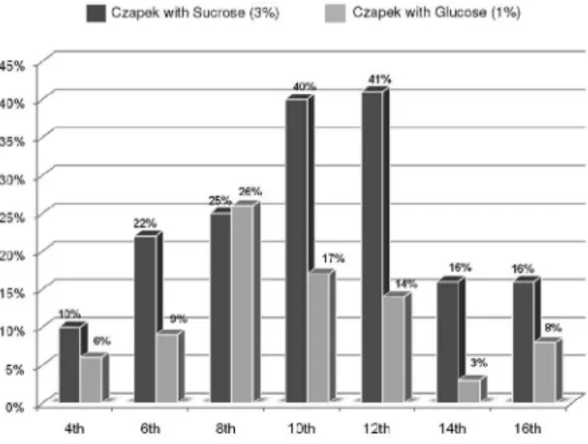
In order to elucidate the biosynthetic origin of the isoprene units in the diterpene aphidicolin, we performed preliminary studies to verify the best conditions for the production of 1
different experiments have been carried out, using 3% sucrose, 3% glucose and 1% glucose in the Czapek medium. The highest level of production of 1 (Figure 2) was found on the 12th day with 3% sucrose based medium exhibiting a yield
of 41% (m/m), followed by the 8th day with 1% glucose based
medium exhibiting a yield of 26 % (m/m). The production
of 1 was not observed in glucose based medium (3%). It is well known that high concentrations of glucose may inhibit the fungal secondary metabolism.16 Once determined the best
conditions for the production of 1 with 1% glucose based medium, N. sphaerica was cultivated with [1-13C]-D-glucose,
a sugar source of 13C-enrichment.
The aphidicolin labeling pattern was determined by quantitative 13C NMR spectroscopy analysis (Figure 3).
Unequivocal 1H and 13C NMR chemical shifts assignments
for aphidicolin were based on the previous data12-13 and
obtained HMQC and HMBC experiments. The 13C NMR
spectrum obtained for 1-13C-labeled showed that signiicant
intensity increase occurred for C-1, C-3, C-5, C-7, C-9, C-12, C-13, C-15, C-17, C-18, C-19 and C-20 signals (Table 1). Analysis of integrals showed relative enrichment higher than 6% for all the enriched carbon signals. A retrobiosynthetic analysis allowed us to establish that C-2, C-4 and C-5 of the isoprene precursor were labeled. This labeling pattern is consistent with the operation of the MVA pathway in the biosynthesis of aphidicolin by N. sphaerica. It had already been proposed in the literature that the aphidicolane skeleton of aphidicolin is the result of further cyclization of a 9β-labdadienyl system with additional rearrangement as shown in Figure 1. The pathway also involves sequential hydroxylation of aphidicolin-16β-ol at C-18, C-3α and C-17, and this oxidation sequence has been established earlier.17,18 However, the number of cytochrome
P450 enzymes is inconsistent with the three necessary hydroxylation reactions in the biosynthesis of aphidicolin. It is possible that one of the two cytochrome P450 enzymes may catalyze more than one hydroxylation reaction, as previously found for the gibberellin biosynthesis.19 Mass
spectrometric based methods have been successfully used as powerful metabolite proiling tools for evaluating metabolic pathways.20 The positive ion, high resolution
electron spray ionization mass spectra, (HRESIMS) of
1-13C-labeled in comparison with the mass spectrum of
1 showed amounts of [M + Na + 1]+ ions m/z 362.2326,
[M + Na + 2]+ ions m/z 363.2358, [M + Na + 3]+ ions m/z
364.2386 and [M + Na + 4]+ ions m/z 365.2413 that were
higher than those of the natural abundance [M + Na]+ ions m/z 361.2349 (Figure S1), indicating that [1-13C]-D-glucose
was incorporated. This result conirms that the endophytic fungus N. sphaerica biosynthesized aphidicolin by the mevalonate pathway.
Conclusions
Our results using 13C-labeled glucose conirmed that N. sphaerica produced aphidicolin via the MVA pathway. These results are in agreement with the literature that suggests that
Table 1.13C NMR data (125 MHz, CD
3OD) and isotopic abundance (%)
of aphidicolin 1-13C-labeled
Carbons d (ppm)a Relative enrichment (%)b
C-1 27.9 9.37
C-2 27.5
-C-3 77.3 7.72
C-4 41.4
-C-5 34.8 8.95
C-6 24.2 0.15
C-7 28.8 10.25
C-8 41.3
-C-9 50.3 6.45
C-10 40.8
-C-11 33.7 0.53
C-12 42.3 7.43
C-13 32.4 7.84
C-14 25.7 0.32
C-15 27.7 8.62
C-16 75.6
-C-17 68.8 6.58
C-18 72.1 7.43
C-19 18.0 8.48
C-20 15.6 8.05
a Referenced to CD 3OD;
b Calculated by comparison of integrals of signals
in the 13C NMR spectrum of 1-13C-labeled, in comparison with integrals
of signals in the 13C NMR spectrum of 1 with 13C at natural abundance.
Figure 2. The optimization of production of 1 on the 12th day with sucrose
majority of fungi and yeasts biosynthesized metabolites through MVA pathway from a single acetyl-CoA,21 and
therefore most probably do not present compartmentation of two different routes (MVA and MEP pathways) to IPP as observed in higher plants. There are few described examples of natural product biosynthesis experiments based on isotopic tracer experiments with endophytic fungi. Recently, biosynthetic studies with a Brazilian endophytic strain showed incorporation of two units of amino acids on brasiliamide structures.22 In addition, to the best of our
knowledge this is the irst report of an experimental evidence for the biosynthetic pathway involved in the formation of the bioactive diterpene aphidicolin.
Supplementary Information
Supplementary data are available free of charge at http://jbcs.sbq.org.br, as PDF ile.
Acknowledgements
The authors are grateful to the Fundação de Amparo Pesquisa do Estado de São Paulo (FAPESP), to the Conselho Nacional de Desenvolvimento Científico e Tecnológico (CNPq), and to the Coordenação de Aperfeiçoamento de Pessoal Coordenação de Pessoal de Nível Superior (CAPES) for their inancial support. This
Figure 3.13C NMR (125 MHz, CD
3OD) spectra of 1 with natural
research is part of the INCT-INBEQMeDI, suppoted by CNPq, MCT and FAPESP, and also part of the CAPES-DGU International Cooperation Project. A. A. L. thanks FAPESP for providing scholarship (2008/01220-6) and M. T. P. is grateful to CNPq for research fellowship. The authors are also grateful to Dr. Nivaldo Boralle from the Instituto de Química de Araraquara, UNESP, for recording the NMR spectra.
References
1. Ajikumar, P. K.; Tyo, K.; Carlsen, S.; Mucha, O.; Phon, T. H.; Stephanopoulos, G.; Mol. Pharmaceutics 2008, 5, 167. 2. Kirby, J.; Keasling, D.; Annu. Rev. Plant Biol. 2009, 60, 335. 3. Walsh, C. T.; Fischbach, M. A.; J. Am. Chem. Soc. 2010, 132,
2469.
4. Rohmer, M.; Pure Appl. Chem. 2003, 75, 375.
5. Rohmer, M.; Seemann, M.; Horbach, S.; Bringer-Meyer, S.; Sahm, H.; J. Am. Chem. Soc.1996, 118, 2564.
6. Rohmer, M.; Nat. Prod. Rep. 1999, 16, 565.
7. Disch, A.; Rohmer, M.; FEMS Microbiol. Lett.1998, 168, 201. 8. Liebermann, B.; Nussbaum, R.; Günther, W.; Teuscher, J.;
Phytochemistry2001, 56, 551.
9. Lopes, A. A.; Baldoqui, D. C.; López, S. N.; Kato, M. J.; Bolzani, V. S.; Furlan, M.; Phytochemistry2007, 68, 2053. 10. Leite, A. C.; Lopes, A. A.; Kato, M.; Bolzani, V. S.; Furlan, M.;
J. Braz. Chem.Soc.2007, 18, 1500.
11. Mahmud, T.; J. Labelled Compd. Radiopharm.2007, 50, 1039. 12. Gallo, M. B. C.; Chagas, F. O.; Almeida, M. O.; Macedo, C. C.; Cavalcanti, B. C.; Barros, F. W. A.; De Moraes, M. O.; Costa-Lotufo, L. V.; Pessoa, C.; Bastos, J. K.; Pupo, M. T.; J. Basic Microbiol. 2009, 49, 142.
13. Bucknall, R. A.; Moores, H.; Simms, R.; Hesp, B.; Antimicrob. Agents Chemother.1973, 4, 294.
14. Oikawa, H.; Toyomasu, T.; Toshima, H.; Ohashi, S.; Kawaide, H.; Kamiya, Y.; Ohtsuka, M.; Shinoda, S.; Mitsuhashi, W.; Sassa, T.; J. Am. Chem. Soc. 2001, 123, 5154.
15. Toyomasu, T.; Nakaminami, K.; Toshima, H.; Mie, T.; Watanabe, K.; Ito, H.; Matsui, H.; Mitsuhashi, W.; Sassa, T.; Oikawa, H.; Biosci. Biotechnol. Biochem.2004, 68, 146. 16. Demain, A. L.; Pure Appl. Chem. 1986, 58, 219.
17. Oikawa, H.; Ohashi, S.; Ichihara, A.; Sakamura, S.; Tetrahedron 1999, 55, 7541.
18. Dewick, P. M.; Nat. Prod. Rep. 2002, 19, 181.
19. Hoffmeister, D.; Keller, N. P.; Nat. Prod. Rep. 2007, 24, 393. 20. Schmidt, J.; Boettcher, C.; Kuhnt, C.; Kutchan, T. M.; Zenk,
M. H.; Phytochemistry2007, 68, 189.
21. Hirai, N.; Yoshida, R.; Todoroki, Y.; Origashi, H.; Biosci. Biotechnol. Biochem. 2000, 64, 1448.
22. Fill, T. P.; Da Silva, B. F.; Rodrigues-Fo, E.; J. Microbiol. Biotechnol. 2010, 20, 622.
Submitted: May 25, 2010
Published online: August 10, 2010
Supplementary Information
S
I
J. Braz. Chem. Soc., Vol. 22, No. 1, S1, 2011. Printed in Brazil - ©2011 Sociedade Brasileira de Química 0103 - 5053 $6.00+0.00
*e-mail: [email protected]
Biosynthesis of Aphidicolin Proceeds via the Mevalonate Pathway in the Endophytic
Fungus
Nigrospora sphaerica
Adriana A. Lopes and Mônica T. Pupo*
Departamento de Ciências Farmacêuticas, Faculdade de Ciências Farmacêuticas de Ribeirão Preto, Universidade de São Paulo, 14040-903 Ribeirão Preto-SP, Brazil
Figure S1. (A) Positive ion HRESIMS mass spectrum of aphidicolin (1); (B) Positive ion HRESIMS mass spectrum of 1 after feeding with [1-13C]-