A
RTIGOO
RIGINAL Revista Brasileira de FisioterapiaAge-graded reductions in quadriceps muscle
strength and peak aerobic capacity in COPD
Reduções graduadas por idade na força muscular do quadríceps e no pico de
consumo de oxigênio na DPOC
Martijn A. Spruit1, Frits M. E. Franssen1, Erica P. A. Rutten1, Scott S. Wagers2, Emiel F. M. Wouters1,3
Abstract
Background: Reductions in quadriceps strength and peak aerobic capacity (VO2) in patients with chronic obstructive pulmonary disease (COPD) have been studied in relatively small samples over a short period. Moreover, results were not corrected for
confounding variables, such as lean muscle mass, gender, and gas transfer capacity of the lungs. Objectives: To compare
quadriceps muscle strength and peak VO2 in women and men while stratifying for age and gas transfer capacity. We then
corrected for lower-limb lean muscle mass to see whether and to what extent the age-graded reduction remained evident.
Methods: Retrospectively, data of 374 women and 593 men with COPD were analyzed: lung function, current drug therapy,
quadriceps strength, peak VO2, lower-limb lean muscle mass, and gas transfer capacity. Results: Quadriceps strength and peak
VO2 were lower in older women and men with a gas transfer capacity of ≤50% predicted, also after adjustment for lower-limb lean
muscle mass. Moreover, quadriceps strength and peak VO2 were lower in older women and men with a gas transfer capacity of
≤50% predicted, also after adjustment for lower-limb lean muscle mass. Moreover, quadriceps strength and peak VO2 were related
to age in COPD, particularly in women and men with a gas transfer capacity of >50% predicted. Yet, counter to our hypothesis, lower-limb lean muscle mass did not show an age-graded reduction and, in turn, could not account for the relationship of age with
quadriceps strength and peak VO2. Conclusions: It is apparent that there is an age-graded reduction in skeletal muscle function
in patients with COPD. Therefore, prevention of an age-graded decline in quadriceps muscle strength and peak VO2 may need
to become an outcome of pulmonary rehabilitation of patients with COPD.
Keywords: Chronic obstructive pulmonary disease; lower-limb lean muscle mass; peak aerobic capacity; quadriceps muscle strength; isokinetic quadriceps peak torque; aging.
Resumo
Contextualização: As reduções da força do quadríceps e do pico de consumo de oxigênio (VO2) em pacientes com doença pulmonar obstrutiva crônica (DPOC) são estudadas em amostras relativamente pequenas e por curto período de tempo. Além disso, os resultados
não são corrigidos por variáveis confundidoras, como conteúdo de massa magra, gênero e capacidade de difusão pulmonar. Objetivos:
Comparar a força muscular do quadríceps e o pico de VO2 em mulheres e homens estratificados por idade e capacidade de difusão
pulmonar e, então, corrigir pela massa magra dos membros inferiores para verificar se e até que ponto a redução graduada por idade
permaneceu evidente. Métodos: Retrospectivamente, foram analisados dados de 374 mulheres e 593 homens com DPOC, referentes a:
função pulmonar, tratamento medicamentoso, força do quadríceps, pico de VO2, massa magra dos membros inferiores e capacidade de
difusão pulmonar. Resultados: A força muscular do quadríceps e o pico de VO2 foram menores em idosos com capacidade de difusão
pulmonar ≤50% do previsto, mesmo após correção pela massa magra dos membros inferiores. Além disso, a força do quadríceps e o pico
de VO2 correlacionaram-se com a idade, especialmente em homens e mulheres com capacidade de difusão >50% do previsto. No entanto,
a massa magra dos membros inferiores não demonstrou redução graduada por idade e não justificou a relação da idade com a força do
1 Program Development Centre, CIRO+ Rehabilitation Network*, Horn, the Netherlands 2 BioSci Consulting, Maasmechelen, Belgium
3 Department of Respiratory Medicine, Maastricht University Medical Centre (MUMC+), Maastricht, the Netherlands
* The CIRO+ Rehabilitation Network consists of: CIRO+, centre of expertise for chronic organ failure in Horn, the Netherlands; Maastricht University Medical Centre (MUMC+) in Maastricht, the Netherlands; St. Jans Gasthuis in Weert, the Netherlands; Laurentius Hospital in Roermond, the Netherlands; Máxima Medical Centre in Veldhoven, the Netherlands; St. Anna Hospital in Geldrop, the Netherlands; Elkerliek Hospital in Helmond, the Netherlands.
Correspondence to: Martijn A. Spruit, Scientific Advisor at Program Development Centre, CIRO+, centre of expertise for chronic organ failure, Hornerheide 1, 6085 NM, Horn, the Netherlands, e-mail: [email protected]
1 1
Introduction
Chronic Obstructive Pulmonary Disease (COPD) is charac-terized by chronic airlow limitation, as assessed by a reduced post-bronchodilator FEV1/FVC ratio <0.7 (where FEV1 is forced expiratory volume in the irst second and FVC is forced vital capacity)1. he prevalence of COPD is estimated at 70 per 1000
population2. Quadriceps muscle strength and peak aerobic
ca-pacity (VO
2) are decreased in patients with COPD compared to
healthy age-matched control subjects3,4. Signiicant reductions
in quadriceps muscle strength and peak VO
2 not only relect a
loss in physical itness, but are also associated with increased dyspnea, fatigue, morbidity, and mortality in patients with COPD4-7.
Aging has been associated with a progressive decline in quadriceps muscle strength and peak VO
2 in healthy
subjects8,9. Quadriceps muscle strength and peak VO 2 have
been demonstrated to decline over time in outpatients with COPD10-12. However, the reductions in quadriceps muscle
strength and peak VO
2 in patients with COPD have only been
studied in relatively small samples over a period of 1 to 5 years. Moreover, results were not corrected for concurrent decline in lean muscle mass. However, lean muscle mass is expected to decrease over time, in particular in the lower limbs13 and it
is a well-known determinant of quadriceps muscle strength and peak VO
2 in patients with COPD
14,15. In addition, gender
and gas transfer capacity of the lungs have both been shown to be determinants of lean muscle mass, quadriceps muscle strength, and peak VO
2 in COPD and should therefore also be
taken into consideration10,14,16-20.
We hypothesized that an age-graded reduction in quadri-ceps muscle strength and peak VO
2 in COPD patients could
largely be attributed to concurrent diminishment in lower-limb lean muscle mass (LL-LMM). In order to address this hypoth-esis, we retrospectively analyzed a large clinical cohort of both women and men with COPD undergoing initial evaluation for pulmonary rehabilitation21. We compared quadriceps muscle
strength and peak VO
2 in women and men while stratifying
for age and gas transfer capacity. Indeed, gas transfer capac-ity is more closely related to peak VO
2 and quadriceps muscle
strength than the degree of airlow limitation in patients with
COPD18,22. We then corrected for LL-LMM to see whether and
to what extent the age-graded reduction remained evident.
Methods
Study subjects and design
We extracted data from the records of 1963 clinically stable patients with the diagnosis of ‘COPD’ who were evaluated at the CIRO+, a centre of expertise for chronic organ failure in Horn (the Netherlands)21 between January 1, 2005 and January 1, 2010. Of
these records, 967 met the following inclusion criteria: presence of all necessary data, a post-bronchodilator FEV1/ FVC ratio ≤0.70, the transfer factor for carbon monoxide (DLCO), peak VO2,
isoki-netic quadriceps peak torque, LL-LMM, and no repeat admission for the same patient. All patients with long-term oxygen therapy were excluded from the analyses due to the lack of a determined peak VO
2. hese retrospective analyses are institutional review
board-exempt due to the use of de-identiied, pre-existing data. All patients were referred by chest physicians from mul-tiple hospitals in 2 southeastern provinces in the Netherlands for a comprehensive pulmonary rehabilitation program21.
Patients used short-acting β2 agonist (33%); short-acting
anti-cholinergic (15%); combination of short-acting β2 agonist and
short-acting anticholinergic in one device (20%); long-acting β2
agonist (21%); long-acting anticholinergic (69%); inhalation corticosteroids (14%); long-acting β2 agonist and inhalation
steroids in one device (71%); theophylline (14%); 36% N-acetyl cystein (34%); maintenance oral steroids (13%); or a combina-tion thereof.
Methods
As part of a 3-day routine baseline assessment21 patients
underwent, amongst other tests, a symptom-limited cardiopul-monary incremental cycle test (+10 watts per minute) where peak VO
2 was determined in accordance with the guidelines
of the American horacic Society and the American College of Chest Physicians23. Routine post-bronchodilator spirometry,
DLCO and arterial blood gas analysis were performed according
quadríceps e o pico de VO2, contrariando a nossa hipótese. Conclusões: Aparentemente, há uma redução graduada por idade na função musculoesquelética
em pacientes com DPOC. Portanto, a prevenção do declínio graduado por idade na força do quadríceps e no pico de VO2 deveria ser um objetivo da
reabilitação pulmonar em pacientes com DPOC.
Palavras-chave: doença pulmonar obstrutiva crônica; massa magra dos membros inferiores; pico de consumo de oxigênio; força muscular do quadrí-ceps; pico de torque isocinético do quadríquadrí-ceps; envelhecimento.
Received: 10/03/2011 – Revised: 11/02/2011 – Accepted: 11/15/2011
to international recommendations24,25. In addition, patients
underwent physical examination by a chest physician (includ-ing assessment of body weight and height) and medical history as described before26. Finally, single-leg isokinetic quadriceps
peak torque and LL-LMM were determined using a BIODEX computerized dynamometer and a Lunar Prodigy dual-energy x-ray absorptiometry scan, respectively13,15.
Statistical analyses
All statistical analyses were carried out using GraphPad Prism 4.03 and SPSS 17.0. Data are presented as mean and standard deviation or proportion, as appropriate. Patients were stratiied by gender and age (age group 1: ≤50 years; age group 2: 51 to 60 years; age group 3: 61 to 70 years; or age group 4: ≥71 years). Moreover, due to the strong correlation in COPD between peak VO
2 and DLCO 22 and D
LCO and
quad-riceps muscle strength18, patients were stratiied based on
the DLCO: ≤50% predicted and >50% predicted27. Please see
Table E1 of the online depository for details on the number of patients per stratum (link to online depository: http://www. ciro-horn.nl/wordpress/wp-content/uploads/2011/11/ONLINE- SUPPLEMENT-Age-graded-reductions-in-quadriceps-muscle-strength-and-peak-aerobic-capacity-in-COPD.pdf).
A two-tailed unpaired t-test was used to determine difer-ences between women and men and between the DLCO strata. Age-graded diferences were assessed using a one-way analysis
of variances. Fisher’s least signiicant diferences test was used as post-hoc test because of an unequal group size. Stepwise multiple regression analyses were done to assess independent contributors to the variance in isokinetic quadriceps peak torque and peak V.O2. A priori, a two-sided level of signiicance was set at p≤0.05.
Results
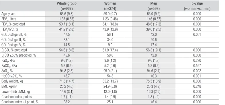
Characteristics
COPD severity ranged from mild to very severe. he aver-age resting blood gas values and body mass index were nor-mal. About half of the patients were current smokers (Table 1). LL-LMM, isokinetic quadriceps peak torque, and peak VO
2
were clearly reduced compared to published data from healthy elderly subjects15,17,28. At peak exercise, patients generally had
little ventilatory reserve and rather high Borg symptom scores for dyspnea and fatigue (Table 2).
Gender differences
On average, the male COPD patients were older, had a worse pulmonary function and a higher score on the Charlson co-morbidity index compared to the female pa-tients. Moreover, men had a significantly higher LL-LMM
Whole group (n=967)
Women (n=374)
Men (n=593)
p-value (women vs. men)
Age, years 63.6 (9.8) 59.9 (9.7) 66.0 (9.2) 0.000
FEV1, liters 1.37 (0.55) 1.23 (0.48) 1.46 (0.57) 0.000 FEV1,% predicted 50.7 (18.1) 54.1 (18.8) 48.6 (17.3) 0.000 FEV1/IVC, % 41.2 (12.8) 43.9 (12.9) 39.6 (12.5) 0.000
GOLD stage I/II, % 47.5 56.1 42.0 0.001
GOLD stage III, % 38.1 34.0 40.6
GOLD stage IV, % 14.5 9.9 17.4
DLCO, % predicted 54.6 (18.6) 51.9 (17.4) 56.3 (19.1) 0.000
DLCO ≤50% predicted, % 45.6 50.0 42.8 0.000
PaO2, kPa 9.6 (1.2) 9.6 (1.2) 9.6 (1.3) 0.290
PaCO2, kPa 5.2 (0.6) 5.2 (0.6) 5.2 (0.6) 0.567
SaO2, % 94.8 (2.3) 95.0 (2.1) 94.6 (2.4) 0.022
HbCO ≥2%, % 45.7 54.3 40.3 0.001
Body weight, kg 71.5 (14.7) 65.2 (13.7) 75.5 (13.9) 0.000
BMI, kg/m2 25.2 (4.6) 24.9 (5.0) 25.3 (4.3) 0.248
Lower-limb LMM, kg 14.6 (3.1) 12.0 (1.8) 16.3 (2.5) 0.000 Charlson index, points 1.7 (1.1) 1.4 (0.9) 1.8 (1.2) 0.000
Charlson index >1 point, % 38.2 25.1 46.4 0.000
Table 1. Characteristics.
Values presented as mean (standard deviation) or as proportion, as appropriate. FEV1: forced expiratory volume in the first second; FM: fat mass; IVC: inspiratory vital capacity; LMM:
lean muscle mass; RV: residual volume; TLC: total lung capacity; DLCO: carbon monoxide transfer factor; %: percentage; PaO2: arterial oxygen tension; PaCO2: arterial carbon monoxide
tension: SaO2: arterial oxygen saturation; kPa: kilo Pascal; BMI: body mass index; m: meters.
(mean difference: 4.3 kg, p<0.01), isokinetic quadriceps peak torque (mean difference: 29.5 Newton-meter, p<0.01), and absolute peak VO
2 (mean difference: 211 ml/min, p<0.01),
while the women had a significantly higher peak VO 2 after
adjustment for LL-LMM (mean difference: 9.8 ml/min/kg LL-LMM, p<0.01). Non-significant gender-related differ-ences were found for peak ventilation (% maximal voluntary ventilation), peak heart rate (% calculated maximum heart rate), Borg symptom scores, and isokinetic quadriceps peak torque after adjustment for LL-LMM (Table 2).
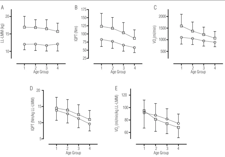
Gender-related differences after stratification for age
As expected, men had a significantly higher mean LL-LMM, isokinetic quadriceps peak torque, and absolute peak VO
2 compared to women also after stratification for
age (Figures 1a to 1c). Moreover, men of age ≥51 years had a significantly higher mean isokinetic quadriceps peak torque after adjustment for LL-LMM compared to women (Figure 1d). In contrast, gender-related differences in absolute peak VO
2 disappeared after adjustment for
LL-LMM. Indeed, mean peak VO
2 adjusted for LL-LMM was
significantly higher in women of age ≥51 years compared to male peers (Figure 1e). See Table E2 of the online deposi-tory for more details on mean differences between groups and p-values.
Age-related difference after stratification for gender
Generally, patients of age ≤50 years had a higher isokinetic quadriceps peak torque and peak VO
2 (absolute values and
after adjustment for LL-LMM) compared to older patients, irrespective of gender (Figure 1). Male patients with age ≥71 years had the lowest LL-LMM compared to younger male pa-tients. No age-graded reductions were found in LL-LMM in female patients (Figure 1a). See Table E3 of the online deposi-tory for more details on mean diferences between groups and p-values.
D
LCO-related differences after stratification for
gender and age
After stratiication for age, women with a DLCO >50%
pre-dicted had a higher mean LL-LMM (in women 51 to 70 years of age; Figure 2a), isokinetic quadriceps peak torque (in women of age ≤50 years; Figure 2b) and absolute peak VO
2 (Figure 2c)
compared to women with a DLCO ≤50% predicted. Moreover,
peak VO
2 remained diferent between both subsets after
ad-justment for LL-LMM (Figure 3b). Isokinetic quadriceps peak torque adjusted for LL-LMM was not diferent between DLCO
strata (Figure 3a). See Table E4 of the online depository for more details on mean diferences between groups and p-values.
A similar pattern was observed in the male patients after stratiication for age and DLCO. Indeed, male patients with a Table 2. Peak exercise performance and quadriceps muscle strength.
Whole group (n=967)
Women (n=374)
Men (n=593)
p-value (women vs. men)
Peak VO
2, ml/min 1138 (347) 1008 (258) 1220 (370) 0.000 Peak VO
2, ml/min/kg BW 16.1 (4.3) 15.8 (4.1) 16.3 (4.4) 0.110 Metabolic equivalents 4.6 (1.2) 4.5 (1.2) 4.7 (1.3) 0.110
Peak VO
2, ml/min/kg LL-LMM 78.6 (19.5) 84.5 (18.8) 74.8 (19.1) 0.000 Peak power output, watts 74.5 (30.6) 65.9 (25.3) 80.0 (32.3) 0.000 Peak VE, liters 45.4 (15.1) 39.9 (12.0) 48.9 (15.8) 0.000 Peak VE, % MVV 86.9 (24.2) 86.0 (24.9) 87.5 (23.8) 0.356 Peak HR, bpm 126.9 (21.4) 130.9 (21.7) 124.3 (21.0) 0.000 Peak HR, % HRmax 81.1 (12.6) 81.7 (12.7) 80.7 (12.5) 0.202 Oxygen pulse, ml/min/bpm 9.1 (2.5) 7.8 (1.9) 9.8 (2.6) 0.000 Peak SaO2, % 91.3 (4.3) 91.8 (4.0) 91.0 (4.4) 0.009
∆ SaO2, % -3.6 (3.7) -3.4 (3.6) -3.7 (3.8) 0.195
Borg score DYS, points 7.0 (2.2) 7.1 (2.1) 6.9 (2.2) 0.085 Borg score FAT, points 5.6 (2.6) 5.7 (2.6) 5.6 (2.0) 0.351
IQPT, Nm 90.1 (32.6) 72.0 (21.6) 101.5 (33.2) 0.000
IQPT, Nm/kg LL-LMM § 12.3 (3.2) 12.1 (3.1) 12.4 (3.3) 0.125
Values presented as mean (standard error). VO
2: VO2; ml/min: milliliter per minute; kg: kilogram; BW: body weight; LMM: lean muscle mass; LL: lower-limbs; bpm: beats per minute;
%: percentage; MVV: maximal voluntary ventilation; HRmax: calculated maximum heart rate (220-age in years); SaO2: transcutaneous oxygen saturation; ∆: change; DYS: dyspnea; FAT:
fatigue; IQPT: isokinetic quadriceps peak torque; Nm: Newton-meter; § : IQPT of the quadriceps muscle has been assessed single-legged, therefore lower-limb lean muscle mass has been divided by 2.
DLCO >50% predicted had a higher mean LL-LMM (in men of age ≥51 years; Figure 2d), isokinetic peak torque (in men ≤70 years of age; Figure 2e) and absolute peak VO
2 (Figure 2f)
com-pared to male patients with a DLCO ≤50% predicted. h ese DLCO-related dif erences remained after adjustment for LL-LMM, in particular for peak VO
2 (Figures 3c and 3d). See Table
E5 of the online depository for more details on mean dif er-ences between groups and p-values.
Age-related differences after stratification for
gender and D
LCO
Women with a DLCO ≤50% predicted showed an age-graded reduction in isokinetic quadriceps peak torque (Figure 2b) and peak VO
2 (Figure 2c). These age-graded
re-ductions remained after adjustment for LL-LMM (Figures 3a and 3b). The age-graded reductions in isokinetic quadriceps
peak torque (Figure 2b) and peak VO
2 (Figure 2c) were even
more explicit in the women with a DLCO >50% predicted. LL-LMM did not show an age-graded reduction in either DLCO group (Figure 2a). A similar pattern was observed for the male patients after stratification for DLCO (Figures 2d, 2e, 2f, 3c, and 3d). See Table E6 of the online depository for more details on mean differences between groups and p-values.
Stepwise multiple regression model
A stepwise multiple regression analysis ascertained LL-LLM, age, peak VO
2, and gender to be significant
deter-minants of isokinetic quadriceps peak torque. This model explained 56.4% of the variance in isokinetic quadriceps peak torque (p=0.01). DLCO (p=0.73), FEV1 (p=0.16), and BMI (p=0.07) did not contribute to the model.
Figure 1. Lower-limb lean muscle mass, isokinetic quadriceps peak torque, and peak aerobic capacity after stratification for age in 374 women and 593 men with COPD.
4
25
500 1000 1500 2000
50 75 100 125 150 175
3 2 Age Group
1 2 3 4
Age Group
1 2 3 4
Age Group 1
10
4 15
LL-LMM (kg) IQPT (Nm) VO
2
(ml/min)
20
A
B
C
4 Age Group
5
60 80 100 120
1 2 3 4
Age Group
1 2 3 4
10 15 20
IQPT (Nm/kg LL-LMM) VO2
(ml/min/kg LL-LMM)
D
E
Lower-limb lean muscle mass (LL-LMM, kg; Figure 1a), isokinetic quadriceps peak torque (IQPT, Newton-meter, Nm; Figure 1b), and peak aerobic capacity (VO
2, ml/min; Figure 1c)
after stratification for age (group 1: ≤50 years; group 2: 51 to 60 years; group 3: 61 to 70 years; group 4: ≥71 years) and gender (women: open circles; men: open squares). See Table
E2 in the online supplement for more details. Isokinetic quadriceps peak torque adjusted for LL-LMM (IQPT/LL-LMM, Nm/kg; Figure 1d) and peak VO
2 adjusted for LL-LMM (VO2/
LL-LMM, ml/min/kg; Figure 1e) after stratification for age and gender (women: open circles; men: open squares). See Table E2 in the online supplement for more details.
10
1 2 3
Age group 4
1 2 3
Age group
4 1 2 3
Age group
4 1 2 3
Age group 4
1 2 3
Age group
4 1 2 3
Age group 4 15
LL-LMM (kg) IQPT (Nm) VO
2
(ml/min)
LL-LMM (kg)
20
175
150
125
100
75
50 500
1000 1500 2000
VO
2
(ml/min)
500 1000 1500 2000 25
IQPT (Nm)
175
150
125
100
75
50
25 10
15 20
A
D
E
F
C
B
Lower-limb lean muscle mass (LL-LMM, kg; Figures 2a and 2d), isokinetic quadriceps peak torque (IQPT, Newton-meter, Nm; Figure 2b and 2e) and peak aerobic capacity (
VO
2, ml/min; Figures 2c and 2f) after stratification for age and DLCO (≤50% predicted, black; >50% predicted, grey) in female and male COPD patients (women: circles;
men: squares), respectively. See Tables E3 and E4 of the online supplement for more details.
Figure 2. Lower-limb lean muscle mass, isokinetic quadriceps peak torque, and peak aerobic capacity after adjustment for age and DLCO in women
and men with COPD.
A stepwise multiple regression analysis ascertained isoki-netic quadriceps peak torque, DLCO, FEV1, BMI, age and
LL-LMM to be signii cant determinants of peak VO
2. h is model
explained 59.3% of the variance in peak VO
2 (p=0.01). Gender
did not contribute to the model (p=0.22).
Discussion
As we expected, quadriceps muscle strength and peak VO 2
were related to age in patients with COPD, particularly in those with DLCO >50% predicted. Yet, counter to our hypothesis
LL-LMM did not account for the relationship of age with quad-riceps muscle strength and peak VO
2. Indeed, an age-graded
reduction in LL-LMM was only present in the older men with
a DLCO >50% predicted. h e results of this study suggest the
presence of age-related qualitative abnormalities in lower-limb muscles in patients with COPD (i.e. a decrease in isokinetic peak torque per unit weight of lower-limb lean muscle mass and/or a decrease in peak VO
2 per unit weight of lower-limb
lean muscle mass).
An age-related decline in quadriceps muscle strength and peak VO
2 is well established in healthy subjects and in cardiac
patients8,9,29. Moreover, small but signii cant reductions in
quad-riceps muscle strength10 and peak
VO 2
11,12 were found in (mostly
male) COPD patients over a 1 to 5 year period, respectively. To the best of our knowledge, this is the i rst study to show an age-graded reduction in quadriceps muscle strength and peak VO
2
in patients with COPD after correction for possible confounding factors, such as gender, LL-LMM, and DLCO. More importantly
this study corroborates the previously identii ed decrease in lower-limb muscle function in patients with COPD3,16. Moreover,
our results also highlight age-graded dif erences between men and women, which also parallels previous i ndings30.
Lower-limb muscle weakness can occur without an overt loss of LL-LMM in patients with COPD suggesting the presence of qualitative skeletal muscle abnormalities16, particularly in
women3. h e present i ndings are in line: skeletal muscle
func-tion can decrease without a decrease in LL-LMM in patients with COPD, in particular in women (Table E2). In healthy el-derly subjects, gender dif erences exist in the contractile prop-erties of lower-limb muscles. Indeed, type I and IIA i bers from older healthy men were generally stronger than similar i bers from older women even after adjusting for size31. Whether and
to what extent gender dif erences in muscle contractile prop-erties are also present in COPD remains currently unknown32.
There are several factors that may explain the overt loss in peak VO
2 (absolute and corrected for LL-LMM) in
COPD. Patients with COPD have a significantly lower mi-tochondrial density33 and activity34, as well as a lower
oxida-tive enzyme activity35 and a lower mechanical efficiency36.
There are also fewer capillaries per muscle fiber in patients with COPD. Lastly, there are more type II muscle fibers in patients with COPD compared to healthy age-matched control subjects37. A worsening of the above-mentioned
intramuscular manifestations over time may explain the age-related differences in peak VO
2. However, most of these
intramuscular changes have been identified in small cross-sectional studies of mostly men, focusing mainly on the vastus lateralis muscle and GOLD stages 3 and 4. Coupled with our results, these mechanistic insights underline the importance of continued research on this phenomenon of loss of lower-limb muscle VO
2. Indeed, there is great
poten-tial for identifying new therapeutic targets38.
Signii cant dif erences in lower-limb muscle function were found in female (Figures 3a and 3b) and male COPD patients
10
5
1 2 3
Age group 4
1 2 3
Age group
IQPT (Nm/kg LL-LMM)
IQPT (Nm/kg LL-LMM) VO
2
(ml/min/kg LL-LMM)
VO
2
(ml/min/kg LL-LMM)
4 1 2 3
Age group 4
1 2 3
Age group 4 15
20
A
C
D
B
10
5 15 20
120
100
80
60
120
100
80
60
Isokinetic quadriceps peak torque adjusted for LL-LMM (IQPT/LL-LMM, Nm/kg; Figures 3a and 3c) and peak VO
2 adjusted for LL-LMM (VO2/LL-LMM, ml/min/kg;
Figures 3b and 3d) after stratification for age and DLCO (≤50% predicted, black; >50% predicted, grey) in female and male COPD patients (women: circles; men: squares),
respectively. See Tables E3 and E4 of the online supplement for more details.
Figure 3. Quadriceps muscle strength and peak aerobic capacity after adjustment for age, lower-limb lean muscle mass, and DLCO in women and men with COPD.
(Figures 3c and 3d) after stratiication for DLCO. his seems to be in line with previous indings of Amann et al.39, who reported that the
high susceptibility to lower-limb muscle fatigue in patients with COPD is in part attributable to insuicient oxygen transport as a consequence of exaggerated arterial hypoxemia and/or excessive respiratory muscle work. Moreover, quadriceps muscle strength was positively related to DLCO in patients with COPD
18.
Age-related reductions in LL-LMM have been reported in healthy women and men17. Hopkinson at al.10 reported a
non-signiicant decline of 0.2 kilogram in fat-free mass during a 1-year follow-up period in 64 COPD patients10. Also in the
current cross-sectional analyses no age-graded reductions in LL-LMM were found, except for the older male COPD patients with a DLCO >50% predicted (Figure 3d and Table E6 of the on-line supplement). It remains diicult to understand the lack of age-graded reductions in LL-LMM in patients with COPD.
he external validity of the present indings is limited to COPD patients without long-term oxygen therapy. hus the present data should not be uncritically applied to GOLD IV pa-tients with long-term oxygen therapy. Obviously, a major limita-tion of the present analysis is the lack of a healthy control group and the lack of information about daily physical activity levels. However, quadriceps muscle strength and peak VO
2 have been
found to decline over time in healthy subjects9,30. Moreover, the
inluence of daily physical inactivity on an age-related decline in peak VO
2 remains a matter of debate
8,30. Patients with COPD
have a reduced level of daily physical activity, which is present even in the earliest stages of the disease40. It is for this reason
that it is not very surprising that quadriceps muscle strength and peak VO
2 in patients with COPD is very low compared to
healthy subjects8,17,30 and not unlike that in patients entering
car-diac rehabilitation29. Accordingly, the majority of subjects with
COPD in our study (98.8%) had a metabolic equivalent below 8, which is an identiied risk factor of death from any cause6. In
ad-dition, an age-related decline in peak VO
2 can explain, at least
in part, the development of disability in patients with COPD41.
Indeed, GOLD stage II patients use a higher proportion of their (reduced) peak VO
2 compared to healthy elderly subjects during
the performance of simple, self-paced domestic activities of daily living4. Finally, large longitudinal studies are needed to
corrobo-rate the current indings.
In summary, we have found that lower-limb muscle qual-ity is lower in older patients with COPD, also after stratiica-tion for gender and gas transfer. herefore, prevenstratiica-tion of an age-graded decline in quadriceps muscle strength and peak
VO
2 may need to become an outcome of the management of
patients with COPD.
References
1 Rabe KF, Hurd S, Anzueto A, Barnes PJ, Buist SA, Calverley P, et al. Global strategy for the diagnosis, management, and prevention of chronic obstructive pulmonary disease: GOLD executive summary. Am J Respir Crit Care Med. 2007;176:532-55.
2 Celli BR, MacNee W; ATS/ERS Task Force. Standards for the diagnosis and treatment of patients with COPD: a summary of the ATS/ERS position paper. Eur Respir J. 2004;23(6):932-46.
3 Seymour JM, Spruit MA, Hopkinson NS, Natanek SA, Man WD, Jackson A, et al. The prevalence of quadriceps weakness in COPD and the relationship with disease severity. Eur Respir J. 2010;36(1):81-8.
4 Vaes AW, Wouters EF, Franssen FM, Uszko-Lencer NH, Stakenborg KH, Westro M, et al. Task-related oxygen uptake during domestic activities of daily life in patients with COPD and healthy elderly subjects. Chest. 2011;140(4):970-9.
5 Decramer M, Gosselink R, Troosters T, Verschulren M, Evers G. Muscle weakness is related to utilization of health care resources in COPD patients. Eur Respir J. 1997;10(2):417-23.
6 Myers J, Prakash M, Froelicher V, Do D, Partington S, Atwood JE. Exercise capacity and mortality among men referred for exercise testing. N Engl J Med. 2002;346(11):793-801.
7 Swallow EB, Reyes D, Hopkinson NS, Man WD, Porcher R, Cetti EJ, et al. Quadriceps strength predicts mortality in patients with moderate to severe chronic obstructive pulmonary disease. Thorax. 2007;62(2):115-20.
8 Fleg JL, Morrell CH, Bos AG, Brant LJ, Talbot LA, Wright JG, et al. Accelerated longitudinal decline of aerobic capacity in healthy older adults. Circulation. 2005;112(5):674-82.
9 Frontera WR, Hughes VA, Fielding RA, Fiatarone MA, Evans WJ, Roubenoff R. Aging of skeletal muscle: a 12-yr longitudinal study. J Appl Physiol. 2000;88(4):1321-6.
10 Hopkinson NS, Tennant RC, Dayer MJ, Swallow EB, Hansel TT, Moxham J, et al. A prospective study of decline in fat free mass and skeletal muscle strength in chronic obstructive pulmonary disease. Respir Res. 2007;8:25.
11 Oga T, Nishimura K, Tsukino M, Sato S, Hajiro T, Mishima M. Exercise capacity deterioration in
patients with COPD: longitudinal evaluation over 5 years. Chest. 2005;128(1):62-9.
12 Oga T, Nishimura K, Tsukino M, Sato S, Hajiro T, Mishima M, et al. Longitudinal deteriorations in patient reported outcomes in patients with COPD. Respir Med. 2007;101(1):146-53.
13 Engelen MP, Schols AM, Does JD, Wouters EF. Skeletal muscle weakness is associated with wasting of extremity fat-free mass but not with airflow obstruction in patients with chronic obstructive pulmonary disease. Am J Clin Nutr. 2000;71(3):733-8.
14 Baarends EM, Schols AM, Mostert R, Wouters EF. Peak exercise response in relation to tissue depletion in patients with chronic obstructive pulmonary disease. Eur Respir J. 1997;10(12):2807-13.
15 Franssen FM, Broekhuizen R, Janssen PP, Wouters EF, Schols AM. Limb muscle dysfunction in COPD: effects of muscle wasting and exercise training. Med Sci Sports Exerc. 2005;37(1):2-9.
16 Bernard S, LeBlanc P, Whittom F, Carrier G, Jobin J, Belleau R, et al. Peripheral muscle weakness in patients with chronic obstructive pulmonary disease. Am J Respir Crit Care Med. 1998;158(2):629-34.
17 Neder JA, Lerario MC, Castro ML, Sachs A, Nery LE. Peak VO2 correction for fat-free mass estimated by anthropometry and DEXA. Med Sci Sports Exerc. 2001;33(11):1968-75
18 Spruit MA, Gosselink R, Troosters T, Kasran A, Gayan-Ramirez G, Bogaerts P, et al. Muscle force during an acute exacerbation in hospitalised patients with COPD and its relationship with CXCL8 and IGF-I. Thorax. 2003;58(9):752-6.
19 Eid AA, Ionescu AA, Nixon LS, Lewis-Jenkins V, Matthews SB, Griffiths TL, et al. Inflammatory response and body composition in chronic obstructive pulmonary disease. Am J Respir Crit Care Med. 2001;164(8 Pt 1):1414-8.
20 Pinto-Plata VM, Celli-Cruz RA, Vassaux C, Torre-Bouscoulet L, Mendes A, Rassulo J, et al. Differences in cardiopulmonary exercise test results by American Thoracic Society/European Respiratory Society-Global Initiative for Chronic Obstructive Lung Disease stage categories and gender. Chest. 2007;132(4):1204-11.
21 Spruit MA, Vanderhoven-Augustin I, Janssen PP, Wouters EF. Integration of pulmonary rehabilitation in COPD. Lancet. 2008;371(9606):12-3.
22 Gosselink R, Troosters T, Decramer M. Peripheral muscle weakness contributes to exercise limitation in COPD. Am J Respir Crit Care Med. 1996;153(3):976-80.
23 American Thoracic Society; American College of Chest Physicians. ATS/ACCP Statement on cardiopulmonary exercise testing. Am J Respir Crit Care Med. 2003;167(2):211-77.
24 Macintyre N, Crapo RO, Viegi G, Johnson DC, van der Grinten CP, Brusasco V, et al. Standardisation of the single-breath determination of carbon monoxide uptake in the lung. Eur Respir J. 2005;26(4):720-35.
25 Miller MR, Hankinson J, Brusasco V, Burgos F, Casaburi R, Coates A, et al. Standardisation of spirometry. Eur Respir J. 2005;26(2):319-38.
26 Spruit MA, Pennings HJ, Janssen PP, Does JD, Scroyen S, Akkermans MA, et al. Extra-pulmonary features in COPD patients entering rehabilitation after stratification for MRC dyspnea grade. Respir Med. 2007;101(12):2454-63.
27 Lamers RJS, Wouters EFM, Kemerink GJ. Emphysema and airflow limitation in patients with advanced chronic obstructive pulmonary disease: a CT study. PhD Thesis (chapter 8) 1994.
28 Gosker HR, Lencer NH, Franssen FM, van der Vusse GJ, Wouters EF, Schols AM. Striking similarities in systemic factors contributing to decreased exercise capacity in patients with severe chronic heart failure or COPD. Chest. 2003;123(5):1416-24.
29 Ades PA, Savage PD, Brawner CA, Lyon CE, Ehrman JK, Bunn JY, et al. Aerobic capacity in patients entering cardiac rehabilitation. Circulation. 2006;113(23):2706-12.
30 Toth MJ, Gardner AW, Ades PA, Poehlman ET. Contribution of body composition and physical activity to age-related decline in peak VO2 in men and women. J Appl Physiol. 1994;77(2):647-52.
31 Frontera WR, Suh D, Krivickas LS, Hughes VA, Goldstein R, Roubenoff R, et al. Skeletal muscle fiber quality in older men and women. Am J Physiol Cell Physiol. 2000;279(3):C611-8.
32 Debigaré R, Côte CH, Hould FS, LeBlanc P, Maltais F. In vitro and in vivo contractile properties
of the vastus lateralis muscle in males with COPD. Eur Respir J. 2003;21(2):273-8.
33 Gosker HR, Hesselink MK, Duimel H, Ward KA, Schols AM. Reduced mitochondrial density in the vastus lateralis muscle of patients with COPD. Eur Respir J. 2007;30(1):73-9.
34 Rabinovich RA, Bastos R, Ardite E, Llinas L, Orozco-Levi M, Gea J, et al. Mitochondrial dysfunction in COPD patients with low body mass index. Eur Respir J. 2006;29(4):643-50.
35 Maltais F, LeBlanc P, Whittom F, Simard C, Marquis K, Belanger M, et al. Oxidative enzyme activities of the vastus lateralis muscle and the functional status in patients with COPD. Thorax. 2000;55(10):848-53.
36 Baarends EM, Schols AM, Akkermans MA, Wouters EF. Decreased mechanical efficiency in clinically stable patients with COPD. Thorax. 1997;52(11):981-6.
37 Gosker HR, Zeegers MP, Wouters EF, Schols AM. Muscle fibre type shifting in the vastus lateralis of patients with COPD is associated with disease severity: a systematic review and meta-analysis. Thorax. 2007;62(11):944-9.
38 Hansen MJ, Gualano RC, Bozinovski S, Vlahos R, Anderson GP. Therapeutic prospects to treat skeletal muscle wasting in COPD (chronic obstructive lung disease). Pharmacol Ther. 2006;109(1-2):162-72.
39 Amann M, Regan MS, Kobitary M, Eldridge MW, Boutellier V, Pegelow DF, et al. Impact of pulmonary system limitations on locomotor muscle fatigue in patients with COPD. Am J Physiol Regul Integr Comp Physiol. 2010;299(1):R314-24.
40 Pitta F, Troosters T, Spruit MA, Probst VS, Decramer M, Gosselink R, et al. Characteristics of physical activities in daily life in chronic obstructive pulmonary disease. Am J Respir Crit Care Med. 2005;171(9):972-7.
41 Guccione AA, Felson DT, Anderson JJ, Anthony JM, Zhang Y, Wilson PW, et al. The effects of specific medical conditions on the functional limitations of elders in the Framingham Study. Am J Public Health. 1994;84(3):351-8.