*Correspondence: Shiva Golmohammadzadeh, School of Pharmacy, Mash-had University of Medical Sciences, MashMash-had, Iran, P.O. Box: 91389-13131. E mail: [email protected]
A
vol. 48, n. 4, oct./dec., 2012
Preparation, characterization and evaluation of moisturizing and
UV protecting effects of topical solid lipid nanoparticles
Shiva Golmohammadzadeh
1,*, Mohsen Mokhtari
1, Mahmoud Reza Jaafari
21Nanotechnology Research Center, School of Pharmacy, Mashhad University of Medical Sciences, Mashhad, Iran, 2Biotechnology Research Center, Nanotechnology Research Center, School of Pharmacy, Mashhad University of Medical
Sciences, Mashhad, Iran
Solid lipid nanoparticles (SLN) were recently proposed as carriers for various pharmaceutical and cosmetic actives. These lipid nanoparticles can act as moisturizers and physical sunscreens on their own. Therefore, the full potential of these carriers has yet to be determined. The present study was aimed to determine and compare moisturizing and UV-protecting effects of different solid lipid nanoparticles (SLN) prepared by different solid lipids including Glyceryl monostearate (GMS), Precirol® (P) and cetyl palmitate (CP)
as carrier systems of moisturizers and sunscreens. The inluence of the size and matrix crystallinity of
the solid lipids on the occlusive factor, skin hydration and UV-protection were evaluated by in vitro and in vivo methods. The SLN were prepared by high-shear homogenization and ultrasound methods. Size, zeta potential and morphological characteristics of the samples were assessed by transmission electron microscopy (TEM) and thermotropic properties with differential scanning calorimetry (DSC) technique.
Results of the assessments showed that SLN-CP signiicantly increases skin hydration and UV-protection,
compared to SLN-GMS and SLN-P. It was demonstrated that the size of SLN, crystallinity index of solid
lipid in SLN and probably other mechanisms besides the occlusive factor can inluence skin hydration and UV-protection indices. Furthermore, indings of the assessments demonstrated signiicant difference
between in vitro and in vivo assessments regarding occlusive factor and moisturizing effects. Findings of the present study indicate that the SLN-CP could be a promising carrier for sunscreens and moisturizers.
Uniterms: Solid lipid nanoparticles/moisturizing effects. Solid lipid nanoparticles/UV protecting.
Nanopartículas lipídicas sólidas (NLS) foram, recentemente, propostas como carreadores de vários ativos cosméticos e farmacêuticos. Essas nanopartículas lipídicas podem atuar como hidratantes e protetores solares físicos por si só. Assim sendo, determinou-se o potencial desses carreadores. Os objetivos do presente estudo foram determinar e comparar os efeitos hidratantes e protetores contra UV das diferentes partículas lipídicas sólidas (NLS) preparadas com diferentes lipídios sólidos, incluindo o monoestearato de gligerila (MSG), Precirol® (P) e palmitato de cetila (PC) como sistemas carreadores de hidratantes
e de protetores solares. A inluência do tamanho e da cristalinidade da matriz dos lipídios sólidos no
fator oclusivo, na hidratação da pele e na proteção ao UV foi avaliada por métodos in vitro e in vivo. As NLS foram preparadas por homogeneização por alto corte e métodos de ultrassom. Tamanho, potencial zeta e características morfológicas das amostras foram determinados por microscopia de transmissão eletrônica (MTE) e as propriedades termotrópicas, com diferentes técnicas de calorimetria diferencial
de varredura (CDV). Os resultados mostraram que NLS-PC aumenta signiicativamente a hidratação
da pele e a proteção ao UV, comparativamente à NLS-MSG e à NLS-P. Demonstrou-se que o tamanho da NLS, índice de cristalinidade do lipídio sólido na NLS e, provavelmente, outros mecanismos além
do fator oclusivo podem inluenciar a hidratação da pele e os índices de proteção ao UV. Além disso, os resultados mostraram diferença signiicativa entre as avaliações in vitro e in vivo com relação ao fator oclusivo e aos efeitos hidratantes. Os resultados do presente estudo indicam que NLS-PC poderia ser um carreador promissor para protetores solares e hidratantes.
INTRODUCTION
Sunlight exposure can be both beneicial and harm -ful for the human body. It has been known for decades that sunscreens are capable of protecting human body of solar radiation-induced harmful effects (Kullavanijaya et al., 2005; Potard et al., 2000). Since sunscreens should act on the surface of the skin, they should penetrate as little as possible into the viable epidermis, the dermis and into the systemic circulation (Potard et al., 2000).
Several factors are involved in the transdermal deliv-ery of drugs and cosmetic actives, from topically-applied formulations. The penetration and the effectiveness of active compounds through the human skin depends on physicochemical properties of the drug, size of the mol-ecule, drug-delivery system, lipophilicity of components,
vehicle and skin hydration which can be inluenced by
occlusive and other compounds (Verma et al., 2003; Zhai et al., 2002).
Solid lipid nanoparticles (SLN) have been intro-duced as a novel drug-delivery systems for pharmaceu-tical drugs and cosmetic active ingredients due to their advantages over conventional formulations (Muller et al., 2000; Wissing et al., 2003a). They are promising carriers as protecting labile active compounds from degradation (Jenning et al., 2001; Muller et al., 2000), releasing active ingredients in a controlled way (Zur Muhlen et al., 1998; Maia et al., 2000), increasing skin water content, (Wiss-ing et al., 2001; Wissing et al., 2002b) and UV-blocking potential as physical sunscreens (Wissing et al., 2001b; Wissing et al., 2002a).
The occlusive property of the SLN is due to its ilm
formation after application through the skin. The extent of the occlusive properties depends on various factors, e.g. particles size, lipid and lipid concentration. The UV
pro-tection is based on the UV relecting and scattering abil -ity like other physical sunscreens (Wissing et al., 2001b; Wissing et al., 2002a; Wissing et al., 2002b).
The occlusive and UV-blocking properties of SLN can introduce them as a promising vehicle for the moistur-izer and sunscreen products.
It was observed by other researches that the oc-clusion factor of lipid microparticles was only 10%, compared to 50% when using lipid nanoparticles of ap-proximately 200 nm (Souto et al., 2008). Meanwhile it was found that among Dynasan 112, Compritol 888 ATO and Softisan 154 as solid lipids in SLN; the highest occlu-sion will be achieved from low melting lipids with highly crystalline particles (Wissing et al., 2003b). These studies do not fully mimic the natural moisture loss conditions and there was no comparison between in vitro and in vivo skin
hydration. SLN have been also introduced as a novel car-rier for sunscreen ingredients. UV protecting of different lipids in SLN formulations were not investigated by SPF determination in vitro method.
In this study, we investigated and compared the
in-luence of size and crystallinity of different lipid composi -tion in SLN formula-tions on the occlusion factor using in vitro method and the skin hydration using corneometer in vivo method. UV protection properties of different lipids in SLN formulations were also investigated by Transpore tape 3Min vitro method.
MATERIAL AND METHODS
Material
Glyceryl palmitostearate (Precirol® ATO 5) and glyceryl monostearate (GMS) were gifted by Gattefossé (Pvt. Ltd., France). Cetyl palmitate (CP) and Tween 80 were purchased from Sigma-Aldrich(Deisenhofen Ger-many). Poloxamer188 was obtained from Uniqema (Ev-erberg, Belgium). All of the original samples were used as their arrival. Water was used a double-distilled water.
Preparation of SLN
The SLN were prepared by high-shear homogeniza-tion and ultrasound method. Precirol® ATO 5, GMS and CP (1 g) were melted by heating at 5 ºC above the melting point of the lipids. The aqueous phase was prepared by dissolving tween 80 (0.5 g) for GMS and CP, or poloxamer 188 (0.5 g) for Precirol® ATO 5 in double-distilled water (10 ml of the solution was produced) and that was heated up to the melting point temperature of the lipid phase. Hot aqueous phase was added to the molten lipid phase and homogenized by Diax 900 homogenizer (Heidolph, Germany) for 2 min at 11,000 rpm. The temperature was kept at 5 ºC above the melting point of the lipid. Coarse hot oil in water emulsion obtained was ultrasonicated by Prob Sonicator (Bransonic, USA). The prob sonication was performed at 6 cycles with 30 seconds of sonication separated by intervals of 15 seconds. The obtained nano-emulsions were cooled to room temperature(Kumar et al., 2007; Venkateswarlu et al., 2004).
Characterization of SLN
Particle size and zeta potentials
and zeta potential of the SLN formulations. All measure-ments were performed in triplicate at a temperature of 25 ºC ± 2 ºC and an angle of 90 ºC to the incident beam. No multi-scattering phenomenon was observed during the assessments.
Transmission electron microscopy (TEM)
TEM assessment (TEM; CEM 902A; Zeiss, Oberkochen, Germany) was performed to characterize the morphology of SLN formulations. The SLN were diluted 50 times with water and then placed on a carbon-coated copper grid for 30 seconds and the excess water was wiped
off by a ilter paper. Then 20 µL of uranyl acetate 2% in
water covered on SLN and after 30 seconds were wiped
off by ilter paper. The grid was dried at room temperature
and then assessed by TEM (Liu et al., 2007).
Differential Scanning Calorimetry (DSC)
Melting and recrystallization behavior of crystalline materials were assessed using Mettler DSC 821e (Mettler Toledo, Gießen, Germany. DSC scans of the bulk lipids and SLN formulations were carried out. An empty alumi-num pan served as reference. Samples were scanned from 25 ºC to 100 ºC (5 ºC/min) under nitrogen atmosphere (20 mL/min); then, the melting point of SLN formulations was compared to the bulk lipid. Before the DSC measurements, the bulk lipids were heated up to 75 ◦C and cooled to the room temperature to imitate the production conditions. Analysis was carried out under nitrogen purge (Jenning et al., 2001; Wissing et al., 2002a).
Occlusive Properties Assessment
For the occlusion test, 100 mL beakers were illed with 50 mL water and covered with ilter paper (cellulose acetate ilter, cutoff size: 4-7 µm) and sealed. Samples were spread on the ilter surface (13.3 mg/cm2) and stored at 32 ºC and 50%-55% Relative Hydration (PH) for 48 h.
Beakers covered with ilter paper with no sample, were
considered as reference. The occlusion factor (F) was cal-culated according to the following Equation (Eq.1) After 24 h and 48 h, where A is the water loss without sample (reference), and B is the water loss with sample. Every experiment was carried out in triplicate (Wissing et al., 2002b; Souto et al., 2004).
F=100× ((A−B)/A) Eq. 1
Skin hydration measurement using Corneometer
The moisture content of the skin was measured by a corneometer (Courage, Khazaka, Cologne, Germany) following application of different SLN formulations. The
test was carried out on 6 volunteers with normal skin at room temperature (ages were between 20 and 35 years). Before the measurements, subjects were given time to adapt to room conditions without covering the measuring sites. On the day of examination, the skin was not washed and nothing was applied to the skin surface. Subjects were instructed not to apply any preparation to the site to be examined one week before investigation. The corneometer CM 820 was used to determine the humidity level of the stratum corneum by measuring electrical capacitance. All the measured values were expressed as the median of three recordings. The measurements were carried out on exactly the same sites. The measuring place was in the middle of
the forearm. In the irst instant, the moisture content of the
skin without any application of the product was measured, and then the measurement was carried out after 30 min, 2, 3, 6 and 10 hours after application of the sunscreens (Sator et al., 2003).
SPF determination of the formulations using Transpore tape in vitro method
The principle of this method is based on the mea-surement of the spectral transmittance of UVR through a sample of a surgical tape which is called Transpore tape with and without the sunscreen applied. This substrate was
introduced irst by Diffey (Diffey, 2002). SLN formula -tions and sunscreen standard were applied on the surface of the TransporeTM tape at 2 mg/cm2. After 15 minutes transmission was measured from 290 nm to 400 nm at
intervals of 5 nm at ive distinct points. The predicted SPF
value was calculated according to the following equation (Eq. 2).
Eq. 2
In this equation: E(λ): CIE Relative Erythemal Spectral Effectiveness, S(λ): Solar Spectral Irradiance
(Wm-2nm-1), T(λ): Spectral Transmittance of the sample (as measured on the UV-1000S). Results were reported as mean SPFs and relative standard deviations as percentage of the mean SPF.
Data analysis
value ± S.D. The statistical data were analyzed using nonparametric with a Tukey-Kramer test. Results with P
< 0.05 were considered statistically signiicant.
RESULTS AND DISCUSSION
Three types of SLN were prepared by high-shear homogenization and ultrasound method. Homogeniza-tion, followed by ultrasonicaHomogeniza-tion, is a simple, reliable and reproducible method for SLN preparation (Venkateswarlu et al., 2004). The process parameters, involved in the preparation of SLN were optimized, including lipid and surfactant (type and concentration), lipid/surfactant ratio, homogenization and sonication time to reduce the size of nanoparticles with narrow size distribution. Physically stable SLN with a narrow size distribution were produced. To obtain stable and smaller SLN, sonication cycles, were varied from 6 to 20 and their effect on particle size was measured. It was obtained that in 12 and 20 cycles, the PI
of the SLN distribution was increased with no signiicant
difference in the mean size of the particles. Therefore, 6 cycles were used for the preparations.
The mean diameter (z-average), PI and zeta potential of the bulk population of the particles, were measured by particle size analyzer (PSA), which are shown in Table I.
Among the lipids used, GMS and Precirol produced
sig-niicantly smaller size of SLN, compared CP. In addition, no signiicant difference of the size between SLN-GMS
and SLN-P were obtained. The difference between the mean particle size and particle size distribution is because
of the difference of the type of lipid and emulsiier in dif -ferent SLN formulations. GMS is a surface active partial glyceride which facilitated emulsification and formed
more rigid surfactant ilms and then improved the
long-term stability of SLN (Hou et al., 2003).
Zeta potential can make a prediction about the stabil-ity of colloid dispersions. A high zeta potential (>30 mV) can provide an electric repulsion to avoid the aggregation of the particles (Levy et al., 1994). The zeta potentials of SLN-GMS and SLN-P were above -35mV which was physically more stable than SLN-CP.
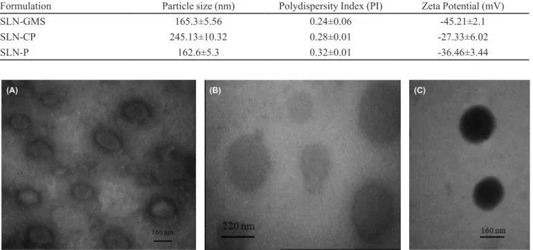
The TEM imaging of GMS, CP and SLN-P SLN are shown in Figure 1. It was observed all of the SLN formulations exhibited nanometric size, spherical shape and had a narrow size distribution. There was no difference between the particle size of from TEM images and PSA.
To determine the extent of crystallinity, the samples were investigated by differential scanning calorimetry (DSC). Samples were scanned from 25 ºC to 100 ºC
TABLE I - The particle size, polydispersity Index (PI) and Zeta potential of different SLN formulations measured by PSA. Data expressed as Mean ± SD (n = 3) (according to the zeta average), * Statistical signiicance with SLN-GMS and SLN-CP (p < 0.05),
not signiicant with SLN-GMS and SLN-P (p > 0.05)
Formulation Particle size (nm) Polydispersity Index (PI) Zeta Potential (mV)
SLN-GMS 165.3±5.56 0.24±0.06 -45.21±2.1
SLN-CP 245.13±10.32 0.28±0.01 -27.33±6.02
SLN-P 162.6±5.3 0.32±0.01 -36.46±3.44
(5 ºC/min) and the melting point of SLN formulations was compared to the bulk lipid. The DSC scans of the formulations and the bulk lipids are shown in Figure 2. The melting peak of pure GMS, Precirol and CP proved the solid character of the lipid matrix at room temperature as they are higher than room temperature. The thermo-gram in Figure 2 (A) and (B) shows that the SLN-GMS and SLN-Precirol depressed the transition temperature of the lipids to a lower temperature and also changed the structure of the peak. The two thermal maxima in the DSC melting of SLN-P curve indicate melting of two different crystalline forms.
The depression of the melting temperature in SLN is due to the lower size and the structure of the SLN. These Figures also showed that the lipid does not recrystallize completely at room temperature however, SLN-CP (Figure 2 (C)) forms highly crystalline lipid nanoparticles and recrystallize completely.
The extent of crystallinity was investigated by DSC. The peak location of SLN-GMS is slightly shifted towards lower temperatures compared to that of the bulk lipid. The recrystallization of the SLN occurred at lower tempera-tures than the bulk material. The decline of the scan can be explained by the small particle sizes of the SLN
formula-tions, their high speciic surface area and the presence of
a surfactant (Saupe et al., 2005). It was demonstrated that when the drug is added into SLN, the lipid crystals in an orderly situation were further disrupted which can reduce the crystallization property of nanoparticles. Therefore, the crystallinity of the lipid gradually declines from GMS to SLN-GMS as was shown in Li et al research (Li et al., 2010).
The dependency of the occlusion factor (F) upon the size and crystanillity of the SLN formulations is shown in Figure 3.
No significant difference of the occlusive factor between SLN formulations was observed after 24 and 48h. Increasing the particle size in CP towards SLN-GMS and SLN-P showed a decrease in occlusive factor although the high crystanillity of SLN-CP may enhance the occlusive factor, therefore, the crytanillity effect can modify the size effect on occlusive factor.
In the current study, it was observed that the oc-clusive factor depends on the size of the particles and crystallinity of the lipids in the SLN formulations. Find-ings of the study conducted by Wissing et al. 2003b), also confirms that, the dependence of occlusion factor on the particle size of SLN-CP. High occlusion fac-tors of 50-60 obtained when the particle size was lower than 400 nm. Lipid micro-particles were only slightly occlusive .
FIGURE 2 - Differential scanning calorimetry thermograms of (A) Red: GMS bulk, Black: SLN-GMS, (B) Red: Precirol, Black: SLN-P and (C) Red: CP, Black: SLN-CP, SLN formulations prepared by high- shear homogenization and ultrasound method.
Skin hydration measurement using Corneometer
In vitro studies do not fully mimic the natural condi-tions. Therefore, the in vivo studies have to be carried out (Souto et al., 2008). It was demonstrated that the occlu-sion factor is dependent upon the sample volume, particle size, crystallinity, lipid concentration and type of colloidal systems (Wissing et al., 2002b; Souto et al., 2004).
and compared to the in vitro results. The skin hydration was measured with a corneometer CM 825 (Courage, Khazaka, Germany).
Figure 4 shows the normalized (RCU, Relative Corneometer Units) hydration values for the readings of the reference site measured with corneometer for the SLN formulations at 1, 3 and 5 hours post application vs. baseline without application (control). CP and
SLN-GMS signiicantly increased the moisture content of the
skin compared to control point times (p<0.01). Figure 4 shows that SLN-CP increases skin hydration higher than SLN-P and SLN-GMS. The trend of the curves in all the treatment groups was nearly the same. At 1 hour applica-tion, the highest water content was observed for SLN-CP; after 3 hours the moisture contents were decreased in all of the formulations.
In this study, the corneometer was used and calibrat-ed to the base line value for each subject before applying
the formulations on the skin. It can be remarked that the extent of hydration of SLN formulations correlates not only with particle size and lipid concentration but also with the degree of crystallinity of the lipid matrix and probably other mechanisms (Wissing et al., 2002b). When applying
lipid particles onto the skin, a ilm layer will be formed,
having a surface area which is dependent on the particle size. In small sizes, the dimensions of the air channels will be much smaller; thus, the hydrodynamic evaporation of water will decrease (Wissing et al., 2003b; Souto et al., 2008). An in vivo study showed that addition of 4% SLN to a conventional o/w cream lead to higher increase of skin hydration compared to the conventional cream after 4 weeks compared to the conventional cream (Wissing et al., 2003b). During the irst 30 minutes after application, the level of water content is usually higher than normal. Measurement of water content of skin at this time may result in erroneous data (Alanen et al., 2004; Golmoham-madzadeh et al., 2007). Therefore, the irst measured time was 30 minute after application. In the current in vivo study it was observed the crystallinity of the lipids has more ef-fective than the size in the sizes below 300 nm. SLN-CP with highly crystalline lipid nanoparticles has shown more hydration on skin than SLN-GMS and SLN-P with does not recrystallize completely.
It was shown that SLN-CP with higher particle sizes and the same occlusive factor demonstrated higher skin hydration. The results of the in vivo study show that the
other mechanisms besides the occlusive factor can inlu -ence on skin hydration; like Lubrication, smoothness, hygroscopic and emolliency in human.
The ability of SLN formulations to UV radia-tion blocking was assessed in vitro with Transpore tape
method. Figure 5 shows the absorption proiles of
SLN-CP, SLN-P and SLN-GMS. It can be clearly seen that the
absorption proiles varied from one type of solid lipid in
SLN formulations to another. The SPF of SLN-GMS, SLN-P and SLN-CP were obtained 1.46 ± 0.03, 2.38 ± 0.09 and 3.31 ± 0.35 respectively.
Regarding the Transpore tape in vitro method
re-FIGURE 3 - The Occlusion factor of SLN-GMS, SLN-CP and SLN-P after 24 h and 48 h as a function of the particle size of the SLN formulations.
FIGURE 4 - Normalized relative hydration values after application of SLN-P, SLN-CP and SLN-GMS vs. control without application of the product (n = 6).
sults, SLN-CP formulations show the highest UV protec-tion abilities because of owing to the high crystallinity of the solid lipid than the other solid lipids. As previously published, increased crystallinity improves UV-blocking effect (Xia et al., 2007). The solid nanoparticles in SLN
formulations are able to scatter and relect UV radiation,
leading to a decrease of the outgoing UV light. Thus, incor-poration a blocker into a carrier system having a UV-blocking effect on its own and thus increasing the overall UV-blocking effect is expected (Wissing et al., 2003a; Wissing et al., 2001a). Cengiz et al showed that t it is pos-sible to obtain a high UV- protection effect even though the solid lipid content or the amount of the sunscreen agent is decreased. Incorporation of TiO2 as a sunscreen agent into SLN formulations gives opportunity to produce stable and safe formulations with reduced amount of the TiO2 but high UV- protection ability (Cengiz et al., 2006).
SLN act as physical sunscreens, therefore, the con-centration of potentially hazardous molecular sunscreen can be decreased while maintaining the sun protection factor. SLN are able to provide a sustained release carrier system, therefore the sunscreen remains longer on the surface of the skin where it is intended to act (Wissing et al., 2003a; Wissing et al., 2002). Thus concentration of UV blockers can be decreased. It was shown that physical and chemical sunscreens can be incorporated to the SLN formulations (Wissing, et al., 2001a). SLN-CP showed better UV protection and introduced as a good carrier for incorporating sunscreens and indicated better moisturizing effect by in vivo method.
CONCLUSION
The results showed that the SLN-CP has more abil-ity to hydrate skin and protect skin against UV irradiation compared to SLN-GMS and SLN-P. The results also dem-onstrated that the crystallinity and occlusive factor of solid
lipid besides some other factors can inluence on skin hydra -tion. Also the crystallinity of solid lipid in SLN formulations is more effective on UV- protection effects than the size of SLN. It was also indicated that the occlusive factor obtained by in vitro study does not simulate the skin hydration by in vivo study. These results showed that the SLN-CP could be a promising carrier for sunscreens and moisturizers.
ACKNOWLEDGMENTS
The authors would like to thank the School of Phar-macy and Nanotechnology Research Center of Mashad
University of Medical Sciences for the inancial support
of this project. Technical assistance of Mrs M. Eskandari
is appreciated.This study was a part of Pharm D thesis of Mohsen Mokhtari.
REFERENCES
ALANEN, E.; NUUTINEN, J.; NICKLEN, K.; LAHTINEN, T.; MONKKONEN, J. Measurement of hydration in the stratum corneum with the moisture meter and comparison with the corneometer. Skin Res. Technol., v.10, n.1, p.32-37, 2004.
CENGIZ, E.; WISSING, S.A.; MULLER, R.H.; YAZAN, Y.
Sunblocking eficiency of various TiO(2)-loaded solid lipid
nanoparticle formulations(1).Int. J. Cosmet Sci., v.28, n.5, p.371-378, 2006.
DIFFEY, B.L. Human exposure to solar ultraviolet radiation. J. Cosmet. Dermatol., v.1, n.3, p.124-130, 2002.
GOLMOHAMMADZADEH, S.; JAAFARI, M.R.; KHALILI, N.; GREENOAK, G. Determination of SPF and moisturizing effects of liposomal and conventional formulations of octyl methoxycinnamte as a sunscreen. Irn. J. Basic Med. Sci., v.10, n.2, p.99-110, 2007.
HOU, D.; XIE, C.; HUANG, K.; ZHU, C. The production and characteristics of solid lipid nanoparticles (SLNs). Biomaterials, v.24, p.1781-1785, 2003.
JENNING, V.; GOHLA, S.H. Encapsulation of retinoids in solid lipid nanoparticles (SLN). J Microencapsulation, v.18, n.2, p.149-158, 2001.
KULLAVANIJAYA, P.H.; LIM, W. Photoprotection. J. Am.
Acad. Dermatol.,v.52, n.6, p.937-958, 2005.
KUMAR, V.V.; CHANDRASEKAR, D.; RAMAKRISHNA, S.; KISHAN, V.; RAO, Y.M.; DIWAN, P.V. Development and evaluation of nitrendipine loaded solid lipid nanoparticles: influence of wax and glyceride lipids on plasma pharmacokinetics. Int. J. Pharm., v.335, n.1-2, p.167-175, 2007.
LEVY, M.Y.; SCHUTZE, W.; FUHRER, C.; BENITA, S. Characterization of diazepam submicron emulsion interface: role of oleic acid. J. Microencapsulation, v.11, n.1, p.79-92, 1994.
LIU, J.; HU, W.; CHEN, H.; NI, Q.; XU, H.; YANG, X. Isotretinoin-loaded solid lipid nanoparticles with skin targeting for topical delivery. Int. J. Pharm., v.328, n.2, p.191-195, 2007.
MAIA, C.S.; MEHNERT, W.; SCHAFER-KORTING, M. Solid lipid nanoparticles as drug carriers for topical glucocorticoids. Int. J. Pharm., v.196, n.2, p.165-167, 2000.
MULLER, R.H.; DINGLER, A. The next generation after the liposomes: solid lipid nanoparticles (SLNe, Lipopearlse) as dermal carrier in cosmetics. Eurocosmetics, v.7, n.8, p.19-26, 2000.
MULLER, R.H.; MADER, K.; GOHLA, S. Solid lipid nanoparticles (SLN) for controlled drug delivery - a review of the state of the art. Eur. J. Pharm Biopharm., v.50, n.1, p.161-177, 2000.
POTARD, G.; LAUGEL, C.; SCHAEFER, H.; MARTY, J.P. The stripping technique: in vitro absorption and penetration
of five UV filters on excised fresh human skin. Skin
Pharmacol. Appl Skin Physiol., v.13, n.6, p.336-344, 2000.
SATOR, P.G.; SCHMIDT, J.B.; HONIGSMANN, H. Comparison of epidermal hydration and skin surface lipids in healthy individuals and in patients with atopic dermatitis. J. Am. Acad. Dermatol., v.48, n.3, p.352-358, 2003.
SAUPE, A.; WISSING, S.A.; LENK, A.; SCHMIDT, C.; MULLER, R.H. Solid lipid nanoparticles (SLN) and nanostructured lipid carriers (NLC) - structural investigations on two different carrier systems. Biomed. Mater. Eng., v.15, n.5, p.393-402, 2005.
SOUTO, E.B.; MULLER, R.H. Cosmetic features and applications of lipid nanoparticles (SLN, NLC). Int. J. Cosmet. Sci., v.30, n.3, p.157-165, 2008.
SOUTO, E.B.; WISSING, S.A.; BARBOSA, C.M.; MULLER, R.H. Development of a controlled release formulation based on SLN and NLC for topical clotrimazole delivery. Int. J. Pharm., v.278, n.1, p.71-77, 2004.
VENKATESWARLU, V.; MANJUNATH, K. Preparation, characterization and in vitro release kinetics of clozapine solid lipid nanoparticles. J. Control Release., v.95, n.3, p.627-638, 2004.
VERMA, D.D.; VERMA, S.; BLUME, G.; FAHR, A. Particle
size of liposomes inluences dermal delivery of substances
into skin. Int. J. Pharm., v.258, n.1-2, p.141-151, 2003.
WISSING, S. A.; LIPPACHER, A.; MULLER, R. Investigations on the occlusive properties of solid lipid nanoparticles (SLN). J. Cosmet. Sci., v.52, n.5, p.313-324, 2001.
WISSING, S.A.; MULLER, R.H. A novel sunscreen system based on tocopherol acetate incorporated into solid lipid nanoparticles. Int. J. Cosmet. Sci., v.23, n.4, p.233-243, 2001a.
WISSING, S.A.; MULLER, R.H. Solid lipid nanoparticles (SLN)-a novel carrier for UV blockers. Pharmazie, v.56, n.10, p.783-786, 2001b.
WISSING, S.A.; MULLER, R.H. Solid lipid nanoparticles as carrier for sunscreens: in vitro release and in vivo skin penetration. J. Control Release., v.81, n.3, p.225-233, 2002a.
WISSING, S.A.; MULLER, R.H. The influence of the crystallinity of lipid nanoparticles on their occlusive properties. Int. J. Pharm., v.242, n.1-2, p.377-379, 2002b.
WISSING, S.A.; MULLER, R.H. Cosmetic applications for solid lipid nanoparticles (SLN).Int. J. Pharm., v.254, n.1, p.65-68, 2003a.
WISSING, S.A.; MULLER, R.H. The inluence of solid lipid
nanoparticles on skin hydration and viscoelasticity--in vivo study. Eur. J. Pharm. Biopharm., v.56, n.1, p.67-72, 2003b.
XIA, Q.; SAUPE, A.; MULLER, R.H.; SOUTO, E.B. Nanostructured lipid carriers as novel carrier for sunscreen formulations. Int. J. Cosmet. Sci., v.29, n.6, p.473-482, 2007.
ZHAI, H.; MAIBACH, H.I. Occlusion vs. skin barrier function. Skin Res. Technol., v.8, n.1, p.1-6, 2002.
ZUR MUHLEN, A.; SCHWARZ, C.; MEHNERT, W. Solid lipid nanoparticles (SLN) for controlled drug delivery--drug release and release mechanism. Eur. J. Pharm. Biopharm., v.45, n.2, p.149-155, 1998.