NEUROFIBROMATOSIS, STROKE AND BASILAR IMPRESSION
CASE REPORT
ELCIO JULIATO PIOVESAN*, ROSANA HERMINIA SCOLA**, LINEU CESAR WERNECK***, VIVIANE H. FLUMIGNAN ZÉTOLA*, EDISON MATOS NÓVAK****,
FABIO MASSAITI IWAMOTO*****, LICIANE MAIA PIOVESAN******
ABSTRACT - Neurofibromatosis type 1 (NF1) can virtually affect any organ, presenting most frequently with “cafe au lait” spots and neurofibromas. Vasculopathy is a known complication of NF1, but cerebrovascular disease is rare. We report the case of a 51-year-old man admitted to the hospital with a history of stroke four months before admission. On physical examination, he presented various “cafe au lait” spots and cutaneous neurofibromas. Neurologic examination demonstrated right-sided facial paralysis, right-sided hemiplegia, and aphasia. Computed tomography scan of head showed hypodense areas in the basal ganglia and centrum semiovale. Radiographs of cranium and cervical spine showed basilar impression. Angiography revealed complete occlusion of both vertebral and left internal carotid arteries, and partial stenosis of the right internal carotid artery. A large network of collateral vessels was present (moyamoya syndrome). It is an uncommon case of occlusive cerebrovascular disease associated with NF1, since most cases described in the literature are in young people, and tend to spare the posterior cerebral circulation. Basilar impression associated with this case may be considered a pure coincidence, but rare cases of basilar impression and NF1 have been described.
KEY WORDS: basilar impression, moyamoya syndrome, neurofibromatosis, stroke
Neurofibromatose, acidente vascular cerebral e impressão basilar: relato de caso
RESUMO - A neurofibromatose tipo 1 (NF1) pode acometer qualquer órgão mas as apresentações mais frequente são manchas café com leite e neurofibromas. O envolvimento de vasos é complicação conhecida da NF1, mas a doença cerebrovascular é rara. Relatamos o caso de paciente do sexo masculino de 51 anos com história de acidente vascular cerebral há quatro meses da admissão. Ao exame físico apresentava várias manchas café com leite e neurofibromas cutâneos. O exame neurológico demonstrou acometimento facial direito, hemiplegia direita e afasia. Tomografia computadorizada de crânio mostrou áreas hipodensas nos gânglios basais e centros semi-ovais. Radiografias do crânio e coluna cervical mostraram impressão basilar. Arteriografia mostrou oclusão de ambas as artérias vertebrais, da artéria carótida interna esquerda e estenose parcial da artéria carótida interna direita. Visualizava-se grande rede de vasos colaterais (síndrome de moyamoya). Este é um caso raro de doença cerebrovascular oclusiva associada a NF1, já que a maioria dos casos descritos na literatura são em pessoas jovens e preservam a circulação cerebral posterior. A impressão basilar associada a este caso pode ser considerada apenas coincidência, mas raros casos de NF1 e impressão basilar já foram descritos.
PALAVRAS-CHAVE: impressão basilar, síndrome de moyamoya, neurofibromatose, acidente vascular cerebral.
Neurofibromatosis type 1 (NF1) is an autosomal dominant disorder characterized by dysplasia of the mesodermal and ectodermal tissues1. Clinical and genetics studies distinguished two different
Division of Neurology, Internal Medicine Department, Hospital de Clinicas of Federal University of Parana: *Neurologist; **Assistant Professor; ***Professor of Neurology; ****Adjunct Professor; *****Medical Student; ******MD. Aceite: 9-fevereiro-1999.
forms of neurofibromatosis: type 1 (von Recklinghausen’s disease) and type 2 (bilateral acoustic neurofibromatosis)2. NF1 can affect virtually any organ, but it most frequently presents with “cafe au
lait” spots and neurofibromas1,2. Vasculopathy is a known complication of NF1, being aortic, celiac,
mesenteric, and renal artery involvement most frequently described3. Cerebrovascular disease is a rare
manifestation of NF1, and it occurs as stenotic/occlusive disease, aneurysm or arteriovenous fistula4.
We report a patient with history of stroke, whose investigation demonstrated severe occlusive disorder of the cerebral arteries associated with NF1, and basilar impression.
CASE REPORT
A 51-year-old man was admitted to the hospital with a history of sudden loss of consciousness four months before admission which evolved to right-sided hemiplegia and aphasia. There was no personal history of systemic arterial hypertension, diabetes mellitus, heart disease, or cigarette smoking. There was no familial history of NF1. On physical examination, the blood pressure and cardiac auscultation were normal. The patient presented many “cafe au lait” spots and cutaneous neurofibromas (Fig 1) and he fulfilled the diagnosis criteria for NF12. Neurologic examination demonstrated right-sided alterations: central facial nerve palsy, hemiplegia,
muscular atrophy, and Babinski sign.
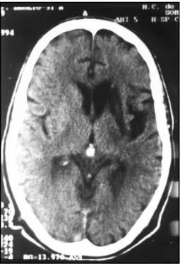
Laboratory investigation with complete blood count, erythrocyte sedimentation rate, sodium, potassium, fasting plasma glucose, BUN, creatinin, prothrombin time, partial thromboplastin time, antithrombin III, serologic test for syphilis (VDRL), cholesterol and triglycerides were within the normal range. Radiographs of cranium and cervical spine showed basilar impression. Computed tomography scan of head showed hypodense areas in the basal ganglia and centrum semiovale (Fig 2). Echocardiography revealed left ventricular hypertrophy, with normal cardiac function.
Angiography revealed proximal occlusion of the left internal carotid artery and stenosis of 50% in the proximal right internal carotid artery. The left carotid system received irrigation from the right internal carotid through the anterior communicant artery. Complete occlusion of both vertebral arteries was observed. A large network of collateral vessels derived from occipital artery, external carotid arteries, and ascendant cervical arteries (moyamoya syndrome) provided the irrigation of the posterior fossa (Fig 3).
After inpatient investigation, treatment with aspirin was started and the patient was referred to physical therapy and speech therapy.
DISCUSSION
NF1 is a relatively common genetic disorder, with an average prevalence of 1 per 3 000 persons5. The pattern of inheritance is
autosomal dominant, but in about 50% of the cases the disease is due to a new mutation and the patient does not present familial history of the disease.The NF1 gene was localized to chromosome 17q11.22. The most frequent clinical
manifestations of NF1 include “cafe au lait” spots, cutaneous neurofibromas, Lisch nodules (pigmented iris hamartomas), inguinal and axillary freckles, and central nervous system tumors, especially optic gliomas1,2.
Vascular involvement is relatively com-mon in NF1, but most patients are asympto-matic3. Virtually any vessel can be affected but
renal arteries are the most frequently involved. Secondary hypertension due to stenosis of the renal arteries is the most common clinical manifestation of the vasculopathy associated with NF16. Vascular alterations of NF1 include
stenosis/occlusion, aneurysm, arteriovenous fistula of medium and large diameter arteries, and moyamoya syndrome. Spontaneous rupture of arteries and infiltration of the adventitious layer by neurofibromas or ganglioneuromas have also been reported4,7-9.
Sobata et al. reviewed the cases of cerebrovascular disorders associated with NF1 and found 33 cases of stenotic/occlusive arterial disorder, six cases of intracranial aneurysm, and four cases with both stenotic/occlusive disorder and aneurysm10. Stenotic/occlusive disorder of intracranial
vascular supply generally presents symptoms in childhood or adolescence, being eventually the first manifestation of NF1 in children11. More rarely, cases in individuals older than 40 were described10.
Fig 2. Computed tomography scan of head showing hypodense area in the basal ganglia.
On the other hand, intracranial aneurysm associated with NF1 tends to occur later, around the fifth decade of life. Most patients with symptomatic occlusive disorder of the cerebral circulation present hemiplegia or seizures. Transitory ischemic attacks, subarachnoid hemorrhage and hematoma due to a ruptured aneurysm can also be manifestations of cerebrovascular disorder associated with NF110.12.
The supraclinoid portion of the internal carotid artery is the segment most frequently involved in the occlusive disorder. Primary involvement of the middle and anterior cerebral arteries is also common5,10. Occlusion of the posterior vessels as presented in our case is much less frequent. Among
37 cases of occlusive cerebrovascular disorder reviewed by Sobata et al., only six had involvement of the posterior circulation10. Extracranial involvement of the carotids and vertebral arteries has also
been described5.
Diagnosis of occlusive cerebrovascular disease of NF1 is achieved through cerebral angiography which presents stenosis or occlusion of cerebral vessels, in general bilaterally10,12. In
most cases, the angiographic pattern is identical to moyamoya disease, an idiopathic occlusive cerebrovascular disease that affects children and young adults in eastern countries10,13.
The natural course of occlusive cerebrovascular disease of NF1 is a gradual stenosis of the arteries with complete occlusion of the vessel. This process is slowly progressive, allowing the development of collateral vessels which provide effective cerebral circulation. The collateral flow of the anterior and middle cerebral arteries is provided by transdural, cortico-cortical and transethmoidal anastomosis12. It allows the survival of these patients, despite the occlusion or stenosis
of the main arteries of the encephalus, in a process similar to the one in moyamoya disease13.
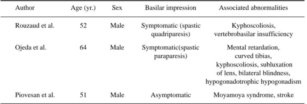
Multiple skeletal defects can also occur in NF1, including pseudoarthrosis of tibia and radium, scoliosis and kyphoscoliosis, sphenoid bone dysgenesis, sella turcica enlargement, delayed development of mastoid, dural ectasia, rib deformities, and enlargement of long bones1,2. These
disturbances seem to be related to the defective development of the mesenchymal tissue. We found only two reports of basilar impression associated with NF114,15. These patients presented other
abnormalities and one of them presented vertebro-basilar insufficiency due to atherosclerosis (Table 1). The association of NF1 and basilar impression may be considered a purely coincidental, due to the relative frequency of the two conditions.
Basilar impression is rarely an isolated find and it is generally accompanied by neurological abnormalities, including pyramidal and cerebellar signs, cranial nerves involvement, and type 1 Chiari malformation. Type 1 Chiari malformation has been recently described in association with NF116-18. In a series of 53 patients with neurofibromatosis, asymptomatic type 1 Chiari malformation
was identified in two patients through magnetic resonance imaging (MRI)17. If associated basilar
impression was present in these cases, it was not reported. In our case, an asymptomatic type 1 Chiari malformation could not be excluded because MRI was not performed.
Table 1. Neurofibromatosis type 1 and basilar impression.
Author Age (yr.) Sex Basilar impression Associated abnormalities
Rouzaud et al. 52 Male Symptomatic (spastic Kyphoscoliosis, quadriparesis) vertebrobasilar insufficiency
Ojeda et al. 64 Male Symptomatic(spastic Mental retardation, paraparesis) curved tibias,
kyphoscoliosis, subluxation of lens, bilateral blindness, hypogonadotrophic hypogonadism
The case described here is especially rare because this patient presents two unusual clinical manifestations of NF1: (1) occlusive cerebrovascular disease in a middle age man with involvement of the posterior cerebral circulation, and (2) asymptomatic basilar impression.
Acknowledgments - The authors would like to thank Dr. Jorge Kanegassuku, Dr. Paulo Cesar de Souza and Dr. Ronei A. Sandrini from the Radiology Division of Hospital de Clinicas of Federal University of Parana for their time and assistance in the interpretation of the neuroradiological investigation performed in this patient.
REFERENCES
1. Riccardi VM. Von Recklinghausen neurofibromatosis. N Engl J Med 1981;305:1617-1626.
2. Mulvihill JJ, Parry DM, Sherman JL, Pikus A, Kaiser-Kupfer MI, Eldridge R. Neurofibromatosis (Recklinghausen disease) and neurofibromatosis 2 (bilateral acoustic neurofibromatosis): an update. Ann Intern Med 1990;113:39-52.
3. Salyer WR, Salyer DC. The vascular lesions of neurofibromatosis. Angiology 1974; 25:510-519.
4. Rizzo JF, Simmons L. Cerebrovascular abnormalities in neurofibromatosis type 1. Neurology 1994;44:1000-1002. 5. Schievink WI, Michels VV, Piepgras DG. Neurovascular manifestations of heritable connective tissue disorders. Stroke
1994;25:889-903.
6. Pellock JM, Kleinman PK, McDonald BM, Wixson D. Childhood hypertensive stroke with neurofibromatosis. Neurology 1980;330:656-659.
7. Muhonen MG, Godersky JC, VanGilder JC. Cerebral aneurysms associated with neurofibromatosis. Surg Neurol 1991;36:470-475.
8. Benatar MG. Intracranial fusiform aneurysms in von Recklinghausen’s disease: case report and literature review. J Neurol Neurosurg Psychiatry 1994; 57:1279-1280.
9. Cluzel P, Pierot L, Leung A, Gaston A, Kieffer E, Chiras J. Vertebral arteriovenous fistulae in neurofibromatosis: report of two cases and review of the literature. Neuroradiology 1994;36:321-325.
10. Sobata E, Ohkuma H, Suzuki S. Cerebrovascular disorders associated with Recklinhausen’s neurofibromatosis: a case report. Neurosurgery 1988;22:544-549.
11. Woody RC, Perrot LJ, Beck SA. Neurofibromatosis cerebral vasculopathy in an infant: clinical, neuroradiographic, and neuropathologic studies. Pediatr Pathol 1992;12:613-619.
12. Tomsick TA, Lukin RR, Chambers AA, Benton C. Neurofibromatosis and intracranial arterial occlusive disease. Neuroradiology 1976;11:229-234.
13. Ikezaki K, Hee-Han D, Kawano T, Kinukawa N, Fukui M. A clinical comparison of definitive Moyamoya disease between South Korea and Japan. Stroke 1997;28:2513-2517.
14. Rouzaud M, Degiovanni E, Kiffer A, Glories P. Von Recklinghausen’s neurofibromatosis, malformation of the cervico-occipital joint, and vertebro-basilar insufficiency. Rev Otoneuroophtalmol 1970;42:201-215.
15. Ojeda E, Clavé E, González MV, Carrera J. Hypogonadism, basillary impression, subluxation of the lenses, and other clinical manifestations in a case of neurofibromatosis Med Clin (Barc) 1981;76:132-135.
16. Parkinson D, Hay R. Neurofibromatosis. Surg Neurol 1986;25:109-113.
17. Bognanno IR, Edward MK, Lee TA, Dunn DW, Ross KL, Klatte EC. Cranial MR imaging in neurofibromatosis. Am J Neuroradiol 1988;9:461-468.