PREPARATION, CHARACTERIZATION AND IN VITRO ANTIMICROBIAL ACTIVITY OF LIPOSOMAL CEFTAZIDIME AND CEFEPIME AGAINST PSEUDOMONAS AERUGINOSA STRAINS
Ieda Maria Sapateiro Torres, Etiene Barbosa Bento, Larissa da Cunha Almeida, Luisa Zaiden Carvalho Martins de Sá, Eliana Martins Lima*
Laboratório de Tecnologia Farmacêutica e Sistema de Liberação de Fármacos, Faculdade de Farmácia, Universidade Federal de
Goias, Goiania, GO, Brasil.
Submitted: August 26, 2011; Approved: June 07, 2012.
ABSTRACT
Pseudomonas aeruginosa is an opportunistic microorganism with the ability to respond to a wide variety of
environmental changes, exhibiting a high intrinsic resistance to a number of antimicrobial agents. This low
susceptibility to antimicrobial substances is primarily due to the low permeability of its outer membrane,
efflux mechanisms and the synthesis of enzymes that promote the degradation of these drugs.
Cephalosporins, particularty ceftazidime and cefepime are effective against P. aeruginosa, however, its
increasing resistance has limited the usage of these antibiotics. Encapsulating antimicrobial drugs into
unilamellar liposomes is an approach that has been investigated in order to overcome microorganism
resistance. In this study, antimicrobial activity of liposomal ceftazidime and cefepime against P. aeruginosa
ATCC 27853 and P. aeruginosa SPM-1 was compared to that of the free drugs. Liposomal characterization
included diameter, encapsulation efficiency and stability. Minimum Inhibitory Concentration (MIC) was
determined for free and liposomal forms of both drugs. Minimum Bactericidal Concentration (MBC) was
determined at concentrations 1, 2 and 4 times MIC. Average diameter of liposomes was 131.88 nm and
encapsulation efficiency for cefepime and ceftazidime were 2.29% end 5.77%, respectively. Improved
stability was obtained when liposome formulations were prepared with a 50% molar ratio for cholesterol in
relation to the phospholipid. MIC for liposomal antibiotics for both drugs were 50% lower than that of the
free drug, demonstrating that liposomal drug delivery systems may contribute to increase the antibacterial
activity of these drugs.
Key words:Pseudomonas aeruginosa, liposomes, cephalosporins.
INTRODUCTION
Pseudomonas aeruginosa is a Gram-negative
opportunistic pathogen, non-fermenting and, unable to produce
energy for cell functioning (14). Non-fermenting bacteria are
ubiquitous in nature, particularly in soil and water, and on
surfaces in contact with soil or water. In the hospital
environment, these microorganisms can be isolated from
humidifiers, ventilators, mattresses and other equipment, as
well as from the skin of health care professionals (16).
Resistance to antimicrobial drugs exhibited by P.
aeruginosa is commonly found, especially in hospitals where
resistant microorganisms are frequently detected in intensive
care units (ICU). Acquired resistance mechanisms for this
pathogen have increased significantly, leading to resistance to
multiple antibacterial agents (5).
Low susceptibility of P. aeruginosa to antimicrobial
substances is primarily due to the low permeability of its outer
membrane, efflux mechanisms and the synthesis of enzymes
that promote the degradation of these drugs (11). The outer
membrane of P. aeruginosa is almost impermeable to many
common antibiotics, such as ampicillin, most cephalosporins
and macrolides, when compared to the outer membrane of
other Gram-negative bacteria (13).
However, bacterial resistance to -lactam antibiotics
caused by low membrane permeability or efflux pumps is quite
frequent for P. aeruginosa, Escherichia coli and Neisseria
gonorrhoeae. Efflux mechanisms result in higher minimum
inhibitory concentrations for penicillins, broad-spectrum
cephalosporins, tetracyclins and fluoroquinolones (12, 30).
Studies performed by Sentry (Antimicrobial Surveillance Program) about the activity of broad-spectrum -lactam
antibiotics against P. aeruginosa showed that about 17 to 34%
of the bacterial samples were resistant to cefepime and
ceftazidime (10, 24).
Since the bacterial outer membrane plays a major role in
the permeability of antibiotics, the use of liposomal carriers is
an interesting approach to enhance the antimicrobial activity of
certain drugs (25). The lipid bilayer of liposomes are able to
fuse with the outer membrane of bacteria (8, 27), altering the
therapeutic index of a drug (20, 28). Drug encapsulation into
liposomes is an effective method to reduce the drug’s toxicity,
prolonging its circulation time after intravenous administration
and enhancing its accumulation in the target site. These
advantages can also improve the efficacy of antibiotics, not
only for the treatment of resistant bacterial strains but also
contributing to overcome bacterial resistance (4, 9, 15).
In this work, liposomal formulations for cefepime and
ceftazidime were prepared and characterized. The antibacterial
effect of liposomal cefepime and ceftazidime against P.
aeruginosa ATCC 27853 and P. aeruginosa SPM-1 (clinical
strain) was investigated by determining the Minimal Inhibitory
Concentration (MIC) and Minimal Bactericidal Concentration
(MBC) in a time-kill study.
MATERIALS AND METHODS
Chemicals
Cefepime was obtained from Alembic Limited,
ceftazidime from Advan Pharma Chem Co. Ltd, soy
phosphatidylcholine (PC) was from Lipoid, Gmb; cholesterol from Avanti Polar Lipids, -tocopherol was purchased from
Sigma Co., all other chemicals and reagents were of analytical
grade or superior.
Microorganisms
Pseudomonas aeruginosa ATCC 27853 was kindly
supplied by INCQS (National Institute for Quality Control in
Health, Rio de Janeiro, Brazil), and P. aeruginosa SPM-1
(clinical strain), was donated by IPTSP (Instituto de Patologia
Tropical e Saúde Pública, Goiás, Brazil). Microorganisms were
kept at 20ºC in trypticase soy broth (Difco), supplemented with
10% (v/v) glycerol. For the experiment, the microorganisms
were inoculated into inclined trypticase soy agar and incubated
for 24 h at 37°C.
Preparation of Liposomes
Liposomes were prepared by the lipid film hydration
method. 40 mM of phosphatidylcholine (PC), 10 or 20 mM of cholesterol (Chol) and 0.04 mM of -tocopherol were
dissolved in 2 mL of chloroform in a round bottom glass tube.
movement of the glass tube promoted the formation of a thin
lipid film on the glass wall. The lipid film was then hydrated
with 4mL of a cefepime solution (54 mg·mL-1) in 0.9% NaCl,
and 8 mL of a ceftazidime solution (37.66 mg·mL-1).After the
hydration, unilamellar liposomes were obtained by sonicating
the lipid dispersion for 10 minutes in pulses of 1 minute with a
Ti-probe sonicator (Misonix, XL 2020). Liposome diameter
was determined by dynamic light scattering (ZetaSizer Nano,
Malvern Instruments).
Encapsulation Efficiency
Non-encapsulated drug was removed from liposomes by
size exclusion chromatography with a Sephadex G-50 column
(10 x 200 mm). Encapsulation efficiency was calculated as the
percentage of encapsulated drug (cefepime or ceftazidime) in
relation to the total amount of drug added to the formulation. In
order to minimize the dilution effect of the column separation
process, liposomal formulations were submitted to
ultracentrifugation at 50,000 rpm for 90 minutes at 4°C and
resuspended in NaCl 0.9% to the desired concentration.
Quantitative determination of cefepime and ceftazidime
was performed by high performance liquid chromatography
(HPLC) using a ProStar 240 Chromatographer (Varian),
equipped with an auto injector and UV detection at 255 nm and
254 nm wavelength, respectively. Separation was made in a
C18 column (250 x 4.6 mm) and mobile phase was
acetonitrile:water (10:90, v/v) for cefepime and
acetonitrile:water (2:98, v/v) for ceftazidime, with an isocratic
flow of 1mL·min-1. Results were calculated based on a
calibration analytical curve prepared with cefepime reference
standard (United States Pharmacopeia) and ceftazidime
reference standard (European Pharmacopoeia), in the range of
0.001 to 0.1 mg·mL-1.
Stability of liposomal formulations
Stability of liposomal formulations was determined in
order to evaluate the rate of drug leakage from liposomes.
After the removal of the non-encapsulated drug, liposome
dispersions were maintained at 4°C for 96 hours and at each 24
hours interval, samples were withdrawn for another step of
separation in the sephadex column. The remaining amount of
entrapped drug was determined by HPLC as described above.
Determination of the Minimum Inhibitory Concentration (MIC)
Antimicrobial activity of cefepime and of ceftazidime
against Pseudomonas aeruginosa ATCC 27853 and P.
aeruginosa SPM-1, was assayed for the free drugs and
liposome entrapped drugs. MIC was determined by the broth
dilution technique, as recommended by the Clinical and
Laboratory Standards Institute (CLSI) (6). Sequential dilutions
of free cefepime and ceftazidime (4096 to 2 µg.mL-1) and
liposomal cefepime and ceftazidime (4096 to 2 µg.mL-1)were
prepared in Mueller-Hinton broth. Immediately after the
preparation, 1 mL of each drug dilution was inoculated with
1mL of the bacterial suspension, for a final bacterial count of 5
x 105 CFU.mL-1. Tubes were incubated for 18 hours at 35°C.
MIC was defined as the lowest concentration at which no
visible microbial growth was detected after 18 hours, following
the addition of 500 µl of an aqueous solution of triphenyl
tetrazolium chloride 0.5%. Positive control tubes were
prepared with the culture medium inoculated with either
bacterial suspension or bacterial suspension and blank
liposomes. Negative (sterility) control tube consisted of culture
medium only. Each test was performed in triplicates, in three
different days to ensure reproducibility.
Determination of Minimum Bactericidal Concentration (MBC)
MBC was also determined by broth dilution technique.
Briefly, overnight cultures of P. aeruginosa ATCC 27853,with
a final bacterial count of 5 x 10⁵ CFU.mL-1 were incubated with
free cefepime and ceftazidime or their liposomal formulations,
at concentrations of 1, 2 and 4 times the previously determined
MIC, and P. aeruginosa SPM-1 was incubated with free and
tubes did not contain drug. Tubes were incubated at 37°C for 2,
6 and 24 hours. At each time point, serial dilutions were
prepared in sterile saline 0.9% and 1 mL of each dilution was
inoculated into Trypticase Soy Agar plates, in triplicates. After
18 hours of incubation, the number of CFUs for each dilution
was counted. Plates with a number of colonies ranging from 30
to 300 were used for counting. MBC was established as the
lowest concentration of either free or liposomal cefepime and
ceftazidime, able to promote a 99.9% reduction of the initial
bacterial inoculums.
RESULTS
Encapsulation Efficiency
The amount of encapsulated drug was 0.150 mg.mL-1 and
0.106 mg.mL-1 for ceftazidime and cefepime, respectively,
following the technique of size exclusion chromatography for
the separation of the free drugs.
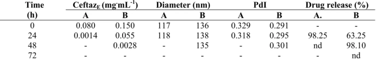
Stability of liposomal cefepime and ceftazidime formulations
Tables 1 and 2 show respectively, the formulation
parameters and drug release data of cefepime and ceftazidime
encapsulated in liposomes, indicating that in preparations of
cefepime with 10mM of cholesterol there was a loss of 97.19%
of the encapsulated drug in the first 24 hours, against 44.91%
observed for the preparation containing 20mM cholesterol. For
liposomal ceftazidime the loss of the encapsulated drug was
98.25% and 63.25%, respectively, from liposomes with 10 mM
and 20 mM of cholesterol, in 24 hours. (Table 1 e 2)
Table 1. Formulation parameters and stability data for liposomal cefepime.
CefepE (mg.mL-1) Diameter (nm) PdI Drug release (%) Time (h)
A B A B A B A B
0 0.0748 0.1002 126 151 0.278 0.313 - -
24 0.0021 0.0552 128 150 0.240 0.319 97.19 44.91
48 - 0.0028 130 158 - 0.330 nd 97.20
72 - - - nd
CefepE: encapsulated cefepime; PdI: polydispersity Index; nd: not detected; Formulation A: 40mM PC, 10mM Chol, 0.04mM -tocopherol; Formulation B: 40mM PC, 20mM Chol, 0.04mM -tocopherol.
Table 2. Formulation parameters and stability data for liposomal ceftazidime.
CeftazE (mg.mL-1) Diameter (nm) PdI Drug release (%) Time
(h) A B A B A B A. B
0 0.080 0.150 117 136 0.329 0.291 - -
24 0.0014 0.055 118 138 0.318 0.295 98.25 63.25
48 - 0.0028 - 135 - 0.301 nd 98.10
72 - - - - nd
CeftzE: encapsulated ceftazidime; PdI: polydispersity Index; nd: not detected; Formulation A: 40mM PC, 10mM Chol, 0.04mM -tocopherol; Formulation B: 40mM PC, 20mM Chol, 0.04mM -tocopherol.
Antimicrobial activity of liposomal cefalosporins
Determination of MIC: MIC for free cefepime and ceftazidime against Pseudomonas aeruginosa ATCC 27853 was 8 g.
mL-1, which is in accordance to CLSI limits (6), while MIC for liposomal cefepime and ceftazidime was 4 g.
mL-1. It
was not possible to determine MIC against P. aeruginosa
SPM-1 for liposomal cefepime due to the low encapsulation
efficiency for this drug, however, the P. aeruginosa SPM-1 had MIC of 1024 g.
mL-1 for free ceftazidime and 512 g.mL-1
for liposomal ceftazidime. Blank liposomes did not have any
effect on bacterial growth.
respectively, the antimicrobial activity of free and liposomal
ceftazidime, and free and liposomal cefepime against P.
aeruginosa ATCC 27853, and figure 3 for the free and
liposomal ceftazidime against P. aeruginosa SPM-1, as a
function of time and concentration.
None of the concentrations of free or liposomal
ceftazidime was able to eliminate 99.9% of the strain of P.
aeruginosa ATCC 27853. Nevertheless, the liposomal
ceftazidime at 4 x MIC (16 µg ml-1) in 24 hours, succeeded in
removing 99.83% of the microorganisms inoculated; whereas
this same concentration of free drug, showed a reduction of
96.5% in 24 hours. (Figure 1)
Free cefepime, against P. aeruginosa ATCC 27853, was
able to kill 99.9% of the microorganisms inoculated with 2 x
MIC (16µg.mL-1) in 6 hours. However, at this concentration,
the drug was not able to maintain this same antimicrobial
performance, with an evident recovery of the bacteria after 24
hours. A reduction of 99.9% of the microorganism after 24
hours was only obtained when the amount of drug was 4 times
higher than the MIC (32µg.mL-1). (Figure 2)
Cefepime encapsulated into liposomes was able to reduce
99.9% of the inoculums of P. aeruginosa ATCC 27853, with
16µg.mL-1 after 24 hours of incubation. In addition, when
liposomal cefepime was used no bacterial recovery was
observed at any concentration or time of incubation used in this
study.
The free ceftazidime was able to kill 99,9% of P.
aeruginosa SPM-1 inoculated with 1 x MIC (1024 µg.mL-1) in
6 hours. However this concentration failed to maintain this
percentage of elimination, with recovery of the microorganisms
within 24 hours of contact. The reduction of 99.9% of the
resistant strain was maintained in only 24 hours of contact with
the free ceftazidime at 2 x MIC (2048 µg mL-1). (Figure 3)
The liposomal ceftazidime proved more effective in
reducing 99.9% of the resistant strain with 1024 µg mL-1 in 24
hours. Moreover, when the liposomal ceftazidime was used, no
bacterial recovery was observed at any concentrations or time
of incubation.
Figure 2. Time-kill curves for P. aeruginosa ATCC 27853 exposed to 1, 2 and 4 times the minimum inhibitory concentration (MIC) of free cefepime (F) and liposomal cefepime (L).
DISCUSSION
Non-fermenting Gram-negative bacteria such as
Pseudomonas aeruginosa are major opportunistic pathogens
involved in severe infections acquired in hospitals, mainly with
patients with nosocomial pneumonia in ICU. The control of
these infections is extremely challenging due to the increased
levels of resistance presented by this microorganism against
most antimicrobial agents (21).
It has been established that encapsulating antibiotic drugs
into liposomes can increase antimicrobial activity while
reducing toxic effects (4, 9, 15).
This study proved that cefepime and ceftazidime can be
successfully encapsulated into liposomes. Natural soy PC was
used for the preparation of liposomes due to its low
immunogenicity and toxicity when compared to other
phospholipids such as cardiolipin, phosphatidylinositol,
phosphatidylglycerol and phosphatidic acid (22, 26). Smaller
amount of cefepime was encapsulated probably due to high
concentrations of L-arginine (725mg.g-1), present in the drug
as pharmaceutical raw material, which is added to control the
pH of the constituted solution at 4,0-6,0.
The encapsulation of hydro soluble drugs like cefepime
and ceftazidime has been studied by several researchers.
Results found in this work are in agreement with those
presented by Antos, Trafny & Grzybowski (3), Omri &
Ravaoarinoro (22) and Drulis-Kawa et al (7), who
demonstrated encapsulation efficiencies from 3 to 7.2% using
several liposome formulations. Conversely, Park et al (23)
were able to encapsulate 75.4% of cefoxitine. However,
liposomes from that study had a mean diameter higher than 600
nm, which despite the larger aqueous internal volume; these
vesicles exhibit a higher content leakage and tendency to
agglomerate.
The marked release of cefepime and ceftazidime from
liposomes prepared with 10mM of cholesterol (97.19% and
98.25%, respectively) against 44.91% from the preparation of
cefepime and of the 63.25% from preparation of ceftazidime,
both with 20mM of cholesterol can be a result of a higher
membrane fluidity in the 10mM formulation. Kinetics of drug
release from liposomes can be controlled by adding different
amounts of cholesterol to the membrane bilayers, which
increases membrane rigidity, reduces permeability to hydro
soluble molecules and promotes better stability of liposome
vesicles in a protein rich medium (2, 31).
Higher concentrations of cholesterol (>30 molar%) can
completely eliminate membrane phase transition and fluidity
(29). In this work, a higher encapsulation efficiency and
stability was obtained with liposome formulations using a
molar ratio for cholesterol of 50 molar% in relation to PC.
Cefepime and ceftazidime leakage from liposomes was
higher than that reported by Drulis-Kawa et al (7) for
meropenem encapsulated into liposomes prepared with natural
and synthetic lipids, with 24% leakage in 24 hours. When
gentamycin was encapsulated into 1,2-dimyristoyl-sn
-glycero-3-phosphocholine (DMPC), 1,2-dipalmitoyl-sn
-glycero-3-phosphocholine (DPPC) and 1,2-distearoyl- sn
-glycero-3-phosphocholine (DSPC) liposomes, approximately 70% of the
drug was retained during the first 48 hours (17). However, both
meropenem and gentamycin are less hydro soluble than the
cephalosporins used in this work. A higher hydrophobicity of
the molecule favors its localization in the lipid bilayer or in the
lipid-water interface due to its partition coefficient, reducing
the leakage of the drug.
Liposomal cefepime and ceftazidime against P.
aeruginosa ATCC 27853 and liposomal ceftazidime against
Pseudomonas aeruginosa SPM-1 exhibited a higher
antibacterial activity when compared to free cefepime and ceftazidime, as indicated by the 4 g/mL and 512 g/mL MIC,
respectively. This 50% reduction was probably due to the
interaction between liposomes and bacterial cells by a fusion
mechanism as previously reported (1, 4, 20), which is capable
of increasing the intracellular concentration of the drug,
researchers showed that liposomal encapsulation was able to
protect piperacylin from hydrolysis promoted by bacterial β
-lactamase, which was probably due to the localization of the
drug in the interior of the liposome where β-lactamase cannot
penetrate (19).
Results from this work are in agreement with those found
by Rukholm et al (25), Mugabe et al (17) and Mugabe et al
(18), demonstrating a remarkable reduction of MIC for
liposomal antibiotics. Liposomal gentamycin formulations
studied by Mugabe et al (17) against resistant P. aeruginosa
strains were effective at concentrations 2 to 256 times lower
than MIC. Rukholm et al (25) did not find a marked difference
between MIC and MBC for liposomal gentamycin against three
strains of P. aeruginosa.
MIC reductions have also been found when amicacin,
netilmycin and tobramycin were encapsulated into liposomes
made of egg yolk PC and cholesterol (7:1 molar ratio).
Amicacin MIC was reduced 4 times for S. aureus ATCC 29213
and E. coli ATCC 25 922 and 8 times for S. faecalis ATCC
29212. Conversely, when amicacin liposomes were tested
against P. aeruginosa ATCC 27853, antimicrobial activity was
lowered 8 times. Netilmycin and tobramycin liposomes were
active against P. aeruginosa ATCC 27853 with a MIC of 2 and
8 times lower than that for the free drug, respectively (22).
Drulis-Kawa et al (7) encapsulated meropenem into
several liposome formulations and evaluated their activity
against eight strains of P. aeruginosa. Cationic liposomes
resulted in a MIC 2 to 4 times lower than the MIC for the free
drug. Anionic liposomes did not increase the antimicrobial
activity, resulting in MICs of equal or higher values than that
of the free drug.
MBC for liposomal cefepime and ceftazidime (16
g·mL-1) was also reduced by 50% when compared to the free drug (32 g·mL-1
). Additionally, when liposomal cefepime was used
against P. aeruginosa ATCC 27853, no bacterial recovery was
observed during the whole period of incubation, demonstrating
a prolonged antibacterial activity of the liposomal formulation
when compared to free cefepime. Similar bactericidal effects
were obtained by Mugabe et al (18) for liposomal amicacin
against P. aeruginosa, where liposomes composed of
DPPC/cholesterol in the molar ratio of 2:1 on average
eradicated clinical strains of P. aeruginosa with a
concentration of 8 mg·mL-1, while the free antibiotic at a
concentration of 256 mg.mL-1, was inactive.
From these results, it can be concluded that encapsulating
cefepime and ceftazidime into liposomes increases their
antibacterial activity against P. aeruginosa ATCC 27853 and
P. aeruginosa SPM1, indicating that liposomal formulations
can be effective alternative for treating infections caused by
these microorganisms and a valid approach against the
development of bacterial resistance.
ACKNOWLEDGEMENTS
Authors would like to thank FINEP/MCT and
FUNAPE/UFG for partially funding this research.
REFERENCES
1. Alipour M.; Halwani M.; Omri A.; Suntres Z.E. (2008). Antimicrobial effectiveness of liposomal polymyxin B against resistant Gram-negative bacterial strains. Int J Pharm. 355, 293-298.
2. Anderson M.; Omri A. (2004). The Effect of Different Lipid Componentes on the In Vitro Stability and Release Kinetics of Liposome Formulations. Drug Deliv. 11, 33-39.
3. Antos M.; Trafny E.A.; Grzybowski J. (1995). Antibacterial Activity of Liposomal Amikacin Against Pseudomonas aeruginosa in vitro.
Pharmacol Res. 32 (nº 1/2), 85-87.
4. Beaulac C.; Sachetelli S.; Lagace J. (1998). In-vitro bactericidal efficacy of sub-MIC concentrations of liposome-encapsulated antibiotic against Gram-negative and Gram-positive bacteria. J Antimicrob Chemother. 41, 35-41.
5. Chastre J.; Trouillet J.L. (2000). Problem pathogens (Pseudomonas aeruginosa and Acinetobacter). Semin Respir Infect. 15, 287-298. 6. Clinical and Laboratory Standards Institute/NCCLS. (2005).
940 West Valley Road, Suite 1400, Wayne, Pennsylvania 19087 – 1898 USA.
7. Drulis-Kawa Z.; Gubernator J.; Dorotkiewicz-Jach A.; Doroszkiewicz W.; Kozubek A. (2006). In vitro antimicrobial of liposomal meropenem against Pseudomonas aeruginosa strains. Int J Pharm. 315, 59-66. 8. Drulis-Kawa Z.; Dorotkiewicz-Jach A.; Gubernator J.; Gula G.; Bocer
T.; Doroszkiewicz W. (2009). The interaction between Pseudomonas aeruginosa cells and cationic PC:Chol:DOTAP liposomal vesicles versus outer-membrane structure and envelope properties of bacterial cell. Int. J Pharm. 367, 211-219.
9. Ellbogen M.H.; Olsen K.M.; Gentry-Nielsen M.J.; Preheim L.C. (2003). Efficacy of liposome encapsulated ciprofloxacin compared with ciprofloxacin and ceftriaxone in rat model of pneumococcal pneumonia.
J. Antimicrob Chemother. 52, 83-91
10. Ferrara A.M. (2006). Potentially multidrug-resistant non-fermentative Gram-negative pathogens causing nosocomial pneumonia. Int J Antimicrob Agents.27, 183-195.
11. Hancock R.E.W.; Speert D.P. (2000). Antibiotic resistance in
Pseudomonas aeruginosa: mechanisms and impact on treatment. Drug Resist Updat. 3, 247-255.
12. Hardman J.G.; Limbird L.E. (2001) Gilmans A.G. Goodman & Gilman’s
The Pharmacological Basis of Therapeutics. 10th ed. McGraw-Hill, New York.
13. Hauser A.R.; Sriram P. (2005). Severe Pseudomonas aeruginosa
infections. Tackling the comundrum of drug resistance. Postgrad Med. 117 (1), 41-48.
14. Japoni A.; Alborzi A.; Kalani M.; Nasiri J.; Hayati M.; Farshad S. (2006). Susceptibility patterns and cross-resistance of antibiotics against
Pseudomonas aeruginosa isolated from burn patients in the South of Iran. Burns. 32, 343-347.
15. Maurer N.; Wong K.F.; Hope M.J.; Cullis P.R. (1998). Anomalous solubility behavior of the antibiotic ciprofloxacin encapsulated in liposomes: a 1H-NMR study. Biochim Biophys Acta. 1374, 9-20. 16. Mcgowan J.E. (2006). Resistance in Nonfermenting Gram-Negative
Bacteria: Multidrug Resistence to the Maximum. Am J Med. 119 (6A), S29-S36.
17. Mugabe C.; Azghani A.O.; Omri A. (2005). Liposome-mediated gentamicin delivery: development and activity against resistant strains of
Pseudomonas aeruginosa isolated from cystic fibrosis patients. J Antimicrob Chemother. 55, 269-271.
18. Mugabe C.; Halwani M.; Azghani R.; Lafrenie R.M.; Omri A. (2006). Mechanism of enhanced Activity of Lipossome-Entrapped Aminoglycosides against Resistant Strains of Pseudomonas aeruginosa.
Antimicrob Agents Chemother. 50 (n° 6), 2016-2022.
19. Nacucchio M.C.; Bellora M.J.G.; Sordelli D.O.; D’Aquino M. (1985).
Enhanced Liposome-Mediated Activity of Piperacillin Against Staphylococci. Antimicrob Agents Chemother. 27 (n°1), 137-139. 20. Nicolosi D.; Scalia M.; Nicolosi V.M.; Pignatello R. (2010).
Encapsulation in fusogenic liposomes broadens the spectrum of action of vancomycin against Gram-negative bacteria. Int J Antimicrob Agents. 35, 553-558.
21. Omri A.; Suntres Z.E.; Shek P.N. (2002). Enhanced activity of lipossomal polymyxin B against Pseudomonas aeruginosa in a rat model of lung infection. Biochem Pharmacol. 64, 1407-1413.
22. Omri A.; Ravaoarinoro M. (1996). Preparation, properties and the effects of amikacin, netilmicin and tobramycin in free and liposomal formulations on Gram-negative and Gram-positive bacteria. Int J Antimicrob Agents. 7, 9-14.
23. Park J.; Suh H.; Sung H.; Han D.; Lee D.H.; Park B.J.; Park Y.H.; Cho B.K. (2003). Liposomal Entrapment of Cefoxitin to Improve Cellular Viability and Function in Human Saphenous Veins. Artif Organs. 27 (7), 623-630.
24. Pfaller M.A.; Sader H.E.; Fritsche T.R.; Jones R.N. (2006). Antimicrobial activity of cefepime tested against ceftazidime-resistant Gram-negative clinical strains from North American Hospitals: report from the SENTRY Antimicrobial Surveillance program (1998-2004).
Diagn Microbiol Infect Dis.56, 63-68.
25. Rukholm G.; Mugabe C.; Azghani A.O.; Omri A. (2006). Antibacterial activity of lipossomal gentamicin against Pseudomonas aeruginosa: a time-kill study. Int J Antimicrob Agents. 27, 247-252.
26. Sachetelli S.; Beaulac C.; Riffon R.; Lagacé J. (1999). Evaluation of the pulmonary and systemic immunogenicity of Fluidosomes, a fluid liposomal-tobramycin formulation for the treatment of chronic infections in lungs. Biochim Biophys Acta. 1428, 334-340.
27. Sachetelli S.; Khalil H.; Chen T.; Beaulac C.; Senechal S.; Lagace J. (2000). Demonstration of a fusion mechanism between a fluid bactericidal liposomal formulation and bacterial cells. Biochim Biophys Acta. 1463, 2554-2566.
28. Schiffelers R.; Storm G.; Bakker-Woudenberg I. (2001). Liposome-encapsulated aminoglycosides in pre-clinical and clinical studies. J Antimicrob Chemother. 48, 333-344.
29. Sharma A.; Sharma U.S. (1997). Liposomes in drug delivery: progress and limitation. Int J Pharm. 154, 123-140.
30. Thomson J.M.; Bonomo R.A. (2005). The threat of antibiotic resistance in Gram-negative pathogenic bacteria: β-lactams in peril! Curr Opin Microbiol. 8, 518-524.