CO-METABOLISM OF DDT BY THE NEWLY ISOLATED BACTERIUM, PSEUDOXANTHOMONAS SP. WAX
Guangli Wang; Ji Zhang; Li Wang; Bin Liang; Kai Chen; Shunpeng Li; Jiandong Jiang*
Department of Microbiology, Key Lab of Microbiological Engineering of Agricultural Environment, Ministry of Agriculture,
College of Life Sciences, Nanjing Agricultural University, 210095, Nanjing, Jiangsu Province, China
Submitted: April 17, 2009; Returned to authors for corrections: October 08, 2009; Approved: February 18, 2010.
ABSTRACT
Microbial degradation of 1,1,1-trichloro-2,2-bis(p-chlorophenyl)ethane (DDT) is the most promising way
to clean up DDT residues found in the environment. In this paper, a bacterium designated as wax, which
was capable of co-metabolizing DDT with other carbon sources, was isolated from a long-term
DDT-contaminated soil sample by an enrichment culture technique. The new isolate was identified as a member
of the Pseudoxanthomonas sp., based on its morphological, physiological and biochemical properties, as
well as by 16S rRNA gene analysis. In the presence of 100 mg l-1 glucose, the wax strain could degrade
over 95% of the total DDT, at a concentration of 20 mg l-1, in 72 hours, and could degrade over 60% of the
total DDT, at a concentration of 100 mg l-1, in 144 hours. The wax strain had the highest degradation
efficiency among all of the documented DDT-degrading bacteria. The wax strain could efficiently degrade
DDT at temperatures ranging from 20 to 37°C, and with initial pH values ranging from 7 to 9. The
bacterium could also simultaneously co-metabolize 1,1-dichloro-2,2-bis(p-chlorophenyl)ethane (DDD),
2,2-bis(p-chlorophenyl)-1,1-dichlorethylene (DDE), and other organochlorine compounds. The wax strain
could also completely remove 20 mg kg-1 of DDT from both sterile and non-sterile soils in 20 days. This
study demonstrates the significant potential use of Pseudoxanthomonas sp. wax for the bioremediation of
DDT in the environment.
Key words: DDT, Co-metabolism, Bioremediation, Pseudoxanthomonas sp.
INTRODUCTION
DDT [1,1,1-trichloro-2,2-bis(p-chlorophenyl)ethane] has
been used extensively as an organochlorine insecticide for the
control of insects on agricultural crops and vector-borne
diseases, such as typhus and malaria (14). DDT is highly
persistent in the environment, with a reported half-life between
2-15 years, and is immobile in most soils. DDT was classified
as a priority persistent organic pollutant by several different
environmental regulatory agencies because of its toxicity,
hydrophobicity and bioaccumulation (7, 9), and was banned in
the 1970s from agricultural use because of its negative impact
on wildlife and its ill effects on human health via the food
chain. However, DDT continues to be used in some developing
countries (primarily tropical countries) to control insect-borne
diseases. Due to its extensive use and recalcitrance, numerous
sites around the world are contaminated with DDT (1, 9, 19),
and many foodstuffs, including processed foods, have been
shown to contain high levels of DDT residue (2, 7, 12).
Moreover, high levels of DDT and its metabolites have also
been detected in human adipose tissue, blood plasma, liver,
brain, placenta, and breast milk (4, 8, 11, 21). Therefore, the
clean-up of DDT pollutants from contaminated sites is of great
importance.
Routes of DDT loss from contaminated sites include
runoff, volatilization, photolysis and biodegradation. However,
microbial degradation of DDT is a promising and cost-effective
method for treating DDT contamination. It was difficult for
microorganisms to degrade DDT when it was used as the sole
carbon source (3), but in the presence of an additional carbon
source, DDT could be co-metabolized by many
microorganisms under aerobic or anaerobic conditions (3, 13,
24). Under anaerobic conditions, the metabolism of DDT was
largely restricted to its alkyl chain (24), but under aerobic
conditions, DDT underwent dioxygenation, followed by ring
cleavage and the formation of p-chlorobenzoate (16, 17). It was
reported that some metabolites, such as
1,1-dichloro-2,2-bis(p-chlorophenyl)ethane (DDD) and
2,2-bis(p-chlorophenyl)-1,1-dichlorethylene (DDE), produced during the co-metabolism of
DDT, were more recalcitrant and toxic than the parent
compound (9). However, few microorganisms have been
reported to be capable of degrading DDT and its metabolites
(DDD and DDE) simultaneously.
In this study, a bacterial strain, Pseudoxanthomonas sp.
wax, which is capable of co-metabolizing DDT, DDD and
DDE, and even some other organochlorine compounds, was
isolated from a long-term DDT-polluted soil sample. The
factors that influence the degradation activities of the wax
strain were evaluated, and the bioremediation of
DDT-contaminated soil by the wax strain was investigated.
MATERIALS AND METHODS
Chemicals and media
Analytical grade DDT (99% pure), purchased from
Sigma-Aldrich Chemical, MO, USA, was used as a standard. The
technical grade DDT (70% pure), pentachloronitrobenzene
(PCNB) (95% pure), cypermethrin (94% pure), cyfluthrin
(90% pure), and bifenthrin (95% pure) were provided by the
Yangnong Agricultural & Chemical Co., Ltd, China.
Chlorothalonil (98% pure) was purchased from the Jiangyin
Suli Fine Chemical Co., Ltd and -hexachlorocyclohexane
(99% pure) was purchased from the Tianjin Dagu Chemical
Factory, China. Acetonitrile, acetone and dichloromethane
were purchased from the Shanghai Chemical Reagent Co., Ltd,
China. All other reagents used in this study were of analytical
grade.
Luria-Bertani (LB) medium and minimal salt medium
(MSM) (1.0 g NH4NO3, 1.5 g K2HPO4, 0.5 g KH2PO4, 0.2 g
MgSO4 and 1.0 g NaCl per liter, pH 7.0) were used in this
study. MSM, supplemented with 20 mg l-1 DDT, was then
called DMM, and DMM, supplemented with 100 mg l-1
glucose was called GMM. Solid media plates were prepared by
adding 1.5% (wt/vol) agar.
Strain isolation and characterization
A soil sample (2.0 g) collected from the long-term DDT
polluted soil in Yangzhou, China, was added to 100 ml of
GMM, and incubated at 30oC for 7 days in a rotary shaker at
150 rpm. Five milliliters of enriched cultures, showing
degradation of DDT, were transferred to 100 ml of fresh
GMM. Three subcultures were performed before the final
isolation of DDT-degrading strains. The final enriched culture
was spread onto GMM plates. The different colonies that
formed were isolated, and then tested for their DDT-degrading
capabilities. One strain, designated as wax, possessed the
highest degradation capacity of DDT, and was selected for
further investigation.
The new isolate was identified based on its morphological,
physiological and biochemical tests, with reference to Bergey’s
Manual of Determinative Bacteriology, combined with a 16S
rRNA gene sequence analysis. The cell morphology was
examined by light microscopy (BH-2; Olympus) and
transmission electron microscopy (TEM) (H-7650, Hitachi
High-Technologies Corporation), using cells from an
exponentially growing culture. The total genomic DNA of wax
strain was prepared by high-salt precipitation (15). The
(Escherichia coli bases 8-27) and 5’-TACCTTGTTACGACT
T-3’ (E. coli bases 1,507-1,492), were used to amplify the 16S
rRNA gene (25). The purified PCR product was sequenced
from both directions, using an ABI 377 sequencer
(Perkin-Elmer Applied Biosystems, Foster City, CA). The 16S rRNA
gene sequence (1,503 bp) of wax strain was deposited in the
GenBank database under the accession number FJ796079.
Alignment of different 16S rRNA gene sequences from
GenBank was performed using Clustal X 1.8.3 (22), with the
default settings. The phylogeny was analyzed with the MEGA
version 3.0 software, and the distance was calculated using the
Kimura 2 parameter distance model. The phylogenetic tree was
built using the neighbor-joining method. Each dataset was
bootstrapped 1,000 times (18).
Analytical methods
The cell density was monitored spectrophotometrically by
measuring the absorbance at 600 nm with a SHIMADZU
UV-Vis Recording spectrophotometer (Shimadzu UV-2410, Japan).
DDT in the cultures was extracted with five times the volume
of dichloromethane, and DDT in the soils was extracted with
an equal volume of hexane and acetone (1:1, v/v) by the
supersonic extraction method. The organic extract was dried
over anhydrous sodium sulfate, and then evaporated using a
vacuum rotary evaporator at room temperature. The residual
extract was dissolved in 5 ml acetonitrile. Twenty microliters
of the resulting solution was analyzed by reverse-phase high
performance liquid chromatography (HPLC) (600 Controller,
Rheodyne 7725i Manual injector, 2487 Dual l Absorbance
Detector; Waters Co., Milford, MA). The separation column
(4.6 mm × 250 mm × 5 µ m) was filled with Kromasil
100-5C18. The mobile phase was acetonitrile/water (70:30, v/v),
with a flow rate of 1.0 ml min-1. The amount of chloride ion
released from DDT was measured spectrophotometrically at
460 nm with mercuric thiocyanate and ferric ammonium
sulfate, according to Iwasaki et al (5). The presence of DDD,
DDE, PCNB, cypermethrin, cyfluthrin, bifenthrinat,
chlorothalonil and -hexachlorocyclohexane was determined
by HPLC, or gas chromatography (GC), as described
previously (10, 23).
Degradation of DDT by wax strain in culture
All aerobic batch cultivations were carried out in 250-ml
bottles, containing 100 ml liquid culture, while being shaken at
150 rpm at 30°C. The wax strain was pre-cultured in LB
medium for 18 hours, was centrifuged at 5,000 g for 10 min,
and the cell pellets were washed three times with fresh MSM.
After adjusting the OD600nm of cell density to 1.0, an inoculum
(5%, v/v) was inoculated into liquid GMM medium unless
otherwise stated. The medium, inoculated with heat-killed wax
cells, was maintained as the control. All treatments were
performed in triplicate. At different time periods, 2 ml aliquots
of the sample were removed for analysis. The degradation
efficiency was determined by an estimation of the removal of
DDT from the culture.
To investigate the effect of alternative carbon source on
DDT biodegradation, 100 mg l-1 of glucose, succinate, starch,
dextrin, or maltose, was added to DMM, respectively. To
investigate the effect of the initial concentration of DDT on
DDT biodegradation, GMM containing 5 mg l-1, 20 mg l-1, 50
mg l-1 and 100 mg l-1 DDT, were used. To investigate the effect
of the initial pH value on DDT biodegradation, the pH values
of the media were adjusted to 4.0, 5.0, 6.0, 7.0, 8.0, 9.0, and
10.0, respectively, by changing the potassium-phosphate buffer
system. To investigate the effect of temperature on DDT
biodegradation, the cultures were incubated at 10°C, 20°C,
25°C, 30°C, 37°C, 40°C, and 45°C, respectively. To
investigate the effect of the initial inoculum size on DDT
biodegradation, 1%, 5%, and 10% (v/v) of wax cells,
respectively, were inoculated into GMM. To investigate the
effect of aeration on DDT biodegradation, 250-ml bottles
containing 30 ml, 60 ml, 100 ml, 120 ml, 150 ml, and 180 ml
of culture were used.
To study whether additional organochlorine compounds
could also be degraded by wax strain, MSM media (containing
100 mg l-1 glucose) supplemented with DDD, DDE, PCNB,
cypermethrin, cyfluthrin, bifenthrinat, chlorothalonil and
were used.
Degradation of DDT in soil by wax strain
Soil samples, from depths of 0 to 20 cm, were taken of a
fine sandy-loam soil in the Jiangsu province, with a pH value
of 7.8. The soil samples were treated as described by Jiang et al
(6). Subsamples (500 g) of sterile and non-sterile soil were
amended with 20 mg l-1 DDT, and inoculated with wax cells at
concentrations of 108 CFU g-1 soil. The control soil samples
were inoculated with heat-killed wax cells that were autoclaved
at 121°C for 15 min. The moisture of the soil was adjusted to
40% (wt/wt) during the experiment, and samples were
incubated in the dark to preclude photolysis reactions (20). At
periodic intervals, 10-g sub-samples were collected and
analyzed immediately. All treatments were performed in
triplicate.
RESULTS
Isolation and characterization of the DDT-degrading wax strain
A DDT-degrading strain, designated as wax, was isolated
from long-term DDT polluted soil by an enrichment culture
technique. The wax strain could degrade over 95% of the total
DDT, at a concentration of 20 mg l-1, in GMM in 72 hours. The
wax strain is a Gram-negative, rod-shaped (0.3-0.5 m ×
1.2-3.0 m), obligate aerobe that is motile via a polarly localized
flagellum (Fig. 1). When growing on LB medium plates, the
colonies of wax were yellow and convex with entire margins.
The wax strain could grow at temperatures ranging from 10°C
to 45°C, and at pH values ranging from 5.0 to 10.0. The
optimal temperature and pH value for wax strain growth was
30°C and 7.0, respectively.
Biochemical tests showed that wax strain was oxidase and
catalase positive, and urease, arginine dihydrolase, and indole
production negative. The wax strain was unable to hydrolyze
casein, cellulose, chitin, tween-40, tween-80 and starch;
however, the wax strain could hydrolyze aesculin, tween-20
and tyrosine. The predominant isoprenoid quinone of wax
strain was Q-8. Nitrite could not be reduced to nitrous oxide by
the wax strain; however, nitrate could be reduced to the
end-product of nitrous oxide. The wax strain was resistant to
streptomycin, nalidixic acid, kanamycin, ampicillin,
gentamicin, fucidin and novobiocin. In addition, the wax strain
could use glucose, maltose, starch and dextrin as carbon
sources, and use yeast extract, peptone, urea, beef extract,
NH4NO3, (NH4)2SO4 and KNO3 as nitrogen sources, and could
grow in the presence of 3% NaCl. The phylogenetic tree of the
16S rRNA gene sequences is shown in Fig. 2. The wax strain is
related to the Pseudoxanthomonas sp. lineage, and is closely
clustered with two types of Pseudoxanthomonas strains,
Pseudoxanthomonas suwonensis 4M1T and
Pseudoxanthomonas broegbernensis B1616T, with sequence
similarity scores of 98.2% and 98.4%, respectively. The result
of the phylogenetic analysis was consistent with the
morphological characteristics and the biochemical tests.
Therefore, wax strain was determined to be a
Pseudoxanthomonas sp.
Figure 1. Micrograph of the strain wax under transmission electronic microscope showing its shape, size, and position of
Figure 2. Phylogenetic tree based on the 16S rRNA gene sequences of the wax strain and related species. The GenBank accession number for each microorganism used in the analysis is shown in parentheses after the species name. The scale bar indicates 0.005
substitutions per nucleotide position. Bootstrap values, obtained with 1,000 resamplings, were indicated as percentages at all
branches.
Degradation of DDT by the wax strain in culture
In GMM, the wax strain degraded 30.6% of the total DDT,
at a concentration of 20 mg l-1 in 24 hours, while over 95% was
degraded in 72 hours. However, in MSM, only 10.2% of the
total DDT was degraded in 72 hours (Fig. 3). These results
demonstrate that the biodegradation of DDT by the wax strain
was greatly affected by the alternative carbon source. When the
wax strain grew in the presence of glucose as the carbon
source, DDT was simultaneously co-metabolized with chloride
ion released from DDT. It was found that other carbon sources,
such as succinate, starch, dextrin and maltose, could also
promote the biodegradation of DDT. Therefore, it is suitable
for the wax strain to degrade DDT in naturally contaminated
sites, where other carbon sources exist.
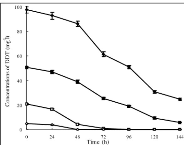
The wax strain could degrade DDT at a wide range of
concentrations, even at very high concentrations. For instance,
over 60% of the total DDT, at a concentration of 100 mg l-1,
was degraded in 144 hours, and specifically 50 mg l-1 of DDT
was nearly completely degraded in this time (Fig. 4). Even
though a high concentration of DDT was toxic to the wax cells,
and there was a delay in biodegradation when the initial
concentration of DDT was over 50 mg l-1, the wax strain
showed an uncommon tolerance of DDT at high
concentrations.
0 5 10 15 20 25
0 12 24 36 48 60 72
T ime (h)
C
o
n
ce
n
tr
at
io
n
s
o
f
D
D
T
(
m
g
l
-1 )
0 20 40 60 80 100 120
R
el
ea
se
d
c
h
lo
ri
d
e
(%
)
Figure 3. Degradation of DDT by Pseudoxanthomonas sp. wax in culture. , DDT control; , DDT biodegradation in DMM
medium; , DDT biodegradation in GMM medium; , chloride
released in DMM medium; , chloride released in GMM medium.
Data are the means of results from triplicate experiments, and error
bars indicate standard errors. Error bars that are not visible fall
within the data point.
Pseudoxanthomonas suwonensis 4M1T (AY927994)
P. taiwanensis CB-226T (AF427039)
P. broegbernensis B1616/1T (AJ012231) strain wax (FJ796079)
P. kaohsiungensis J36T (AY650027)
P.koreensis TR7-09T (AY550263)
P. daejeonensis TR6-08T (AY550264)
P. mexicana AMX 26BT (AF273082)
P. japonensis 12-3T (AB008507)
P. kalamensis JA40T (AY686710)
P. dokdonensis DS-16T (DQ178977)
P. spadix IMMIB AFH-5T (AM418384)
P. sacheonensis BD-c54T (EF575564)
P. yeongjuensis GR12-1T (DQ438977)
99 100
65
60 98
83 63 93
26 32
52
0 20 40 60 80 100
0 24 48 72 96 120 144
Time (h)
C
o
n
ce
n
tr
at
io
n
s
o
f
D
D
T
(
m
g
l
-1)
Figure 4. Effect of the initial DDT concentration on biodegradation of DDT by the wax strain in GMM. , 100 mg
l-1; , 50 mg l-1; , 20 mg l-1; , 5 mg l-1. Data are the means of
results from triplicate experiments, and error bars indicate
standard errors. Error bars that are not visible fall within the
data point.
The wax strain could efficiently degrade DDT at
temperatures ranging from 20 to 37°C, with an optimal
degradation temperature of 30°C. The biodegradation of DDT
was completely inhibited when the temperature was above
45°C or below 10°C. The wax strain could rapidly degrade
DDT, at initial pH values ranging from 7 to 9, with an optimal
pH value of 7.5; biodegradation of DDT was inhibited when
the initial pH value was below 4.0. The degradation of DDT
increased with an increase in inoculum size, for instance, when
the inoculum size was 10%, 100 mg l-1 of DDT was degraded
to a non-detectable level in 144 hours. The effect of aeration on
DDT biodegradation was complex. Under significant aerobic
conditions (30 ml culture in the 250-ml bottle), growth of the
wax strain was optimal, however, the rate of DDT degradation
was reduced. On the other hand, the highest rate of DDT
degradation was found when the 250-ml bottle contained 100
ml culture, albeit the growth of the wax strain was not the most
optimal.
Interestingly, the wax strain had a broad
degrading-substrate range of organochlorine compounds. The degradation
time course of some organochlorine compounds by the wax
strain is presented in Fig. 5. It was found that 20 mg l-1 DDD
and DDE were nearly completely degraded in 144 hours, and
that PCNB, cypermethrin, cyfluthrin and bifenthrinat were
degraded to various extents. However, chlorothalonil and
-hexachlorocyclohexane could not be degraded, and thus it was
deduced that the side chains could also affect the
biodegradation of organochlorine compounds.
0 5 10 15 20 25
0 48 96 144
Time (h)
C
o
n
ce
n
tr
at
io
n
s
(m
g
l
-1 )
Figure 5. Degradation of different organochlorine compounds by the wax strain in liquid MSM supplemented with 100 mg l-1
glucose. , cyfluthrin; , bifenthrinat; , cypermethrin; ,
DDD; , DDE; ×, PCNB; , chlorothalonil; ,
hexachlorocyclohexane. Data are the means of results from
triplicate experiments, and error bars indicate standard errors.
Error bars that are not visible fall within the data point.
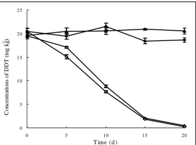
Degradation of DDT in soil by the wax strain
It was found that 20 mg kg-1 of DDT was completely
removed from both sterile and non-sterile soil samples by wax
cells, at a concentration of 108 CFU g-1, in 20 days (Fig. 6),
while there was nearly no decline in the DDT concentration in
the control soil samples, indicating that the wax cells were
solely responsible for DDT removal. The high removal
efficiency of DDT in non-sterile soil showed that the wax
strain is potentially useful for the bioremediation of
DDT-contaminated soil, even though there was competition between
0 5 10 15 20 25
0 5 10 15 20
Time (d )
C
o
n
ce
n
tr
at
io
n
s
o
f
D
D
T
(
m
g
k
g
-1 )
Figure 6. Degradation of DDT in sterile and non-sterile soils by the wax strain at a concentration of 108 CFU g-1. , DDT
control in sterile soil; , DDT control in non-sterile soil; ,
DDT degradation in sterile soil; , DDT degradation in
non-sterile soil. Data are the means of results from triplicate
experiments, and error bars indicate standard errors. Error bars
that are not visible fall within the data point.
DISCUSSION
DDT is highly persistent in the environment, and was
banned in most of the advanced countries in the 1970s.
Numerous sites around the world are contaminated with DDT.
The microbial degradation of DDT shows a promising way to
clean up the DDT-contaminated sites. Even though isolation of
a microorganism capable of utilizing DDT as a sole carbon
source is difficult, the co-metabolism of DDT by
microorganisms with other carbon sources has been well
documented, and the major metabolism products have been
identified as DDD and DDE (1). To eliminate these
contaminants from the environment, attempts have been made
to isolate microorganisms that are capable of simultaneous
degradation of DDT, DDD and DDE; however, few
microorganisms are reported to have this capability.
In the present study, a DDT degrading strain, designated
as wax, was isolated from long-term DDT-contaminated soils.
The wax strain could degrade over 95% of total DDT at 20 mg
l-1 in 72 hours in GMM. Based on the morphological,
physiological and biochemical tests, combined with 16S rRNA
gene sequence analysis, the wax strain was identified to be a
Pseudoxanthomonas sp. of bacteria. Bacteria from the genus
Pseudoxanthomonas are metabolically versatile, and this plays
an important role in the biodegradation of organic xenoboitics.
However, to our knowledge, this has been the first isolation of
a strain of Pseudoxanthomonas species that could degrade
DDT efficiently. Moreover, wax strain could not only degrade
DDT, but could also degrade DDE, DDD, and other
organochlorine compounds, which emphasizes the importance
of the wax strain for the bioremediation of sites that are
contaminated with multiple organochlorine pesticides. The wax
strain could efficiently degrade DDT at temperatures ranging
from 20 to 35°C, and at initial pH values ranging from 7 to 9,
while the wax strain could even tolerate and degrade high
concentrations of DDT (100 mg l-1). To our knowledge, the
wax strain had the highest degradation efficiency among all of
the documented DDT-degrading bacteria. The inoculation of
wax cells at 108 CFU g-1 into both sterile and non-sterile soils,
resulted in the complete removal of 20 mg kg-1 of DDT in 20
days. Due to broad substrate specificity, a strong degradation
ability and adaptability to temperature variation, the wax strain
is a promising candidate for the bioremediation of
DDT-contaminated sites; although, the biochemical degradation
pathway of DDT by wax strain has not yet been elucidated.
Therefore, further research will be needed to clarify the
degradation pathway and the properties of the key enzymes
involved in the biodegradation of DDT.
ACKNOWLEDGMENTS
This work was supported by grants from the National
Programs for High Technology Research and Development of
China (2007AA10Z405) and the Key Technology R&D
Program of Jiangsu Province (BE2008669, BE2009670).
REFERENCES
1. Aislabie, J.M.; Richards, N.K.; Boul, H.L. (1997). Microbial degradation of DDT and its residues-a review. NZ. J. Agric. Res. 40(2), 269-282. 2. Appaiah, K.M. (1988). Insecticide residues in foods. A review of work
3. Beunink, J.; Rehm, H.J. (1988). Synchronous anaerobic and aerobic degradation of DDT by an immobilized mixed culture system. Appl. Microbiol. Biotechnol. 29, 72-80.
4. Dale, W.E.; Copel, M.F.; Hayes, W.J. (1985). Chlorinated pesticides in the body fat of people of India. Bull. World Health Org. 33, 471-477. 5. Iwasaki, I.; Utsumi, S.; Ozawa, T. (1952). New colorimetric
determination of chloride using mercuric thiocyanate and ferric ion. Bull. Chem. Soc. Japan. 25(3), 226-226.
6. Jiang, J.; Zhang, R.; Li, R.; Gu, J.; Li, S. (2007). Simultaneous biodegradation of methyl parathion and carbofuran by a genetically engineered microorganism constructed by mini-Tn5 transposon. Biodegradation. 18, 403-412.
7. Kannan, K.; Tanabe, S.; Ramesh, A.; Subramanian, A.; Tatsukawa, R. (1992). Persistent organochlorine residues in food stuffs from India and their implications on human dietary exposure. J. Agric. Food Chem. 40, 518-524.
8. Kannan, K.; Tanabe, S.; Williams, R.J.; Tatsukawa, R. (1994). Persistent organochlorine residues in foodstuffs from Australia, Papua New Guinea and the Solomon Islands: Contamination levels and dietary exposure. Sci. Total. Environ. 153, 29-49.
9. Kelce, W.R.; Stone, C.R.; Laws, S.C.; Gray, L.E.; Kemppainen, J.A.; Wilson, E.M. (1995). Persistent DDT metabolite p, p-DDE is a potent an-drogen receptor antagonist. Nature. 375, 581-585.
10. Kim, Y.M. (2007). Cloning and characterization of bacterial gene involved in chlorothalonil-biotransformation. J. Nation. Fish University. 56(1), 153-160.
11. Kunhi, A.A.M.; Ajithkumar, P.V.; Ahamad, P.Y.A.; Chandrashekaraiah, D.H.; Reddy, N.S. 1995. A novel enrichment technique for the development of microbial consortia capable of degrading alpha-, beta-, gamma- and delta-isomers of hexachlorocyclohexane. Patent 2448/DEL/95 dt.
12. Lal, R.; Dhanraj, P.S.; Narayanarao, V.V.S. (1989) Residues of organochlorine insecticides in Delhi vegetables. Bull. Environ. Contam. Toxicol. 42, 45-49.
13. Masse, R.; Lalanne, D.; Messier, F.; Sylvestre, M. (1989). Characterization of new bacterial transformation products of 1,1,1-trichloro-2,2-bis(4-chlorophenyl)ethane (DDT) by gas chromatography/mass spectrometry. Biomed. Environ. Mass. Spectrom.
18, 741-752.
14. Metcalf, R.L. (1973). A century of DDT. J. Agric. Food Chem. 21, 511-519.
15. Miller, S.A.; Dykes, D.D.; Polesky, H.F. (1998). A simple salting out procedure for extracting DNA from human nucleated cells. Nucleic. Acids. Res. 16, 1215.
16. Nadeau, L.J.; Menn, F.M.; Breen, A.; Sayler, G.S. (1994). Aerobic degradation of 1,1,1-trichloro-2,2-bis(4-chlorophenyl) ethane(DDT) by Alcaligenes eutrophus A5. Appl. Environ. Microbiol. 60, 51-55. 17. Nadeau, L.J.; Sayler, G.S.; Spain, J.C. (1998). Oxidation of
1,1,1-trichloro-2,2-bis(4-chlorophenyl) ethane (DDT) by Alcaligens eutrophus A5. Arch. Microbiol. 171, 44-49
18. Saitou, N.; Nei, M. (1987). The neighbor-joining method: a new method for reconstructing phylogenetic trees. Mol. Biol. Evol. 4, 406-425. 19. Simonich, S.L.; Hites, R.A. (1995). Global distribution of persistent
organochlorine compounds. Science. 269,1851-1854.
20. Singh, B.K.; Walker, A.; Morgan, J.A.W.; Wright, D.J. (2003). Effects of soil pH on the biodegradation of chlorpyrifos and isolation of a chlorpyrifos-degrading bacterium. Appl. Environ. Microbiol. 69, 5198-5206.
21. Tanabe, S.; Gondaira, F.; Subramanian, A.; Ramesh, A.; Mohan, D.; Kumaran, P.; Venugopalan, V.K.; Tatsukawa, R. (1990). Specific pattern of persistent organochlorine residues in human breast milk from South India. J. Agric. Food Chem. 18, 899-903.
22. Thompson, J.D.; Gibson, T.J.; Plewniak, F.; Jeamougin, F.; Higgins, D.G. (1997). The Clustal_X windows interface: flexible strategies for multiple sequence alignment aided by quality analysis tools. Nucleic. Acids. Res. 25, 4876-4882.
23. Torres, R.M.; Grosset, C.; Alary, J. (2000). Liquid chromatographic analysis of PCNB and its metabolites in soil. Chromatographia. 51(9), 526-530.
24. Wedemeyer, G. (1967). Dechlorination of 1,1,1-trichloro-2,2-bis(p-chlorophenyl)ethane by Aerobacter aerogenes. Appl. Microbiol. 15, 569-574.