Mediterranean Lineage, Wrongly Identified as
Mycobacterium
pinnipedii
(Spoligotype International Type 863 [SIT863]), Causing
Active Tuberculosis in South Brazil
Elis R. Dalla Costa,aSidra E. G. Vasconcelos,bLeonardo S. Esteves,a,cHarrison M. Gomes,b Lia L. Gomes,bPedro Almeida da Silva,d João Perdigão,eIsabel Portugal,eMiguel Viveiros,fRuth McNerney,gArnab Pain,hTaane G. Clark,g,iNalin Rastogi,jGisela Unis,k Maria Lucia R. Rossetti,a,cPhilip Noel Suffysb,l
Fundação Estadual de Produção e Pesquisa em Saúde (FEPPS), Porto Alegre, Brazila; Laboratório de Biologia Molecular Aplicada a Micobactérias, Instituto Oswaldo Cruz, Fiocruz, Rio de Janeiro, Brazilb; Universidade Luterana do Brasil (ULBRA/RS), Porto Alegre, Brazilc; Núcleo de Pesquisa em Microbiologia Médica, Faculdade de Medicina, Universidade Federal do Rio Grande, Rio Grande do Sul, Brazild; Instituto de Investigação do Medicamento (iMed.ULisboa), Faculdade de Farmácia da Universidade de Lisboa, Lisbon, Portugale; Grupo de Micobactérias, Unidade de Microbiologia Médica, Global Health and Tropical Medicine (GHMT), Instituto de Higiene e Medicina Tropical (IHMT), Universidade Nova de Lisboa (UNL), Lisbon, Portugalf; Faculty of Infectious and Tropical Diseases, London School of Hygiene & Tropical Medicine, London, United Kingdomg; Biological and Environmental Sciences and Engineering Division, King Abdullah University of Science and Technology, Thuwal, Kingdom of Saudi Arabiah; Faculty of Epidemiology and Population Health, London School of Hygiene & Tropical Medicine, London, United Kingdomi; Unité de la Tuberculose et des Mycobactéries, Institut Pasteur de Guadeloupe, Les Abymes, Guadeloupe, Francej; Hospital Sanatório Partenon (HSP), Porto Alegre, Brazilk; Unit of Mycobacteriology at the Institute of Tropical Medicine, Antwerp, Belgiuml
We recently detected the spoligotype patterns of strains ofMycobacterium pinnipedii, a species of theMycobacterium tuberculo-siscomplex, in sputum samples from nine cases with pulmonary tuberculosis residing in Porto Alegre, South Brazil. Because this species is rarely encountered in humans, we further characterized these nine isolates by additional genotyping techniques, in-cluding 24-locus mycobacterial interspersed repetitive-unit–variable-number tandem-repeat (MIRU-VNTR) typing, verification of the loci TbD1, RD9,pks15/1, RDRio, andfbpC, the insertion of IS6110at a site specific to theM. tuberculosisLatin American
Mediterranean (LAM) lineage, and whole-genome sequencing. The combined analysis of these markers revealed that the isolates are in factM. tuberculosisand more specifically belong to the LAM genotype. Most of these isolates (nⴝ8) were shown to be multidrug resistant (MDR), which prompted us to perform partial sequencing of therpoA,rpoB,rpoC,katG, andinhAgenes. Seven isolates (77.8%) carried the S315T mutation inkatG, and one of these (11%) also presented the C(ⴚ17)T single-nucleotide polymorphism (SNP) ininhA. Interestingly, six of the MDR isolates also presented an undescribed insertion of 12 nucleotides (CCA GAA CAA CCC) in codon 516 ofrpoB. No putative compensatory mutation was found in eitherrpoAorrpoC. This is the first report of anM. tuberculosisLAM family strain with a convergentM. pinnipediispoligotype. These spoligotypes are ob-served in genotype databases at a modest frequency, highlighting that care must be taken when identifying isolates in theM. tu-berculosiscomplex on the basis of single genetic markers.
T
uberculosis (TB) is a disease caused by organisms belonging to theMycobacterium tuberculosiscomplex (MTBC), which are known to infect humans and domestic and wild animals. The MTBC complex includesM. tuberculosis,M. africanum,M. mi-croti,M. bovis,M. bovisbacillus Calmette-Guérin (BCG),M. ca-prae,M. pinnipedii,M. orygis,M. mungi, andM. suricattae(1–8). M. tuberculosisis the predominant cause of human TB worldwide, butM. africanumandM. bovisremain important agents of human disease in certain geographical regions (9). The MTBC species share identical 16S rRNA sequences, and recent studies have im-proved our knowledge on the genetic diversity, host range, epide-miological aspects, and differences in pathogenicity and virulence among the species of the complex (10,11). Based on the various genotyping techniques, like spoligotyping (12), restriction frag-ment length polymorphism (RFLP) (13), mycobacterial inter-spersed repetitive-unit–variable-number tandem-repeat (MIRU-VNTR) typing (14), and whole-genome sequencing (15), M. tuberculosisstrains have been subdivided into lineages and fami-lies. The most geographically widespread family worldwide (spo-ligotyping) is the Latin American Mediterranean (LAM) lineage, which is part of the heterogeneous Euro-American lineage, one ofsevenM. tuberculosislineages (16). The LAM family strains are widespread across all five continents, with a marked incidence in
Received24 July 2015 Returned for modification1 September 2015
Accepted16 September 2015
Accepted manuscript posted online23 September 2015
CitationDalla Costa ER, Vasconcelos SEG, Esteves LS, Gomes HM, Gomes LL, Almeida da Silva P, Perdigão J, Portugal I, Viveiros M, McNerney R, Pain A, Clark TG, Rastogi N, Unis G, Rossetti MLR, Suffys PN. 2015. Multidrug-resistant
Mycobacterium tuberculosisof the Latin American Mediterranean lineage, wrongly identified asMycobacterium pinnipedii(spoligotype international type 863 [SIT863]), causing active tuberculosis in South Brazil. J Clin Microbiol 53:3805–3811.doi:10.1128/JCM.02012-15.
Editor:G. A. Land
Address correspondence to Elis R. Dalla Costa, dallacostaer@gmail.com, or Philip Noel Suffys, psuffys@gmail.com.
E.R.D.C. and S.E.G.V., and M.L.R.R. and P.N.S., contributed equally to this article.
Supplemental material for this article may be found athttp://dx.doi.org/10.1128 /JCM.02012-15.
South America, and they account for 17% of all strains in the SITVITWEB database (17).
In a previous work, nine drug-resistant isolates sourced from Porto Alegre (Rio Grande do Sul, Brazil) presented with the spo-ligotype international type 863 (SIT863), a strain type previously described by Perizzolo et al. (18). This spoligotype, according to the SITVITWEB database, is representative ofM. pinnipediiand, more specifically, the PINI2 clade (17). This species was originally described as the etiological agent of TB in seals and sea lions and in some terrestrial mammals, while a single report related putative transmission to a zoo caretaker but without evolution to active disease (6,19–21). There is a paucity of information concerning M. pinnipediias a cause of human disease and on its pathogenicity/ virulence, drug resistance, and epidemiology. The nine strains with the PINI2 spoligopattern were therefore submitted to more extensive genetic characterization and evaluation of their drug susceptibility patterns.
MATERIALS AND METHODS
Mycobacteriumstrains.Nine MTBC isolates were derived from sputum samples from individuals who were diagnosed in 2006 (n⫽1) and 2010 (n⫽8) with pulmonary TB as part of routine diagnosis at the Hospital Sanatório Partenon, the reference center for drug-resistant TB in Porto Alegre, the capital of Rio Grande do Sul, South Brazil. The sputum sam-ples from these patients were processed for acid-fast bacilli microscopy detection on Ziehl-Neelsen-stained slides and cultured in Lowenstein-Jensen medium. The isolates were submitted to standard bacteriological and biochemical tests for differentiation of species within the MTBC and nontuberculous mycobacteria, including biochemical testing for niacin, para-nitrobenzoic acid, and tiofeno-2-carboxylic acid hydrazine (22). In addition, they were submitted to drug susceptibility testing using the pro-portion method on Lowenstein-Jensen solid medium (22,23).
Nucleic acid extraction and genotyping controls.For genotyping, nucleic acids were extracted as described by van Soolingen et al. (24). All PCR-based genotyping reactions included negative controls (ultrapure water) and positive controls, composed of DNA fromM. pinnipedii (strains 76 and 8; kindly provided from the collection of the National Institute for Public Health and the Environment-RIVM) (25),M.
tuber-culosisH37Rv (ATCC 27294), and twoM. tuberculosisisolates of the LAM
genotype from Brazil, as defined by spoligotyping and 24-locus MIRU-VNTR typing (26).
Genotyping.All PCRs were performed on a Veriti thermal cycler (Ap-plied Biosystems, Foster City, CA). Spoligotyping was performed using the commercially available kit from Ocimum Biosolutions (Hyderabad, India), as described by Kamerbeek et al. (12). For 24-locus MIRU-VNTR typing, amplification of loci was performed by using a commercial typing kit (Genoscreen, Lille, France) and automated MIRU-VNTR analysis, performed as previously described (27). The fragment sizes of the ampli-cons were analyzed on an ABI3730 DNA sequence analyzer (Applied Bio-systems), and the number of copies of each locus was determined by automated assignment using the GeneMapper 4.0 software (Applied Bio-systems). In the case of doubtful results, the sizes of the repeats were double checked by size estimation compared to a DNA ladder (50 and 100 bp) and the positive control (H37Rv) on agarose gel and by comparing to a reference table, as described previously (14).
Comparison of genotypes with spoligotype and MIRU-VNTR databases. The spoligopatterns of the strains were compared with the SITVITWEB database (http://www.pasteur-guadeloupe.fr:8081/SITVIT _ONLINE) for defining the spoligotype international type (SIT) and distri-bution frequency on a global level (17). The spoligotype and 24-locus MIRU-VNTR profiles were also compared with the MIRU-VNTRplus da-tabase (http://www.miru-vntrplus.org/MIRU/index.faces#) (28). The defini-tion of lineages was based on 24-locus MIRU-VNTR typing using best-match analysis and tree-based identification using the categorical index.
Detection of a LAM-specific IS6110insertion.Briefly, the LAM fam-ily was confirmed based on the verification of the presence or not of an IS6110element at position 932204 of the H37Rv genome (GenBank ac-cession no.NC_000962.2), as described by Sampson et al. (29).
Verification of RDRiostatus.Verification of RDRiostatus was
per-formed as described by Lazzarini et al. (30) using a multiplex PCR proto-col. The amplified products of 1,175 bp or 530 bp in the presence or absence of the deletion, respectively, were analyzed in a 1.5% agarose gel. Characterization of thefbpC103SNP.Characterization of thefbpC103
SNP was performed as described by Vasconcellos et al. (31). The amplified products of 519 bp were analyzed on 2% agarose gels after staining with ethidium bromide. Partial sequencing was performed using the BigDye Terminator kit (PE Applied Biosystems) on an ABI 3730 DNA analyzer (Programa de Desenvolvimento Technológico em Insumos para Saúde [PDTIS] DNA sequencing platform at Fiocruz [http://www.dbbm.fiocruz .br/PDTIS_Genomica/]), and the results were analyzed with SeqScape software version 3.0 (Applied Biosystems, CA, USA), as previously de-scribed (32,33).
Detection of the insertion inpks15/1and of RD9 and TbD1.Six- or 7-bp insertions were detected by partial sequencing of thepks15/1locus. The initial amplifications were performed as described by Huard et al. (33). The sequencing was performed using sequencing kits and an ana-lyzer, as described above; the results were interpreted as previously de-scribed (32,33). For analysis of the RD9 and TbD1 loci, we used 3-primer combinations (site specific), as described by Vasconcellos et al. (31). The 3-primer PCRs were each designed to amplify a product of one size when the target locus is intact or to produce a different band size when a known long-sequence polymorphism is present.
Sequencing ofrpoA,rpoB,rpoC,katG, andinhA.ForrpoB,katG, and inhA, the PCR-based sequencing was performed as described by Ra-masoota et al. (34), de Oliveira et al. (35), and Dalla Costa et al. (36), while forrpoAandrpoC, the conditions were as described by de Vos et al. (37). Sequencing and analysis ofrpoA,rpoB, andrpoCgenes was per-formed at Fiocruz-RJ, as described above, while thekatGandinhA genes were sequenced and analyzed at the sequencing platform at Cen-tro de Desenvolvimento Científico e Tecnológico (CDCT)-Fundação Estadual de Produção e Pesquisa em Saúde (FEPPS).
Whole-genome sequencing.Three isolates (M. tuberculosis RG74, RG112, and RG621257) were submitted to paired-end sequencing (105 bp) using the Illumina HiSeq 2500 platform at the King Abdullah Univer-sity of Science and Technology (KAUST), Saudi Arabia. Raw read data were mapped to the reference genome ofM. tuberculosisH37Rv (GenBank accession no. NC_000962.3) using the Burrows-Wheeler Aligner tool, and variants were called using the SAMtools package (38, 39). The obtained mean fold coverages were 249.51, 251.44, and 221.94 for RG74, RG112, and RG621257, respectively. Comma-separated files containing all detected variants are available in Tables S1 to S3 in the supplemental material. A script was written to extract the nucleotide cov-erage at each reference genomic position, and an R script was developed to classify each strain according to the recentM. tuberculosisSNP barcode typing system (15) (see Files S1 and S2 in the supplemental material).
Nucleotide sequence accession number.All sequencing data have been submitted to the European Nucleotide Archive (http://www.ebi.ac .uk/ena/) under study accession no. PRJEB10715.
RESULTS
Bacteriological identification.The Ziehl-Neelsen-stained micro-scopic slides showed the presence of acid-fast bacilli, while the Lowenstein-Jensen cultures presented rough colonies without pigmentation after 3 to 4 weeks of incubation at 37°C. The isolates scored positive for niacin andpara-nitrobenzoic acid but were nitratase negative, which is characteristic of organisms of the MTBC.
Lowenstein-Jensen solid medium demonstrated that eight isolates were mul-tidrug resistant (MDR), while one isolate was resistant to isoniazid (INH) and another to rifampin (RIF) only; all were susceptible to ethambutol (EMB) and streptomycin (SM). The sequencing re-sults corroborated resistance to INH by presenting the S315T mu-tation inkatGin seven isolates, with one of these also carrying the C(⫺17)T SNP ininhA; one strain did not show genetic evidence for resistance to INH. Among the nine isolates that were resistant to RIF, six presented a duplication of 12 nucleotides (CCA GAA CAA CCC) located before the last nucleotide of codon 516 of rpoB, and all these also presented a substitution of GAC to GGC in codon 516, causing the amino acid change D516V, besides the insertion of the glutamine, asparagine, isoleucine, and proline (QNIP). One isolate (M. tuberculosis100285) that did not carry the 12-nucleotide (nt) duplication presented the GAC to GTC SNP, which was responsible for the amino acid change D513V. No other mutations were observed in the studied part ofrpoBin this set of isolates, including the RIF-susceptible isolate (M. tuberculo-sis100056), and one isolate (RG621257) that was RIF resistant and was submitted to whole-genome sequencing presented the wild-typerpoBallele. Finally, none of the isolates presented missense mutations in the SNPs inrpoAandrpoCdescribed by de Vos et al. (37).
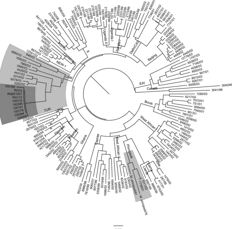
Genotyping for lineage classification.All nine isolates pre-sented SIT863, which is characteristic ofM. pinnipedii(Fig. 1). Upon 24-locus MIRU-VNTR typing, eight isolates presented the same MIRU pattern, while one isolate presented five fewer copies of MIRU21 (see Table S4 in the supplemental material). In a com-parison of these MIRU patterns to those present in the SITVIT2 database, they were clearly different from the patterns that are characteristic ofM. pinnipedii. Using the neighbor-joining-based phylogenetic tree building tool of MIRU-VNTRplus, the patterns of these isolates were organized within the MIRU patterns char-acteristic of LAM9 (Fig. 2). In addition, when constructing a neighbor-joining tree together with 24-locus MIRU patterns from a recent sample set from Brazil (26), the MIRU patterns were closest to those of LAM strains (Fig. 2). Upon analysis of the pres-ence of otherM. tuberculosismarkers characteristic of LAM, their LAM nature was confirmed, and all were TbD1 negative and RD9 positive while not presenting the RDRiogenotype.
Whole-genome sequencing-based typing.The raw sequence reads for RG74, RG112, and RG621257 were aligned to the H37Rv reference sequence, and 869, 877, and 881 SNPs were identified, respectively. Upon excluding SNPs present in proline-glutamate (PE)/proline-proline-glutamate (PPE) genes and other repeat-rich regions, the three isolates shared 816 SNPs, each strain bear-ing between one and five unique SNPs (Fig. 1). In addition, 72 (RG74), 74 (RG112), and 87 (RG621257) short indels (⬍100 bp) were present. According to the 62-SNP barcoding scheme recently proposed by Coll et al. (15), the three strains were classified as belonging to lineage 4.3.3, part of the Euro-American lineage (lin-eage 4), confirming their LAM classification. Whole-genome quencing confirmed the mutations observed by conventional se-quencing of the hot spot region of rpoB, including the 12-nt insertion at genomic position 761123 (WGS codon 435). The iso-late without the insertion did not present an SNP in the rest of rpoB(see Table S4 in the supplemental material). In addition, the absence of a mutation inrpoAandrpoCwas confirmed, except for the presence of a synonymous SNP (GCC to GCG) at position 763370 (amino acid position 542).
DISCUSSION
M. pinnipedii, formally known as the “seal bacillus,” was described in 2003 on the basis of a characterization of isolates ofM. tuber-culosis-like organisms obtained from seals (6) and being the caus-ative agent of TB in this mammal host. Later, the involvement of M. pinnipediiin TB transmission to humans was observed in a European zoo, where sea lion keepers had been infected but did not develop active TB during the time of the study (20). ThisM. pinnipediiisolate sourced from the animals was susceptible to the main antituberculosis drugs, isoniazid and rifampin, and strepto-mycin, ethambutol, andpara-aminosalicylic acid (6).
In the spolDB4 database, the spoligotype profile obtained for the isolates under analysis in the present study corresponded to SIT863, initially described as characteristic of theM. tuberculosisU family (40) and later on redefined and presented by SITVITWEB as PINI2 (17). Isolates with the genotype SIT863 were reported in three earlier studies carried out in Porto Alegre, the first reported by Cafrune et al. (41), who observed six human isolates between 2005 and 2007. Subsequently, Perizzolo et al. (18) identified two isolates between 2004 and 2006, while Khuleis et al. (42) reported five additional isolates in a study carried out between 2007 and 2008. Outside Brazil, there have been reports of a single case in the United States and two more in Venezuela (43,
44). At SITVITWEB, four cases were reported, and three of these were isolated in Pelotas (Rio Grande do Sul, Brazil) but without information about the isolation data; the other case was isolated in the United States in 2003. These four SIT863 strains were classified as PINI2 based on revised rules for PINI, PINI1, and PINI2 (unpublished data). Accordingly, PINI2 (to which SIT863 belongs) is characterized by the obligate pres-ence of spacer 25 only; therefore, the SIT863 pattern (prespres-ence of spacers 25 to 28) is compatible with the PINI2 definition and does not match any other signature in our updated SITVIT2 database. A review of the SITVIT2 database showed the pres-ence of a total of 159 isolates with spoligotypes characteristic of M. pinnipedii(PINI1,n⫽33; PINI2,n⫽105; PINI-like,n⫽7) but with the same number of isolates with the SIT863 pattern as in the earlier version of the database (data not shown). These data FIG 1Venn diagram depicting the number of SNPs shared by the threeM.
suggest that human infection rates forM. pinnipedii might be higher than has been suggested in earlier studies (20).
However, the misclassification of lineages ofM. tuberculosisby spoligotyping has been reported, and it is now widely accepted that this genotyping method does not present the same level of discrimination of M. tuberculosis isolates as that with MIRU-VNTR typing (45). Convergent evolution has been described as relatively common when using the direct repeat (DR) region as a genetic marker, and incorrect classification of lineages by spoligo-typing was recently evidenced in Brazil (26) and more frequently when the deletion of large blocks of spacers occurs, such as causing
misclassification ofM. tuberculosisof the Beijing type (46). There-fore, our observation of human isolates with the SIT863 spoligo-type characteristic of PINI2, together with the lack of notification ofM. pinnipediias a cause of human TB, lead us to believe that we might be dealing with cases of misclassification.
The pattern we observed in the nine isolates was clearly different from that obtained with the twoM. pinnipediicontrol strains, as can be observed inFig. 2, and different from those characteristic of M. pinnipedii in SITVITWEB (256324222321, 216424222322, 256424222321, and 226424253522). In addition, the construction of a neighbor-joining tree using MIRU-VNTRplusdemonstrated that our MIRU-VNTR profile was close to that of LAM genotypes (Fig. 2), and this was confirmed when we compared the genotype with that obtained in a recent study in Rio de Janeiro (data not shown).
The LAM lineage of the isolates was confirmed by the addi-tional genotyping procedures specific for LAM strains, including the presence of IS6110at a particular site of the genome (48). Another marker is present in the polyketide synthase (pks)15/1 locus, reported to be polymorphic among members of the MTBC (49). This genetic region has an intact open reading frame in Indo-Oceanic, East Asian, and East African-Indian lineages. However, the Euro-American lineage contains a typical 7-bp deletion in the pks15/1gene, while other MTBC species, such asM. pinnipedii, contain a 6-bp deletion (31,33,49–51). Presently, a 7-bp insertion instead of the 6-bp insert specific forM. pinnipediiwas observed. A third marker, thefbpCSNP, which changed nucleotide G to A at codon 103 (E103N), differentiates LAM strains from non-LAM strains (52); this also confirmed the LAM nature of the present isolates. Finally, we verified the absence of TbD1, which is charac-teristic ofM. tuberculosis, and the presence of RD9, which is char-acteristic ofM. africanumand MTBC species that infect mostly animals (53). Interestingly, the phenotype of the isolates also cor-roborated them beingM. tuberculosisand notM. pinnipedii, since all were niacin positive, andM. pinnipediiis niacin negative.
While allM. pinnipediiisolates reported so far are susceptible to antibiotics, seven of the nine isolates were MDR; INH resistance was due to S315T inkatGin six cases, while one isolate presented S315T and an additional T-to-G transition at position⫺17 of inhA. Interestingly, in seven of the nine isolates, a duplication of 12 nucleotides was observed in codon 516, which was reported earlier in Porto Alegre by Perizzolo et al. (18), causing the inser-tion of four amino acids. Because isolate RG621257 did not pres-ent a mutantrpoBallele, we aimed to discover if mutations in the rest of the genome and not present in RG74 and RG112 might explain resistance to RIF. We observed some SNPs in genes asso-ciated with efflux pumps and impermeability of cell membranes (data not shown), and whether these mutations are directly asso-ciated with resistance to RIF is under investigation.
Because of the size of this insertion and its possible putative influence on the structure of the beta polymerase unit (unpub-lished data), we expected to observe compensatory mutations in eitherrpoAorrpoC, as described recently inM. tuberculosisstrains (37,54,55). Surprisingly, we did not observe any non-synony-mous mutations in therpoAandrpoCregions covered by our sequencing approach, and this might mean that (i) the duplication does not interfere with the biological function of the polymerase, which seems unlikely due to the size of the duplication; (ii) un-known and undescribed compensatory mutations are present in other regions of the genome; or (iii) this is a recent evolutionary event for which no compensatory mutation has yet been acquired and is fixed in the population that contains the clinical isolate. Interestingly, in a RIF-resistant isolate ofM. smegmatis, an inser-tion of six amino acids was observed at codons 434 and 435 of rpoB, which led to growth attenuation (56).
To classify our strains to the lineage level, confirm their LAM nature also based on the SNP barcode (15), and better understand their similarity on a genetic level, three strains were submitted to whole-genome sequencing. The observed genetic distances be-tween the three isolates whose genomes have been sequenced also point toward recent transmission of this strain. The three isolates were found to be within 5 to 11 SNPs in distance, which strongly indicates an epidemiological link between the patients, as reported by Walker et al. (56); the strongest link was between isolates RG112 and RG621257, with a genome difference of only 5 SNPs. The remaining isolate (RG74), which presented differences of 8 or 11 SNPs from the other strains, falls into the indeterminate inter-val (6 to 12 SNPs) (56) for the delineation of TB outbreaks using WGS data. This demonstrated that independent of the lineage nature of these isolates, they are part of an active transmission chain of MDR-TB in Porto Alegre over a period of several years.
Earlier outbreaks of MDR strains have been reported in the city of Porto Alegre, including a LAM5 strain reported by Perizzolo et al. (18) and a recent MDR-TB LAM2 strain (57), both being of the RDRiogenotype; the nine isolates described here, however, were not of the RDRiogenotype.
This work further reinforces the extreme care needed to be taken when using databases for comparisons of genotype identi-fications of localM. tuberculosisisolates, and it highlights the need to use multiple markers for correct species and/or lineage assigna-tion of isolates with infrequent spoligotypes.
ACKNOWLEDGMENTS
Miguel Viveiros was supported by “Ciências em Fronteiras/Professor Visitante Especial” (no. 88881.064961/2014-01) (Jose R. Lapa e Silva/ UFRJ coordinator) by CAPES/MEC/Brazil. This study was partially sup-ported by a research grant from the European Society of Clinical Micro-biology and Infectious Diseases, for which we would like to acknowledge the Study Group for Mycobacterial Infections; the Fundação para a Ciên-cia e Tecnologia, Portugal (project reference no. PTDC/SAU-EPI/122400/ 2010); a postdoctoral fellowship (SFRH/BPD/95406/2013) awarded to João Perdigão; and by CNPq (441499/2014-7 MCTI/CNPQ/Universal 14/ 2014).
REFERENCES
1.van Soolingen D, van der Zanden AG, de Haas PE, Noordhoek GT, Kiers A, Foudraine NA, Portaels F, Kolk AH, Kremer K, van Embden JD.1998. Diagnosis ofMycobacterium microtiinfections among humans by using novel genetic markers. J Clin Microbiol36:1840 –1845. 2.Niemann S, Richter E, Rüsch-Gerdes S.1999. Biochemical and genetic
evidence for the transfer ofMycobacterium tuberculosissubsp.caprae Aranaz et al. to the speciesMycobacterium bovisKarlson and Lessel 1970 (approved lists 1980) asMycobacterium bovissubsp.capraecomb. nov. Int J Syst Evol Microbiol52:433– 436.http://dx.doi.org/10.1099/00207713-52 -2-433.
3.Garnier T, Eiglmeier K, Camus JC, Medina N, Mansoor H, Pryor M, Duthoy S, Grondin S, Lacroix C, Monsempe C, Simon S, Harris B, Atkin R, Doggett J, Mayes R, Keating L, Wheeler PR, Parkhill J, Barrell BG, Cole ST, Gordon SV, Hewinson RG.2003. The complete genome sequence ofMycobacterium bovis. Proc Natl Acad Sci U S A100:7877– 7882.http://dx.doi.org/10.1073/pnas.1130426100.
4. Cousins DV, Williams SN, Reuter R, Forshaw D, Chadwick B, Coughran D, Collins P, Gales N.1993. Tuberculosis in wild seals and characterization of the seal bacillus. Aust Vet J70:92–97.http://dx.doi.org /10.1111/j.1751-0813.1993.tb03284.x.
6.Cousins DV, Bastida R, Cataldi A, Quse V, Redrobe S, Dow S, Duignan P, Murray A, Dupont C, Ahmed N, Collins DM, Butler WR, Dawson D, Rodríguez D, Loureiro J, Romano MI, Alito A, Zumarraga M, Ber-nardelli A.2003. Tuberculosis in seals caused by a novel member of the Mycobacterium tuberculosiscomplex:Mycobacterium pinnipediisp. nov. Int J Syst Evol Microbiol.53:1305–1314.http://dx.doi.org/10.1099/ijs.0 .02401-0.
7.van Ingen J, Rahim Z, Mulder A, Boeree MJ, Simeone R, Brosch R, van Soolingen D.2012. Characterization ofMycobacterium orygisasM. tuber-culosiscomplex subspecies. Emerg Infect Dis18:653– 655.http://dx.doi .org/10.3201/eid1804.110888.
8.Parsons SD, Drewe JA, Gey van Pittius NC, Warren RM, van Helden PD.2013. Novel cause of tuberculosis in meerkats, South Africa. Emerg Infect Dis19:2004 –2007.http://dx.doi.org/10.3201/eid1912.130268. 9.de Jong BC, Antonio M, Gagneux S.2010.Mycobacterium africanum–
review of an important cause of human tuberculosis in West Africa. PLoS Negl Trop Dis4:e744.http://dx.doi.org/10.1371/journal.pntd.0000744. 10. Van Soolingen D.2001. Molecular epidemiology of tuberculosis and other
mycobacterial infections: main methodologies and achievements. J Intern Med249:1–26.http://dx.doi.org/10.1046/j.1365-2796.2001.00772.x. 11. Gagneux S.2013. Genetic diversity inMycobacterium tuberculosis. Curr
Top Microbiol Immunol 374:1–25. http://dx.doi.org/10.1007/82.2013 .329.
12. Kamerbeek J, Schouls L, Kolk A, van Agterveld M, van Soolingen D, Kuijper S, Bunschoten A, Molhuizen H, Shaw R, Goyal M, van Embden J.1997. Simultaneous detection and strain differentiation of Mycobacte-rium tuberculosisfor diagnosis and epidemiology. J Clin Microbiol35:
907–914.
13. van Embden JD, Cave MD, Crawford JT, Dale JW, Eisenach KD, Gicquel B, Hermans P, Martin C, McAdam R, Shinnick TM, Small PM.
1993. Strain identification ofMycobacterium tuberculosisby DNA finger-printing: recommendations for a standardized methodology. J Clin Mi-crobiol31:406 – 409.
14. Supply P, Mazars E, Lesjean S, Vincent V, Gicquel B, Locht C.2000. Variable human minisatellite-like regions in theMycobacterium tubercu-losis genome. Mol Microbiol 36:762–771. http://dx.doi.org/10.1046/j .1365-2958.2000.01905.x.
15. Coll F, McNerney R, Guerra-Assunção JA, Glynn JR, Perdigão J, Vivei-ros M, Portugal I, Pain A, Martin N, Clark TG.2014. A robust SNP barcode for typingMycobacterium tuberculosiscomplex strains. Nat Com-mun5:4812.http://dx.doi.org/10.1038/ncomms5812.
16. Gagneux S, DeRiemer K, Van T, Kato-Maeda M, de Jong BC, Naray-anan S, Nicol M, Niemann S, Kremer K, Gutierrez MC, Hilty M, Hopewell PC, Small PM.2006. Variable host-pathogen compatibility in Mycobacterium tuberculosis. Proc Natl Acad Sci U S A103:2869 –2873. http://dx.doi.org/10.1073/pnas.0511240103.
17. Demay C, Liens B, Burguière T, Hill V, Couvin D, Millet J, Mokrousov I, Sola C, Zozio T, Rastogi N.2012. SITVITWEB–a publicly available international multimarker database for studyingMycobacterium tubercu-losisgenetic diversity and molecular epidemiology. Infect Genet Evol12:
755–766.http://dx.doi.org/10.1016/j.meegid.2012.02.004.
18. Perizzolo PF, Dalla Costa ER, Ribeiro AW, Spies FS, Ribeiro MO, Dias CF, Unis G, Almeida da Silva P, Gomes HM, Suffys PN, Rossetti ML.
2012. Characteristics of multidrug-resistantMycobacterium tuberculosisin southern Brazil. Tuberculosis (Edinb) 92:56 –59. http://dx.doi.org/10 .1016/j.tube.2011.09.008.
19. Moser I, Prodinger WM, Hotzel H, Greenwald R, Lyashchenko KP, Bakker D, Gomis D, Seidler T, Ellenberger C, Hetzel U, Wuennemann K, Moisson P.2008.Mycobacterium pinnipedii: transmission from South American sea lion (Otaria byronia) to Bactrian camel (Camelus bactrianus bactrianus) and Malayan tapirs (Tapirus indicus). Vet Microbiol127:399 – 406.http://dx.doi.org/10.1016/j.vetmic.2007.08.028.
20. Kiers A, Klarenbeek A, Mendelts B, Van Soolingen D, Koëter G.2008. Transmission ofMycobacterium pinnipediito humans in a zoo with ma-rine mammals. Int J Tuberc Lung Dis12:1469 –1473.
21. Jurczynski K, Scharpegge J, Ley-Zaporozhan J, Ley S, Cracknell J, Lyashchenko K, Greenwald R, Schenk JP.2011. Computed tomographic examination of South American sea lions (Otaria flavescens) with sus-pectedMycobacterium pinnipediiinfection. Vet Rec169:608.http://dx.doi .org/10.1136/vr.100234.
22. Ministério da Saúde Brasil.2011. Manual de recomendações para o controle da tuberculose no Brasil, Secretaria de Vigilância em Saúde/ Programa Nacional de Controle da Tuberculose, Ministério da Saúde
Bra-sil, Brasília, Brazil.http://portalsaude.saude.gov.br/images/pdf/2015/jun ho/30/MANUAL-DE-RECOMENDACOES-PARA-O-CONTROLE-DA-TUBERCULOSE-NO-BRASIL.pdf.
23. Canetti G, Fox W, Khomenko A, Mahler HT, Menon MK, Mitchison DA, Rist N, Šmelev NA.1969. Advances in techniques of testing myco-bacterial drug sensitivity, and the use of sensitivity tests in tuberculosis control programmes. Bull World Health Organ41:21– 43.
24. van Soolingen D, de Haas PE, Hermans PW, van Embden JD.1994. DNA fingerprinting ofMycobacterium tuberculosis. Methods Enzymol
235:196 –205.http://dx.doi.org/10.1016/0076-6879(94)35141-4. 25. Kremer K, van Soolingen D, Frothingham R, Haas WH, Hermans PW,
Martín C, Palittapongarnpim P, Plikaytis BB, Riley LW, Yakrus MA, Musser JM, van Embden JD.1999. Comparison of methods based on different molecular epidemiological markers for typing ofMycobacterium tuberculosiscomplex strains: interlaboratory study of discriminatory power and reproducibility. J Clin Microbiol37:2607–2618.
26. Vasconcellos SE, Acosta CC, Gomes LL, Conceição EC, Lima KV, de Araujo MI, Leite Mde L, Tannure F, Caldas PC, Gomes HM, Santos AR, Gomgnimbou MK, Sola C, Couvin D, Rastogi N, Boechat N, Suffys PN.
2014. Strain classification ofMycobacterium tuberculosisisolates in Brazil based on genotypes obtained by spoligotyping, mycobacterial inter-spersed repetitive unit typing and the presence of large sequence and single nucleotide polymorphism. PLoS One 9:e107747. http://dx.doi.org/10 .1371/journal.pone.0107747.
27. Supply P, Gaudin C, Raze D.2014. Optimization of standard 24-locus variable-number tandem-repeat typing ofMycobacterium tuberculosis iso-lates: a multicenter perspective. J Clin Microbiol52:3518 –3519.http://dx .doi.org/10.1128/JCM.01790-14.
28. Weniger T, Krawczyk J, Supply P, Niemann S, Harmsen D. 2010. MIRU-VNTRplus: a Web tool for polyphasic genotyping of Mycobacte-rium tuberculosiscomplex bacteria. Nucleic Acids Res38:W326 –W331. http://dx.doi.org/10.1093/nar/gkq351.
29. Sampson SL, Warren RM, Richardson M, van der Spuy GD, van Helden PD.1999. Disruption of coding regions by IS6110insertion in Mycobacterium tuberculosis. Tuber Lung Dis79:349 –359.http://dx.doi .org/10.1054/tuld.1999.0218.
30. Lazzarini LCO, Huard RC, Boechat NL, Gomes HM, Oelemann MC, Kurepina N, Shashkina E, Mello FC, Gibson AL, Virginio MJ, Marsico AG, Butler WR, Kreiswirth BN, Suffys PN, Lapa e Silva JR, Ho JL.2007. Discovery of a novelMycobacterium tuberculosislineage that is a major cause of tuberculosis in Rio de Janeiro, Brazil. J Clin Microbiol45:3891– 3902.http://dx.doi.org/10.1128/JCM.01394-07.
31. Vasconcellos SE, Huard RC, Niemann S, Kremer K, Santos AR, Suffys PN, Ho JL.2010. Distinct genotypic profiles of the two major clades of Mycobacterium africanum. BMC Infect Dis10:80.http://dx.doi.org/10 .1186/1471-2334-10-80.
32. Huard RC, Lazzarini LCO, Butler W, van Soolingen D, Ho JL.2003. PCR-based method to differentiate the subspecies of theMycobacterium tuberculosiscomplex on the basis of genomic deletions. J Clin Microbiol
41:1637–1650.http://dx.doi.org/10.1128/JCM.41.4.1637-1650.2003. 33. Huard RC, Fabre M, de Haas P, Lazzarini LCO, van Soolingen D,
Cousins D, Ho JL.2006. Novel genetic polymorphisms that further de-lineate the phylogeny of theMycobacterium tuberculosiscomplex. J Bacte-riol188:4271– 4287.http://dx.doi.org/10.1128/JB.01783-05.
34. Ramasoota P, Pitaksajjakul P, Phatihattakorn W, Pransujarit V, Boonyasopun J.2006. Mutations in therpoBgene of rifampicin-resistant Mycobacterium tuberculosisstrains from Thailand and its evolutionary im-plication. Southeast Asian J Trop Med Public Health37:136 –147. 35. de Oliveira MM, da Silva Rocha A, Cardoso Oelemann M, Gomes HM,
Fonseca L, Werneck-Barreto AM, Valim AM, Rossetti ML, Rossau R, Mijs W, Vanderborght B, Suffys P.2003. Rapid detection of resistance against rifampicin in isolates ofMycobacterium tuberculosisfrom Brazilian patients using a reverse-phase hybridization assay. J Microbiol Methods
53:335–342.
36. Dalla Costa ER, Ribeiro MO, Silva MSN, Arnold LS, Rostirolla DC, Cafrune PI, Espinoza RC, Palaci M, Telles MA, Ritacco V, Suffys PN, Lopes ML, Campelo CL, Miranda SS, Kremer K, da Silva PEA, Fonseca LS, Ho JL, Kritski AL, Rossetti MLR.2009. Correlations of mutations in katG,oxyR-ahpCandinhAgenes andin vitrosusceptibility in Mycobacte-rium tuberculosisclinical strains segregated by spoligotype families from tuberculosis prevalent countries in South America. BMC Microbiol9:39. http://dx.doi.org/10.1186/1471-2180-9-39.
RM, Gagneux S, Victor TC.2012. Putative compensatory mutations in therpoCgene of rifampicin-resistantMycobacterium tuberculosisare associated with ongoing transmission. Antimicrob Agents Chemother
57:827– 832.
38. Li H, Durbin R.2009. Fast and accurate short read alignment with Bur-rows-Wheeler transform. Bioinformatics 25:1754 –1760. http://dx.doi .org/10.1093/bioinformatics/btp698.
39. Li H, Handsaker B, Wysoker A, Fennell T, Ruan J, Homer N, Marth G, Abecasis G, Durbin R, 1000 Genome Project Data Processing Subgroup.
2009. The Sequence Alignment/Map format and SAMtools. Bioinformatics
25:2078 –2079.http://dx.doi.org/10.1093/bioinformatics/btp352.
40. Brudey K, Driscoll JR, Rigouts L, Prodinger WM, Gori A, Al-Hajoj SA, Allix C, Aristimuño L, Arora J, Baumanis V, Binder L, Cafrune P, Cataldi A, Cheong S, Diel R, Ellermeier C, Evans JT, Fauville-Dufaux M, Ferdinand S, Garcia de Viedma D, Garzelli C, Gazzola L, Gomes HM, Guttierez MC, Hawkey PM, van Helden PD, Kadival GV, Kre-iswirth BN, Kremer K, Kubin M, Kulkarni SP, Liens B, Lillebaek T, Ho ML, Martin C, Martin C, Mokrousov I, Narvskaïa O, Ngeow YF, Naumann L, Niemann S, Parwati I, Rahim Z, Rasolofo-Razanamparany V, Rasolonavalona T, Rossetti ML, Rüsch-Gerdes S, Sajduda A, Samper S, Shemyakin IG, et al.2006.Mycobacterium tuberculosiscomplex genetic diversity: mining the fourth international spoligotyping database (SpolDB4) for classification, population genetics and epidemiology. BMC Microbiol6:23.http://dx.doi.org/10.1186/1471-2180-6-23.
41. Cafrune PI, Possuelo LG, Ribeiro AW, Ribeiro MO, Unis G, Jarczewski CA, Rossetti ML, Zaha A.2009. Prospective study applying spoligotyping directly to DNA from sputum samples of patients suspected of having tuberculosis. Can J Microbiol55:895–900.http://dx.doi.org/10.1139/W09 -033.
42. Kuhleis D, Ribeiro AW, Costa ER, Cafrune PI, Schmid KB, Costa LL, Ribeiro MO, Zaha A, Rossetti ML.2012. Tuberculosis in a southern Brazilian prison. Mem Inst Oswaldo Cruz107:909 –915.http://dx.doi.org /10.1590/S0074-02762012000700012.
43. Borsuk S, Dellagostin MM, Madeira Sde G, Lima C, Boffo M, Mattos I, Almeida da Silva PE, Dellagostin OA.2005. Molecular characterization ofMycobacterium tuberculosisisolates in a region of Brazil with a high incidence of tuberculosis. Microbes Infect7:1338 –1344.http://dx.doi.org /10.1016/j.micinf.2005.05.009.
44. Sequera CM, Delgado SV, Araque MW, Torrealba OM, Núñez MR, da Mato JO, Abadia PE, Takiff H, de Waard J. 2008. Mycobacterium tuberculosis: espoligotipos en el Estado Carabobo, Venezuela. Rev Chi In-fect25:362–367. doi.org/10.4067/S0716-10182008000500009.
45. Gagneux S, Deriemer K, Van T, Kato-Maeda M, de Jong BC, Naray-anan S, Nicol M, Niemann S, Kremer K, Gutierrez MC, Hilty M, Hopewell PC, Small PM.2006. Variable host-pathogen compatibility in Mycobacterium tuberculosis. Proc Natl Acad Sci U S A103:2869 –2873. http://dx.doi.org/10.1073/pnas.0511240103.
46. Fenner L, Malla B, Ninet B, Dubuis O, Stucki D, Borrel Sonia Borrel Huna T, Bodmer T, Egger M, Gagneux S. 2011. ‘‘Pseudo-Beijing’’: evidence for convergent evolution in the direct repeat region of
Mycobac-terium tuberculosis. PLoS One 6:e24737. http://dx.doi.org/10.1371
/journal.pone.0024737.
47. Oelemann MC, Diel R, Vatin V, Haas W, Rüsch-Gerdes S, Locht C, Niemann S, Supply P.2007. Assessment of an optimized mycobacterial interspersed repetitive-unit–variable-number tandem-repeat typing sys-tem combined with spoligotyping for population-based molecular epide-miology studies of tuberculosis. J Clin Microbiol45:691– 697.http://dx .doi.org/10.1128/JCM.01393-06.
48. Gagneux S, Small PM.2007. Global phylogeography ofMycobacterium tuberculosisand implications for tuberculosis product development. Lancet Infect Dis7:328 –337.http://dx.doi.org/10.1016/S1473-3099(07)70108-1. 49. Constant P, Perez E, Malaga W, Lanéelle MA, Saurel O, Daffé M,
Guilhot C.2002. Role of thepks15/1gene in the biosynthesis of phenolg-lycolipids in theMycobacterium tuberculosiscomplex. Evidence that all strains synthesize glycosylatedp-hydroxybenzoic methyl esters and that strain devoid of phenolglycolipids harbor a frameshift mutation in the pks15/1gene. J Biol Chem277:38148 –38158.http://dx.doi.org/10.1074 /jbc.M206538200.
50. Reed MB, Domenech P, Manca C, Su H, Barczak AK, Kreiswirth BN, Kaplan G, Barry CE, III.2004. A glycolipid of hypervirulent tuberculosis strains that inhibits the innate immune response. Nature431:84 – 87.http: //dx.doi.org/10.1038/nature02837.
51. Gibson AL, Huard RC, Gey van Pittius NC, Lazzarini LC, Driscoll J, Kurepina N, Zozio T, Sola C, Spindola SM, Kritski AL, Fitzgerald D, Kremer K, Mardassi H, Chitale P, Brinkworth J, Garcia de Viedma D, Gicquel B, Pape JW, van Soolingen D, Kreiswirth BN, Warren RM, van Helden PD, Rastogi N, Suffys PN, Lapa e Silva J, Ho JL.2008. Appli-cation of sensitive and specific molecular methods to uncover global dis-semination of the major RDRio
sublineage of the Latin American-MediterraneanMycobacterium tuberculosisspoligotype family. J Clin Microbiol46:1259 –1267.http://dx.doi.org/10.1128/JCM.02231-07. 52. Comas I, Borrell S, Roetzer A, Rose G, Malla B, Kato-Maeda M,
Galagan J, Niemann S, Gagneux S.2012. Whole-genome sequencing of rifampicin-resistantMycobacterium tuberculosisstrains identifies com-pensatory mutations in RNA polymerase genes. Nat Genet44:106 –110. http://dx.doi.org/10.1038/ng.1038.
53. Brosch R, Gordon SV, Marmiesse M, Brodin P, Buchrieser C, Eiglmeier K, Garnier T, Gutierrez C, Hewinson G, Kremer K, Parsons LM, Pym AS, Samper S, van Soolingen D, Cole ST.2002. A new evolutionary scenario for theMycobacterium tuberculosiscomplex. Proc Natl Acad Sci U S A99:3684 –3689.http://dx.doi.org/10.1073/pnas.052548299. 54. Perdigão J, Silva H, Machado D, Macedo R, Maltez F, Silva C, Jordao
L, Couto I, Mallard K, Coll F, Hill-Cawthorne GA, McNerney R, Pain A, Clark TG, Viveiros M, Portugal I.2014. UnravelingMycobacterium tuberculosisgenomic diversity and evolution in Lisbon, Portugal, a highly drug resistant setting. BMC Genomics15:991.http://dx.doi.org/10.1186 /1471-2164-15-991.
55. Malshetty V, Kurthkoti K, China A, Mallick B, Yamunadevi S, Sang PB, Srinivasan N, Nagaraja V, Varshney U.2010. Novel insertion and dele-tion mutants of RpoB that renderMycobacterium smegmatisRNA poly-merase resistant to rifampicin-mediated inhibition of transcription. Mi-crobiology156:1565–1573.http://dx.doi.org/10.1099/mic.0.036970-0. 56. Walker TM, Ip CL, Harrell RH, Evans JT, Kapatai G, Dedicoat MJ, Eyre
DW, Wilson DJ, Hawkey PM, Crook DW, Parkhill J, Harris D, Walker AS, Bowden R, Monk P, Smith EG, Peto TE. 2013. Whole-genome sequencing to delineateMycobacterium tuberculosisoutbreaks: a retro-spective observational study. Lancet Infect Dis13:137–146.http://dx.doi .org/10.1016/S1473-3099(12)70277-3.
57. Dalla Costa ER, Lazzarini LC, Perizzolo PF, Díaz CA, Spies FS, Costa LL, Ribeiro AW, Barroco C, Schuh SJ, da Silva Pereira MA, Dias CF, Gomes HM, Unis G, Zaha A, Almeida da Silva PE, Suffys PN, Rossetti ML.2013.Mycobacterium tuberculosisof the RDRiogenotype is the